257
Horner syndrome was observed to be associated with lesions at the lower cervical level2). In general, motor weakness predomi-nates over sensory abnormalities6). Although symptoms mani-fest in a progressive manner, remission and fluctuation have been reported in 30% of cases2). Also, exacerbation of symp-toms due to the Valsalva maneuver and the head-up position has been observed6).
We have encountered an unusual presentation of an extradu-ral arachnoid cyst located in the retroperitoneal space. The pa-tient complained of abdominal discomfort without any evi-dence of neurological compromise. The lesion was placed in retroperitoneal space without compression of neither spinal thecal sac nor nerve root.
CASE REPORT
A 26-year-old man presented with abdominal discomfort mainly over the right lower quadrant for 4 months. Although the patient did not have any significant medical or surgical past history, L4 hemivertebra had been incidentally found during a
INTRODUCTION
Extradural arachnoid cysts are uncommon enlarging lesions of the spinal cord which originate from small diverticula of the subarachnoid space3). Although termed “arachnoid cysts”, the inner arachnoid lining has been shown to be frequently absent, and the term has been used interchangeably with “extradural meningeal cyst”9). Epidemiologically, these lesions demonstrate a male predominance and a peak incidence in the second de-cade of life1). These cysts occur most frequently in the mid to lower thoracic region, followed by the lumbosacral and thora-columbar regions. Cervical extradural arachnoid cysts are rela-tively rare6).
Being a space-occupying lesion, the clinical presentation of an extradural arachnoid cyst is usually due to spinal cord and/ or nerve root compression. Patients with thoracic cysts tend to suffer from spastic or flaccid paraparesis, while patients with lumbar and lumbosacral cysts complain of lower back pain, ra-diculopathy, or dysfunction of bladder and bowel2). Cervical cysts have been reported to cause spastic quadriparesis, and the
Retroperitoneal Spinal Extradural Arachnoid Cyst
Combined with Congenital Hemivertebrae
Se-Hwan Park, M.D.,1 Sung-Uk Kuh, M.D., Ph.D.,1 Beom Jin Lim, M.D., Ph.D.2
Department of Neurosurgery,1 Yonsei University College of Medicine, Spine and Spinal Cord Institute, Gangnam Severance Hospital, Seoul, Korea Department of Pathology,2 Yonsei University College of Medicine, Seoul, Korea
Spinal extradural arachnoid cysts usually cause symptoms related to spinal cord or nerve root compression. Here, we report an atypical presenta-tion of a spinal extradural arachnoid cyst combined with congenital hemivertebra which was presented as a retroperitoneal mass that exerted mass effects to the abdominal organs. On image studies, the communication between the cystic pedicle and the spinal arachnoid space was indistinct. Based on our experience and the literature of the pathogenesis, we planned anterior approach for removal of the arachnoid cyst in order to focus on mass removal rather than ligation of the fistulous channel. In our estimation this was feasible considering radiologic findings and also essential for the symptom relief. The cyst was totally removed with the clogged ‘thecal sac-side’ end of the cystic pedicle. The patient was free of abdominal dis-comfort by one month after the surgery.
Key Words : Abdominal discomfort · Cystic pedicle ligation · Extradural arachnoid cyst · Retroperitoneal mass.
Case Report
•Received : January 11, 2012 •Revised : July 25, 2012 •Accepted : August 29, 2012 •Address for reprints : Sung-Uk Kuh, M.D., Ph.D.
Department of Neurosurgery, Yonsei University College of Medicine, Spine and Spinal Cord Institute, Gangnam Severance Hospital, 211 Eonju-ro, Gangnam-gu, Seoul 135-720, Korea
Tel : +82-2-2019-3404, Fax : +82-2-3461-9229, E-mail : [email protected]
•This is an Open Access article distributed under the terms of the Creative Commons Attribution Non-Commercial License (http://creativecommons.org/licenses/by-nc/3.0)
which permits unrestricted non-commercial use, distribution, and reproduction in any medium, provided the original work is properly cited.
J Korean Neurosurg Soc 52 : 257-260, 2012
http://dx.doi.org/10.3340/jkns.2012.52.3.257
Copyright © 2012 The Korean Neurosurgical Society
PrintISSN 2005-3711 On-line ISSN 1598-7876
www.jkns.or.kr
258 J Korean Neurosurg Soc 52 | September 2012
the cystic pedicle was cone-shaped and appeared to be clogged (Fig. 3).
Although in usual circumstances removal of a spinal extradu-ral arachnoid cyst focuses on exploration and ligation of the fis-tulous channel1), a direct anterior retroperitoneal approach to the cyst was planned in this case, considering that removal of the main mass in the retroperitoneal space was essential for symptom relief, and that the communication between the cyst and the spinal subarachnoid space was unlikely. This decision was based on our experience with spinal extradural arachnoid cysts without identifiable communication points on imaging, in which detachment from the spinal cord was feasible and did not require ligation of the cystic origin. The literature also sup-ported this concept of noncommunicating spinal extradural arachnoid cysts5,12).
The patient was placed in a “three-quarter lateral” position, and an incision was made along the lateral margin of the rectus abdominis muscle. After retraction of peritoneum and dissec-minor fall-off event in his teenage years (Fig. 1). He denied any
symptom related to L4 hemivertebra including lower back pain and never had sought any treatment for such abnormality. In the examination, he did not complain any pain in his leg and lower back or any difficulty in defecation, voiding, and walking. We could not find any deficit in the neurologic examination, either.
Symptoms of the patient were refractory to common diges-tive medicines and gastrointestinal motility modulating drugs. During the course of treatment, he underwent a series of stud-ies, and a large cystic mass was first noted on abdominal sonog-raphy. For further diagnosis, he underwent abdominal comput-ed tomography (CT) and lumbar spine magnetic resonance imaging (MRI), in turn. A large (6.2×4.0×8.0 cm) cystic mass originating from the right L4 spinal nerve root sleeve and ex-tending to the prevertebral space of L2, L3, and L4 through the associated bony defect in the L4 vertebral body was revealed (Fig. 2). MRI demonstrated a homogenous signal, hypointense on T1-weighted imaging and hyperintense on T2-weighted im-aging that was not enhanced by gadolinium contrast media. On conventional lumbar MRI and magnetic resonance (MR) my-elography, a long pedicle of the cyst was identified, but the com-munication between the cystic space and the spinal subarach-noid space was indistinct. In addition, the ‘thecal sac-side’ end of Fig. 1. Lumbar radiographs. A : Antero-posterior view showing L4
hemi-vertebra. B : Lateral view showing mild spondylolisthesis L4 on L5. B A
Fig. 2. Magnestic resonance myelography. A : Three dimensional view
showing large cystic mass with a pedicle to the spinal thecal sac. B : Coronal view showing the end of cystic pedicle which is clogged.
B A
Fig. 3. T2-weighted magnetic resonance images. A : Sagittal view,
im-ages are in medial-to-lateral order. B : Axial view, imim-ages are in foot-to-head order going clockwise from the left upper image. Note the ‘cone-shaped’ cystic pedicle.
A
259
Retroperitoneal Spinal Extradural Arachnoid Cyst | SH Park, et al.
posed considering xanthochromia of the cystic contents. The osmotic pressure of xanthochromic fluid is higher than that of tissue fluid3). More feasible explanation comes from the pulsa-tile nature of CSF. Intrathecal CSF dynamics change greatly by elevation of intra-abdominal pressure and this is far more influ-ential than pressure change during the respiratory cycle. The change in intra-abdominal pressure result in enlargement of the cystic sac and persistent CSF pulsation cause continuous growth of the cyst under the law of Leplace11). One-way valvular mech-anism was followed in order to complement the relationship between imposed hydrostatic pressure and continuous cystic growth. Folds of meninges at the ostium of the cyst can act as a flap-like one-way valve, or rather slit-like communication with the subarachnoid space results in one-way valve11). More recent-ly, a ‘ball-valve’ theory was proposed to explain the valvular mechanism. When intrathecal pressure surges, the spinal arach-noid space is communicated with the cystic space and fluid flows tion through the retroperitoneal fat, the right kidney was noted.
The cyst was identified below the right kidney and was placed on the surface of psoas major muscle (Fig. 4). The cyst was dis-placing the inferior vena cava ventrally. For convenient dissec-tion, internal decompression was performed; the cyst contained a clear liquid. The pedicle of the cyst originated from the inter-vertebral foramen, and the remaining cystic pedicle could be removed en bloc by undermining into the intervertebral fora-men and gently drawing it. After the removal was completed we explored the cystic pedicle of the specimen with a blunt probe and observed that the ‘thecal sac-side’ end was clogged (Fig. 5). The final tissue pathology diagnosis was reported to be consis-tent with an arachnoid cyst (Fig. 6).
During the postoperative period, the patient did not complain of any symptoms other than postoperative pain. Flatus was passed on the first postoperative day, and the patient was dis-charged on the sixth postoperative day without any problem. A postoperative lumbar MRI was obtained one month after sur-gery and demonstrated no residual cyst and intact thecal sac without evidence of cerebrospinal fluid (CSF) leakage (Fig. 7). After the subsidence of postoperative pain, the patient was free of any abdominal discomfort.
DISCUSSION
Although the pathogenesis of spinal extradural arachnoid cysts has not yet been clarified, extradural arachnoid cysts are thought to be diverticula of the arachnoid membrane due to a dural defect which can be either congenital5) or acquired fol-lowing events such as spinal surgery, trauma, infection6,11), or percutaneous procedure7). The location of diverticula is known to be most commonly occurring at the junction of the theca and the nerve root sleeve followed by dorsal midline and the nerve root sleeve itself11).
At first the cysts must be merely small diverticula of arach-noid space and should get enlarged to cause any symptom. Sev-eral mechanisms have been postulated in order to explain the progressive nature of spinal extradural arachnoid cysts. Active fluid secretion from the lining cells of the cyst was proposed. But this could not explain observation that secretory cells were frequently absent in the lining3). Osmosis of water was also
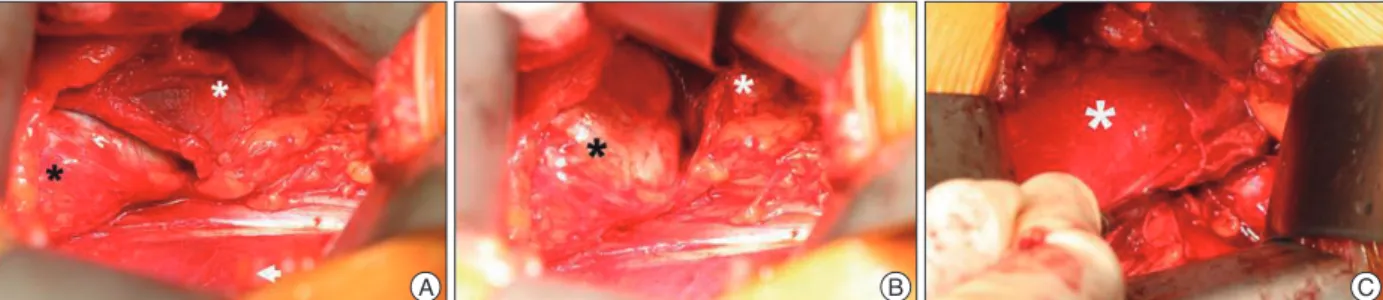
pro-A B C
Fig. 4. Intraoperative view. A : Right kidney (white asterisk) and the arachnoid cyst (black asterisk) were exposed after the peritoneum (white arrow
head) was retracted. B : Exposure of the arachnoid cyst after further retraction of right kidney. C : The arachnoid cyst was removed and the blunt dis-sector indicated the origin of the arachnoid cyst.
Fig. 5. The specimen. Note that the ‘thecal sac-side’ end was clogged.
Fig. 6. Histopathological examination reveals a cyst composed of
thick-ened arachnoid membrane with attenuated epithelial cells (Hematoxylin and Eosin, A : ×40, B : ×100, respectively).
B A
260 J Korean Neurosurg Soc 52 | September 2012
tectable on imaging studies, the proba-bility of communication disruption is high, so that, when deciding on the surgical plan, it is possible to disregard ligation of a fistulous channel in the cystic pedicle. In the current case, com-munication of the cystic pedicle with the spinal subarachnoid space was not detected on MR myelography, and the cystic pedicle was confirmed to be clogged.
CONCLUSION
This is an atypical presentation of a spinal extradural arach-noid cyst which extended into the retroperitoneal space, and was combined with a congenital vertebral malformation. The cystic pedicle was not communicating with the spinal arach-noid space on image study and the cyst was removed in en bloc manner with clogged ‘thecal sac-side’ end of the cystic pedicle by an anterior approach. This case supports the hypothesis that, when the cystic pedicle is not identified on image study, remov-al of the cyst without ligating the fistulous channel can be possi-bly done.
References
1. Choi JY, Kim SH, Lee WS, Sung KH : Spinal extradural arachnoid cyst.
Acta Neurochir (Wien) 148 : 579-585; discussion 585, 2006
2. Cloward RB : Congenital spinal extradural cysts : case report with re-view of literature. Ann Surg 168 : 851-864, 1968
3. Gortvai P : Extradural cysts of the spinal canal. J Neurol Neurosurg
Psychiatry 26 : 223-230, 1963
4. Kulkarni AG, Goel A, Thiruppathy SP, Desai K : Extradural arachnoid cysts : a study of seven cases. Br J Neurosurg 18 : 484-488, 2004 5. Lake PA, Minckler J, Scanlan RL : Spinal epidural cyst : theories of
pathogenesis. Case report. J Neurosurg 40 : 774-778, 1974
6. Liu JK, Cole CD, Kan P, Schmidt MH : Spinal extradural arachnoid cysts : clinical, radiological, and surgical features. Neurosurg Focus 22 : E6, 2007
7. Mao HQ, Yang HL, Geng DC, Bao ZH, Tang TS : Spinal extradural arachnoid cyst following percutaneous vertebroplasty. Eur Spine J 20
Suppl 2 : S206-S210, 2011
8. McCrum C, Williams B : Spinal extradural arachnoid pouches. Report of two cases. J Neurosurg 57 : 849-852, 1982
9. Nabors MW, Pait TG, Byrd EB, Karim NO, Davis DO, Kobrine AI, et al. : Updated assessment and current classification of spinal meningeal cysts.
J Neurosurg 68 : 366-377, 1988
10. Neo M, Koyama T, Sakamoto T, Fujibayashi S, Nakamura T : Detection of a dural defect by cinematic magnetic resonance imaging and its selec-tive closure as a treatment for a spinal extradural arachnoid cyst. Spine
(Phila Pa 1976) 29 : E426-E430, 2004
11. Rohrer DC, Burchiel KJ, Gruber DP : Intraspinal extradural meningeal cyst demonstrating ball-valve mechanism of formation. Case report. J
Neurosurg 78 : 122-125, 1993
12. Sakellaridis N, Panagopoulos D, Mahera H : Sacral epidural noncom-municating arachnoid cyst. Case report and review of the literature. J
Neurosurg Spine 6 : 473-478, 2007 into the cyst. As intrathecal pressure goes down, the cyst body
exerts a force to impede the cystic pedicle following the law of Leplace as the cyst has the larger radius and therefore has the greater wall tension than the cystic pedicle. This is actually a two-way system of unequal flow, however, CSF is trapped in the cystic space6,11).
Various imaging modalities have been used to identify tissue communication points; however, CT myelography using water-soluble contrast media has been the study of choice for illumi-nating the location of communication points between the cyst and the spinal thecal sac6). More recently, there has been a re-port using cinematic MRI for detecting dural defects10). There have been cases in which the cyst was not filtrated by contrast media, or the pedicle was unidentifiable6). In our case, the pa-tient has brought MR myelography from the referring hospital and refused additional CT myelography. The cystic pedicle and its communication with the spinal arachnoid space can be de-termined using MRI and MR myelography6).
To date, removal of extradural arachnoid cyst has focused on obliterating the fistulous channel, that is, the pedicle1). Choi et al.1) reported a case of spinal extradural arachnoid cyst which was removed following ligation of the fistulous channel, and re-viewed 17 additional cases of spinal extradural arachnoid cyst which was either excised or ligated in a similar manner. Kulkar-ni et al.4) reported that, in their 7 cases of spinal extradural arach-noid cysts, they could not identify any connection between the cyst and the spinal arachnoid space in the operative field. In contrast, Cloward2) reported that communication was verified in 43 of 92 cases of congenital spinal extradural arachnoid cysts. Although there was no identifiable communication with the spinal arachnoid space, the characteristics of the cyst contents match that of CSF in the majority of cases4). McCrum and Wil-liams8) explained the formation of these cysts without commu-nication using osmosis or an active secretion mechanism previ-ously mentioned.
It has been proposed that the communication between extra-dural arachnoid cysts and the subarachnoid space gradually de-creases as the pressure gradient fades and eventually becomes nonexistent5). In other words, spinal extradural arachnoid cysts may or may not communicate with the spinal subarachnoid space depending on the stage of evolution. What can be duced from this theory is that, if the communication is not
de-Fig. 7. Postoperative T2-weighted magnetic resonance images. Sagittal view, axial view and