Vijay P Joshi et al.
202 Asian Spine J 2014;8(2):202-205
Copyright Ⓒ 2014 by Korean Society of Spine Surgery
This is an Open Access article distributed under the terms of the Creative Commons Attribution Non-Commercial License (http://creativecommons.org/licenses/by-nc/3.0/) which permits unrestricted non-commercial use, distribution, and reproduction in any medium, provided the original work is properly cited.
Asian Spine Journal • pISSN 1976-1902 eISSN 1976-7846 • www.asianspinejournal.org
Received Dec 4, 2012; Revised Aug 4, 2013; Accepted Aug 8, 201 Corresponding author: Amit Agrawal
Department of Neurosurgery, Narayna Medical College Hospital, Chinthareddypalem, Nellore-524003, Andhra Pradesh, India Tel: +91-8096410032, E-mail: [email protected]
Cervical Perineural Cyst Masquerading as a Cervical Spinal Tumor
Vijay P Joshi
1, Atul Zanwar
2, Anuradha Karande
3, Amit Agrawal
41Department of Neurosurgery, Ashwini Sahakari Rugnalaya and research center, Solapur, Maharashtra, India
2Ashwini Sahakari Rugnalaya and Research Center, Solapur, Maharashtra, India
3Department of Anesthesia, Ashwini Sahakari Rugnalaya and research center, Solapur, Maharashtra, India
4Department of Neurosurgery, Narayna Medical College Hospital, Nellore, India
Tarlov (perineural) cysts of the nerve roots are common and usually incidental findings during magnetic resonance imaging of the lum- bosacral spine. There are only a few case reports where cervical symptomatic perineural cysts have been described in the literature.
We report such a case where a high cervical perineural cyst was masquerading as a cervical spinal tumor.
Keywords: Tarlov’s cysts; Perineural cyst; Spinal cyst; spine; Cervical spine
Case Report Asian Spine J 2014;8(2):202-205 • http://dx.doi.org/10.4184/asj.2014.8.2.202
ASJ A SJ
Asian Spine Journal Asian Spine Journal
Introduction
Tarlov (perineural) cysts of the nerve roots were first de- scribed in 1938 [1] and are common and usually inciden- tal findings during magnetic resonance imaging of the lumbosacral spine [2-4]. There are only a few case reports where cervical symptomatic perineural cysts have been described in the literature [5-7]. We report such a case where a high cervical perineural cyst was masquerading as a cervical spinal tumor.
Case Report
A male patient presented with mild ill-defined neck pain and progressive weakness of all four limbs of six months duration. He had difficulty in walking and holding objects in both upper limbs. There was no history of headache, cough, difficulty in speech or difficulty in swallowing.
Bowel and bladder functions were normal. His general
and systemic examination was unremarkable. Higher mental functions and cranial nerves were normal. There was increased tone in the upper and lower limbs. Motor power was grade 4+/5 in all groups. There was weakness of bilateral hand grip. Planters were bilateral extensor.
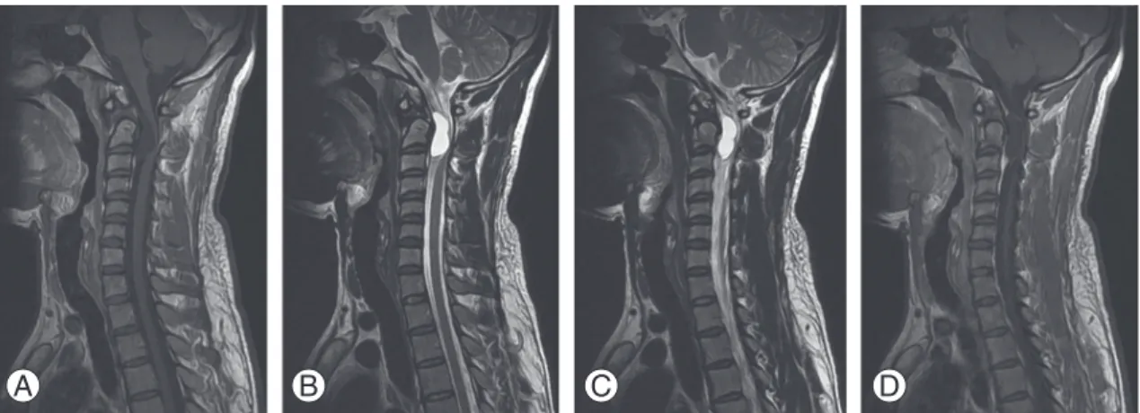
Magnetic resonance imaging (MRI) of the cervical spine revealed a large well-defined cystic lesion pushing the cord to the right at the C1−C2 level, that was hypointense on T1W images and hyperintense on T2W and fluid- attenuated inversion recovery sequence images with pe- ripheral enhancement after contrast administration (Figs.
1, 2). Based on the imaging findings, a diagnosis of cystic schwannoma was suspected. The patient underwent C−
C2 laminectomy. The lesion was located near the dorsal root ganglion of C2; once the lesion was opened, clear fluid came out of the lesion. The cyst could be excised completely while keeping the nerve root intact. Histo- pathological examination revealed inflamed layers of meninges and the presence of neural elements (Fig. 3).
Cervical perineural cyst
Asian Spine Journal
Asian Spine Journal
203Copyright Ⓒ 2014 by Korean Society of Spine Surgery
This is an Open Access article distributed under the terms of the Creative Commons Attribution Non-Commercial License (http://creativecommons.org/licenses/by-nc/3.0/) which permits unrestricted non-commercial use, distribution, and reproduction in any medium, provided the original work is properly cited.
Asian Spine Journal • pISSN 1976-1902 eISSN 1976-7846 • www.asianspinejournal.org
The patient improved after surgery and was doing well at follow-up.
Discussion
Tarlov cysts (perineurial cysts) are defined as cysts formed within the nerve root sheath at the dorsal root ganglion [8], and most of them are clinically insignificant [1,7,9]. The estimated incidence is approximately 5%
(symptomatic cases are rare, constituting less than 1%
of the total) [10], and on MRI it has been estimated that Tarlov cysts are present in 4.6% to 9% of the population, with an estimated 10% becoming symptomatic at some point during life [10]. Commonly Tarlov cysts are found
in the lumbo-sacral region [1,4,7,9], with the S2/S3 nerve roots most commonly affected [4,7]. The exact etiology of perineurial cysts remains unclear. It was proposed that hemosiderin deposition caused by blockage of the venous drainage of the perineurium and epineurium after local trauma can lead to the development of these cysts [11], or that congenital arachnoid proliferation along the exit- ing nerve roots can result in the formation of perineurial cysts [12], and it has also been suggested that the ball- valve mechanism is responsible for the entry of cerebro- spinal fluid into the cyst during systolic pulsations but that the cerebrospinal fluid is unable to exit through the same portal during diastole [7,9,11]. A histological char- acteristic of the Tarlov cyst is the presence of nerve fibers
Fig. 1. Magnetic resonance imaging of the cervical spine axial T2 weighted images showing a large cystic lesion pushing the cord to the right. (A) T2 weighted and (B) fluid-attenuated inversion Recovery sequence images.
A B
Fig. 2. Magnetic resonance imaging cervical spine sagittal images showing an isointense lesion on the T1W image (A), becoming hyperintense on T2W (B) and fluid-attenuated inversion recovery sequence images (C), with peripheral rim of enhancement after contrast administration (D).
A B C D
Vijay P Joshi et al.
204 Asian Spine J 2014;8(2):202-205
in the cyst wall [1,7-9]. Symptomatic Tarlov cysts are rare and clinical symptoms depend on the location of the cyst;
symptoms range from backache, perineal pain or sciatica to overt cauda equina syndrome [4,9]. The symptoms are mostly exacerbated by maneuvers that elevate the intra- spinal cerebrospinal fluid pressure, including coughing, walking, change of posture, and the Valsalva maneuver [13]. In the present case, the patient had features of com- pressive cervical myelopathy because of the location of the cyst. MRI is an effective way to investigate these le- sions as it will provide better details such as showing the extent of the lesion and its relationship to surrounding structures [3,6,10]. For symptomatic cases microsurgical excision of the cysts is curative and has a good outcome
[3,4,7]. In the present case, because of the rarity of the lesion, we did not suspect a Tarlov cyst at first; however, the complete microsurgical excision resulted in a good outcome.
Conflict of Interest
No potential conflict of interest relevant to this article was reported.
References
1. Tarlov IM. Perineurial cysts of the spinal nerve roots.
Arch Neurol Psychiatry 1938;40:1067-74.
Fig. 3. (A) Section shows a cyst wall lined by flattened to cuboidal lining epithelium. The subepithelial tissue shows a nerve bundle and fibrocollagenous tissue with congested blood vessels (H&E, ×50). (B) Section shows a cyst wall lined by flattened to cuboidal lining epithelium which is thrown into papillae in one foci. The subepithelial tissue shows a nerve bundle and fibrocol- lagenous tissue with congested blood vessels (H&E, ×50). (C) Section shows a cyst wall lined by flattened to cuboidal lining epi- thelium which is thrown into papillae in one focus. The subepithelial tissue shows a nerve bundle and fibrocollagenous tissue with congested blood vessels (H&E, ×100). (D) Section shows a cyst wall lined by low columnar epithelium with cells that have round to oval elongated nuclei. The subepithelial tissue showing a nerve bundle (H&E, ×400).
A B
C D
Cervical perineural cyst
Asian Spine Journal
Asian Spine Journal
2052. Sen RK, Goyal T, Tripathy SK, Chakraborty S. Tarlov cysts: a report of two cases. J Orthop Surg (Hong Kong) 2012;20:87-9.
3. Singh PK, Singh VK, Azam A, Gupta S. Tarlov cyst and infertility. J Spinal Cord Med 2009;32:191-7.
4. Chaiyabud P, Suwanpratheep K. Symptomatic Tarlov cyst: report and review. J Med Assoc Thai 2006;89:1047-50.
5. Bayrakli F, Kurtuncu M, Karaarslan E, Ozgen S. Peri- neural cyst presenting like cubital tunnel syndrome.
Eur Spine J 2012;21 Suppl 4:S387-9.
6. Kim K, Chun SW, Chung SG. A case of symptomatic cervical perineural (Tarlov) cyst: clinical manifesta- tion and management. Skeletal Radiol 2012;41:97- 101.
7. Seo JY, Ha KY. Chronic suppurative inflammatory cyst in the sacrum. Eur J Orthop Surg Traumatol 2012;22:5-8.
8. Goyal RN, Russell NA, Benoit BG, Belanger JM. In-
traspinal cysts: a classification and literature review.
Spine (Phila Pa 1976) 1987;12:209-13.
9. Tanaka M, Nakahara S, Ito Y, et al. Surgical results of sacral perineural (Tarlov) cysts. Acta Med Okayama 2006;60:65-70.
10. Paulsen RD, Call GA, Murtagh FR. Prevalence and percutaneous drainage of cysts of the sacral nerve root sheath (Tarlov cysts). AJNR Am J Neuroradiol 1994;15:293-7.
11. Tarlov IM. Spinal perineurial and meningeal cysts. J Neurol Neurosurg Psychiatry 1970;33:833-43.
12. Fortuna A, La Torre E, Ciappetta P. Arachnoid diver- ticula: a unitary approach to spinal cysts communi- cating with the subarachnoid space. Acta Neurochir (Wien) 1977;39:259-68.
13. Mummaneni PV, Pitts LH, McCormack BM, Corroo JM, Weinstein PR. Microsurgical treatment of symp- tomatic sacral Tarlov cysts. Neurosurgery 2000;47:74- 8.