© 2016 The Korean Ophthalmological Society
This is an Open Access article distributed under the terms of the Creative Commons Attribution Non-Commercial License (http://creativecommons.org/licenses /by-nc/3.0/) which permits unrestricted non-commercial use, distribution, and reproduction in any medium, provided the original work is properly cited.
Original Article
Age-related macular degeneration (AMD) is the most common cause of irreversible visual impairments [1,2].
Choroidal neovascularization (CNV) and subsequent vas- cular leakage is the main cause of severe visual impair- ment in AMD [3] and increased levels of vascular endothe- lial growth factor (VEGF) is the most important factor contributing to the development of CNV [4,5]. To date,
many studies including the ANCHOR and MARINA trials have shown good efficacy with conventional dose an- ti-VEGF (CDAV) treatment of CNV in AMD [6-11].
Pigment epithelial detachment (PED) is a pathological process in which the retinal pigment epithelium (RPE) separates from the underlying Bruch’s membrane [12,13].
PED is a frequent finding in patients with AMD. The asso- ciation between PED and neovascular AMD is notable as it is a marker of disease severity, progression, and in some cases, resistance to treatment. Despite several therapeutic efforts, PED represents a significant cause of visual mor- bidity in patients with neovascular AMD and remains a significant treatment challenge. The therapeutic effect for
High Dose Intravitreal Bevacizumab for Refractory Pigment Epithelial Detachment in Age-related Macular Degeneration
Dong Kyu Lee, Soon Hyun Kim, Yong Sung You, Oh Woong Kwon
Retina Center, Nune Eye Hospital, Seoul, Korea
Purpose: Intravitreal anti-vascular endothelial growth factor (anti-VEGF) is the first choice of treatment for age-related macular degeneration. However, quite a few eyes treated using conventional dose anti-VEGF (CDAV) have persistent pigment epithelial detachment (PED) on optical coherence tomography. This study investigated the efficacy and safety of high dose anti-VEGF (HDAV) for refractory PED.
Methods: In this retrospective study, 31 eyes of neovascular age-related macular degeneration patients with persistent PED findings despite six or more intravitreal injections of CDAV (bevacizumab 1.25 mg or ranibi- zumab 2.5 mg) were analyzed. Changes in visual outcome, central foveal thickness, and PED height were compared before and after HDAV (bevacizumab 5.0 mg) for these refractory PED cases.
Results: The mean age of patients was 67.7 years. The number of CDAV injections was 12.1. The number of HDAV injections was 3.39. Best-corrected visual acuity in logarithm of the minimum angle of resolution be- fore and after HDAV was 0.49 and 0.41 (p < 0.001), respectively. Central foveal thickness before and after HDAV was 330.06 and 311.10 µm (p = 0.125), respectively. PED height before and after HDAV was 230.28 and 204.07 µm (p = 0.014), respectively. There were no serious adverse reactions in all the eyes.
Conclusions: Increasing the dose of bevacizumab in refractory PED may be a possible treatment option.
Key Words: Age-related macular degeneration, Bevacizumab, Retinal pigment epithelial detachment, Vascular endothelial growth factor therapy
Received: March 24, 2015 Accepted: September 25, 2015
Corresponding Author: Oh Woong Kwon, MD, PhD. Retina Center, Nune Eye Hospital, #404 Seolleung-ro, Gangnam-gu, Seoul 06198, Ko- rea. Tel: 82-2-2086-7752, Fax: 82-2-2086-7779, E-mail: owkwon0301@
yuhs.ac
PED is not consistent among previous studies and a de- crease in visual acuity of more than 15 letters is still seen in 5% to 15% of treated patients [7,11,14]. The reason for the inconsistent therapeutic effect may be due to the less than optimal dosage of CDAV in PED treatment. Increas- ing the dosage of anti-VEGF may be required for PED non-responsive to CDAV. An animal study performed on monkeys supports the evidence for high dose anti-VEGF (HDAV) intended to reach high concentrations, in which the intravitreal maximal concentration of ranibizumab was 3.6 fold higher in the 2.0 mg dosage group compared to the 0.5 mg dosage group [15].
This study sought to determine the effects of high dose bevacizumab for refractory PED previously, but unsuc- cessfully treated with CDAV. Specifically, the changes in visual acuity, central foveal thickness, and PED height af- ter CDAV and HDAV were investigated.
Materials and Methods
The charts of patients diagnosed with neovascular AMD patients from the Retina Center of Nune Eye Hospital were reviewed. This study was approved by the institutional re- view board of the Nune Eye Hospital.
The following were the inclusion criteria used to identify subjects: patients diagnosed with neovascular AMD at an age of more than 50 years; and patients with persistent PED on spectral domain-optical coherence tomography (SD-OCT; Spectralis OCT, Heidelberg Engineering, Hei- delberg, Germany) despite having six or more prior injec- tions at 4- to 6-week intervals with CDAV (bevacizumab [Avastin, 1.25 mg/0.05 mL; Genentech, South San Francis- co, CA, USA] or ranibizumab [Lucentis, 0.5 mg/0.05 mL;
Genentech]). Patients with diabetic retinopathy, retinal vein occlusion, and other disorders invading the macular region were excluded. Those who had a history of ophthalmologi- cal surgery within the previous 3 months were also exclud- ed.
The data from visual acuity tests using an Early Treat- ment Diabetic Retinopathy Study chart, intraocular pressure measurements, slit lamp examinations, fundus examina- tions using indirect ophthalmoscope, fundus photography, fluorescein angiography, and SD-OCT were reviewed on all included subjects. Central foveal thickness was mea- sured through a program in SD-OCT. The PED height was
measured with a computerized ruler from the base to the top of the PED in the sub fovea.
In the vitreous cavity of each eye, a dosage of increased bevacizumab 5.0 mg/2.0 mL was injected. Proparacain was dropped in the eye and wiped off with 5% betadine.
Opening the eyelids with a speculum, a few drops of 5%
betadine were added. After a 90-second wait to allow for the 5% betadine to dry, the pars plana was punctured 3 to 4 mm away from the limbus using an insulin syringe and a 30-guage needle and the prepared bevacizumab was slowly injected. The injected site was pressed with a cotton swab to prevent withdrawal of the medication. All subjects were given a drop of moxifloxacin 0.5% eyedrop (Viga- mox; Alcon Laboratories, Fort Worth, TX, USA).
On the first day, seventh day, and fourth week of injec- tions, subjects visited the clinic and underwent slit lamp examinations, visual acuity tests, and intraocular pressure measurements. Visual acuity testing and SD-OCT at 1-month intervals and angiography at 3-month intervals were performed. The changes in visual acuity, central fo- veal thickness, and PED height before and after CDAV and HDAV were analyzed using the paired t-test (SPSS ver.
15.0; SPSS Inc., Chicago, IL, USA).
Results
A total of 22 males and 9 females were included and 31 eyes analyzed. The mean age was 67.7 ± 6.4 years (range, 56 to 82 years). Before HDAV, the mean number and dura- tion of CDAV was 12.1 ± 8.6 times (range, 7 to 34 times) and 26.1 ± 21.2 months (range, 9 to 60 months). The mean number and duration of HDAV (bevacizumab 5.0 mg) was 3.4 ± 0.8 times (range, 3 to 7 times) and 4.1 ± 1.0 months (range, 3 to 7 months) (Table 1).
After CDAV, the best-corrected visual acuity in loga- rithm of the minimum angle of resolution changed from 0.45 ± 0.48 to 0.49 ± 0.43 (mean best-corrected visual acui- ty change, 0.05 ± 0.24; p = 0.278) with no significant dif- ference. After HDAV, the best-corrected visual acuity was 0.41 ± 0.42 showing a significant improvement with a mean of 0.08 ± 0.12 (p < 0.001) (Fig. 1).
After CDAV, the mean central foveal thickness increased from 321.03 ± 90.01 to 330.06 ± 106.01 µm with no signifi- cant difference (mean thickness change, 9.03 ± 77.48 µm; p
= 0.521). After HDAV, the mean central foveal thickness
decreased from 330.06 ± 106.01 to 311.10 ± 112.73 µm with no significant difference (mean thickness change, 18.79 ± 66.83 µm; p = 0.125) (Fig. 2).
After CDAV, the mean PED height decreased from 277.46 ± 199.44 to 230.28 ± 134.36 µm with no significant difference (mean height change, 47.17 ± 39.44 µm; p = 0.529). After HDAV, the mean PED height significantly decreased from 230.28 ± 134.36 to 204.07 ± 142.28 µm (mean height change, 18.79 ± 66.83 µm; p = 0.014) (Fig. 3).
Quantitative OCT analysis showed a decrease in the PED
height of more than 50 µm in 51.6% (16 / 31) of eyes. Com- plete resolution of PED was seen in two eyes.
During the follow-up period, side effects such as a con- junctival hemorrhage were seen in 9.4% of patients, vitre- ous floaters in 6.5%, increased intraocular pressure in
Table 1. Patient demographics
Variable Value
No. of eyes 31
Mean age (yr) 67.7 ± 6.4 (56-82)
Male / female 22 / 9
No. of CDAV* 12.1 ± 8.6 (7-34)
No. of HDAV† 3.4 ± 0.8 (3-7)
Duration of CDAV* (mon) 26.1 ± 21.2 (9-60) Duration of HDAV† (mon) 4.1 ± 1.0 (3-7) Values are presented as number or mean ± standard deviation (range).
CDAV = conventional dose anti-vascular endothelial growth fac- tor; HDAV = high dose anti-vascular endothelial growth factor.
*Ranibizumab 0.5 mg or bevacizumab 1.25 mg; †Bevacizumab 5.0 mg.
Fig. 1. Change in best-corrected visual acuity (BCVA) in loga- rithm of the minimum angle of resolution. BCVA was 0.45 ± 0.48 and 0.49 ± 0.43 before and after conventional dose anti-vascular endothelial growth factor (CDAV), respectively. BCVA showed a mean decrease of 0.05 ± 0.24 logarithm of the minimum angle of resolution (p = 0.278) despite CDAV. BCVA was 0.41 ± 0.42 after high dose anti-vascular endothelial growth factor (HDAV).
BCVA showed a significant improvement of 0.08 ± 0.12 (*p <
0.001).
Base CDAV HDAV
1.00 0.80 0.60 0.40 0.20 0.00
0.45 ± 0.48 0.49 ± 0.43
0.41 ± 0.42
*
Fig. 2. Change in central foveal thickness (CFT). CFT was 321.03
± 90.01 and 330.06 ± 106.01 µm before and after convention- al dose anti-vascular endothelial growth factor (CDAV; mean change of CFT, 9.03 ± 77.48 µm; p = 0.521), respectively. CFT de- creased to 311.10 ± 112.73 µm after high dose anti-vascular endo- thelial growth factor (HDAV; mean change of CFT, 18.79 ± 66.83 µm; p = 0.125).
Base CDAV HDAV
400
300
200
100
0
321.03 ± 90.01 330.06 ± 106.01
311.10 ± 112.73 (μm)
Fig. 3. Change in height of pigment epithelial detachment (PED).
The height of the PED was 227.46 ± 199.44 and 230.28 ± 134.36 µm after conventional dose anti-vascular endothelial growth fac- tor (CDAV) treatment, respectively. The PED height decreased by a mean of 47.17 ± 39.44 µm with no statistical significance (p = 0.529). In contrast, eyes in the high dose anti-vascular endothelial growth factor (HDAV) group were found to have a final PED height of 204.07 ± 142.28 µm, which was significantly decreased by a mean of 18.79 ± 66.83 µm (*p = 0.014).
Base CDAV HDAV
300
200
100
0
277.46 ± 199.44
230.28 ± 134.36
204.07 ± 142.28
*
(μm)
3.2%, and a foreign body sensation in 12.9%. However, systemic problems were not noted (Table 2).
Case 1
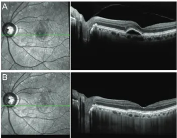
A 64-year-old man with AMD complicated by PED in the right eye and a visual acuity of 20 / 100 was injected with CDAV 28 times (ranibizumab 0.5 mg) during a 40-month period. Subfoveal-vascularized PED persisted with no improvement in visual acuity (Fig. 4A). After- wards, bevacizumab 5.0 mg was injected monthly. Three months later, the PED height on SD-OCT was found to be decreased and visual acuity improved to 20 / 80 (Fig. 4B).
Case 2
A 72-year-old woman with AMD complicated by PED in the left eye was injected with CDAV 10 times (ranibi- zumab 0.5 mg × 3, bevacizumab 1.25 mg × 7) during a 10-month period. After several injections of CDAV, visual acuity slightly improved from 20 / 125 to 20 / 100, but PED findings on SD-OCT persisted (Fig. 5A-5C). After two monthly injections of bevacizumab 5.0 mg, PED was found to be significantly decreased and visual acuity im- proved to 20 / 50 (Fig. 5D).
Discussion
The pathogenesis of PED formation in AMD is not com- pletely understood, but likely involves the penetration of CNV through Bruch’s membrane into the sub-RPE space with secondary extravasation of fluid or blood, and an in- crease in hydrostatic pressure that separates the RPE from
the underlying Bruch’s membrane [12].
The clinical implication of PED is seen in the worsening of visual acuity during its natural course. Nearly 50% of patients with newly diagnosed untreated PED experienced a loss of more than 15 letters during a mean observation period of 1 year [16]. Casswell et al. [17] reported function- al worsening in most of their patients with PED after 1 year of observation. However, there is no consensus on the therapeutic effect or even on the need for active treatment for PED. There have been few studies reporting on the ef- fect of anti-VEGF drugs for PED, and of those that have been done, results show varying levels of effectiveness [18- 22].
The optimal dosage of anti-VEGF with the lowest toxici- ty and highest therapeutic effect for PED treatment is not established. In clinical practice, patients with PED and no improvement on CDAV (bevacizumab 1.25 mg or ranibi- zumab 0.5 mg) are commonly encountered. According to the report of the CATT Research Group [9], among pa- tients who received ranibizumab 0.5 mg monthly, 50%
showed persistent fluid and 7% had residual PED. It is un- known why some eyes with neovascular AMD dry up an- atomically with fewer injections of anti-VEGF, but up to half have OCT finding of disease activity even with con-
Table 2. Adverse reactions (n = 31)
Adverse reaction Number (%)
Conjunctival hemorrhage 3 (9.4)
Vitreous floaters 2 (6.5)
Intracocular pressure increased 1 (3.2)
Foreign body sensation 4 (12.9)
Tear of retinal pigment epithelium 0
Excessive inflammation 0
Systemic reactions (e.g., thromboembolic event) 0
Fig. 4. (A) A 64-year-old man with pigment epithelial detach- ment (PED) secondary to age-related macular degeneration in right eye with subfoveal choroidal neovascularization was inject- ed with conventional dose anti-vascular endothelial growth factor 28 times (ranibizumab 0.5 mg) during a 40-month period. Sub- foveal PED persisted with no improvement of visual acuity. (B) Afterwards, bevacizumab 5.0 mg was injected monthly. Three months later, the marked PED noted on spectral domain-optical coherence tomography had resolved.
A
B
tinuous monthly injection [9].
It is possible that some patients may require a higher concentration of VEGF blockade to achieve disease quies- cence or may have faster clearance of anti-VEGF drug
from their vitreous cavity. A higher dosage of anti-VEGF drug should theoretically address both hypothetical mech- anisms.
The effect of a higher dosage of anti-VEGF drug for AMD is inconsistent across many studies. Modarres et al.
[23] reported that bevacizumab 2.5 mg has the same thera- peutic effect as bevacizumab 1.25 mg, but with more tox- icity such as vitreous reactions. Wu et al. [24] studied 25 eyes and concluded that bevacizumab 1.25 and 2.5 mg showed similar effects. In the HARBOR study, ranibizum- ab 2.0 mg did not show improvements in visual acuity compared with conventional dosages [25].
On the other hand, Costa et al. [26] reported improve- ments in CNV secondary to AMD with bevacizumab 1.5 mg and 2.0 mg compared with 1.0 mg. Chan et al. [27] saw dramatic improvements in vascularized PED patients with ranibizumab 2.0 mg. Recently, the super dose anti-VEGF trials [28] for recalcitrant neovascular AMD patients showed that monthly injected ranibizumab 0.5 mg was as- sociated with anatomical improvements and visual recov- ery after ranibizumab 2.0 mg. In the PEARL2 trial (un- published data; Kokame G. Macula Society annual meeting, 2012 Jun, Israel), good results were observed with monthly ranibizumab 2.0 mg for 6 months.
In this study, a decrease in the PED height of more than 50 µm after HDAV treatment was seen in 16 / 31 (51.6%) patients, compared to CDAV treatment (mean 12.1 injec- tions in a mean duration of 26.1 months). However, a sub- stantial proportion of eyes represented by the remainder of the study population (15 / 31, 49.4%) showed an insufficient response. This suggests that PED may represent a common pathway in AMD, rather than a response to reflect the dis- ease’s activity, and HDAV may still be an important treat- ment option in the future for a subset of patients who are more likely to benefit.
There are some reports on the safety of HDAV. Rosen- feld et al. [29] reported that increasing doses up to 2.0 mg ranibizumab were well tolerated and demonstrated a bene- ficial clinical effect. Manzano et al. [30] injected more than 5.0 mg of bevacizumab in rabbit eyes and observed vitre- ous inflammation but no signs of retinal toxicity, electro- physiologically or histologically. In several animal studies [30-32], rabbits injected with bevacizumb with doses rang- ing from 1.25 to 5.0 mg showed no toxicity on eletroretino- gram or histological examination.
There are some limitations of the present study. First, Fig. 5. (A) A 72-year-old woman with sub-retinal fluid and pig-
ment epithelial detachment (PED) secondary to age-related mac- ular degeneration in left eye was injected with conventional dose anti-vascular endothelial growth factor nine times (ranibizumab 0.5 mg × 3, bevacizumab 1.25 mg × 7) during a 10-month period.
(B) No change in PED after three injections of ranibizumab 0.5 mg. (C) No change in PED after seven injections of bevacizumab 1.25 mg. (D) Significant improvement in PED after 2 monthly injections of bevacizumab 5.0 mg.
A
B
C
D
because this was not a prospective controlled trial, the in- jections intervals were not consistent in the subjects. Sec- ond, this study included a sample size of only 31 eyes and the findings may not be applied generally. Third, the fol- low-up period of the subjects was relatively short.
This study indicates significant efficacy of HDAV for PED treatment without systemic side effects. The study subjects were a homogenous ethnic group of Koreans. To date, no large studies on the efficacy of HDAV for PED treatment have been conducted and the promising results from the present study may encourage prospective studies in a larger group.
In conclusion, the administration of higher anti-VEGF dosages for PED in AMD patients who do not respond to conventional dosages is an option worth considering. Pro- spective case control studies should be done to validate this treatment.
Conflict of Interest
No potential conflict of interest relevant to this article was reported.
References
1. Klein R, Peto T, Bird A, Vannewkirk MR. The epidemiolo- gy of age-related macular degeneration. Am J Ophthalmol 2004;137:486-95.
2. Bressler NM, Bressler SB, Congdon NG, et al. Potential public health impact of Age-Related Eye Disease Study re- sults: AREDS report no. 11. Arch Ophthalmol 2003;121:
1621-4.
3. Ferris FL 3rd, Fine SL, Hyman L. Age-related macular de- generation and blindness due to neovascular maculopathy.
Arch Ophthalmol 1984;102:1640-2.
4. Amin R, Puklin JE, Frank RN. Growth factor localization in choroidal neovascular membranes of age-related macu- lar degeneration. Invest Ophthalmol Vis Sci 1994;35:3178- 88.
5. Kliffen M, Sharma HS, Mooy CM, et al. Increased expres- sion of angiogenic growth factors in age-related maculopa- thy. Br J Ophthalmol 1997;81:154-62.
6. Avery RL, Pieramici DJ, Rabena MD, et al. Intravitreal bevacizumab (Avastin) for neovascular age-related macular
degeneration. Ophthalmology 2006;113:363-72.e5.
7. Fung AE, Lalwani GA, Rosenfeld PJ, et al. An optical co- herence tomography-guided, variable dosing regimen with intravitreal ranibizumab (Lucentis) for neovascular age-re- lated macular degeneration. Am J Ophthalmol 2007;143:
566-83.
8. Chan CK, Meyer CH, Gross JG, et al. Retinal pigment epi- thelial tears after intravitreal bevacizumab injection for neovascular age-related macular degeneration. Retina 2007;27:541-51.
9. CATT Research Group, Martin DF, Maguire MG, et al.
Ranibizumab and bevacizumab for neovascular age-related macular degeneration. N Engl J Med 2011;364:1897-908.
10. Brown DM, Kaiser PK, Michels M, et al. Ranibizumab versus verteporfin for neovascular age-related macular de- generation. N Engl J Med 2006;355:1432-44.
11. Rosenfeld PJ, Brown DM, Heier JS, et al. Ranibizumab for neovascular age-related macular degeneration. N Engl J Med 2006;355:1419-31.
12. Gass JD. Serous retinal pigment epithelial detachment with a notch: a sign of occult choroidal neovascularization. Reti- na 1984;4:205-20.
13. Gass JD. Pathogenesis of tears of the retinal pigment epi- thelium. Br J Ophthalmol 1984;68:513-9.
14. Brown DM, Michels M, Kaiser PK, et al. Ranibizumab versus verteporfin photodynamic therapy for neovascular age-related macular degeneration: two-year results of the ANCHOR study. Ophthalmology 2009;116:57-65.e5.
15. Gaudreault J, Fei D, Rusit J, et al. Preclinical pharmacoki- netics of Ranibizumab (rhuFabV2) after a single intravitre- al administration. Invest Ophthalmol Vis Sci 2005;46:726- 33.
16. Pauleikhoff D, Loffert D, Spital G, et al. Pigment epithelial detachment in the elderly: clinical differentiation, natural course and pathogenetic implications. Graefes Arch Clin Exp Ophthalmol 2002;240:533-8.
17. Casswell AG, Kohen D, Bird AC. Retinal pigment epitheli- al detachments in the elderly: classification and outcome.
Br J Ophthalmol 1985;69:397-403.
18. Inoue M, Arakawa A, Yamane S, Kadonosono K. Variable response of vascularized pigment epithelial detachments to ranibizumab based on lesion subtypes, including polypoi- dal choroidal vasculopathy. Retina 2013;33:990-7.
19. Parodi MB, Iacono P, Papayannis A, et al. Intravitreal ran- ibizumab for pigment epithelium detachment with subfo- veal occult choroidal neovascularization: a prospective
24-month case series. Am J Ophthalmol 2013;155:103-8.e2.
20. Bolz M, Michels S, Geitzenauer W, et al. Effect of systemic bevacizumab therapy on retinal pigment epithelial detach- ment. Br J Ophthalmol 2007;91:785-9.
21. Chen E, Kaiser RS, Vander JF. Intravitreal bevacizumab for refractory pigment epithelial detachment with occult choroidal neovascularization in age-related macular degen- eration. Retina 2007;27:445-50.
22. Ach T, Hoeh AE, Ruppenstein M, et al. Intravitreal bevaci- zumab in vascular pigment epithelium detachment as a re- sult of subfoveal occult choroidal neovascularization in age-related macular degeneration. Retina 2010;30:1420-5.
23. Modarres M, Naseripour M, Falavarjani KG, et al. Intravit- real injection of 2.5 mg versus 1.25 mg bevacizumab (Avastin) for treatment of CNV associated with AMD. Ret- ina 2009;29:319-24.
24. Wu L, Arevalo JF, Roca JA, et al. Comparison of two doses of intravitreal bevacizumab (Avastin) for treatment of mac- ular edema secondary to branch retinal vein occlusion: re- sults from the Pan-American Collaborative Retina Study Group at 6 months of follow-up. Retina 2008;28:212-9.
25. Busbee BG, Ho AC, Brown DM, et al. Twelve-month effi- cacy and safety of 0.5 mg or 2.0 mg ranibizumab in pa- tients with subfoveal neovascular age-related macular de- generation. Ophthalmology 2013;120:1046-56.
26. Costa RA, Jorge R, Calucci D, et al. Intravitreal bevaci- zumab for choroidal neovascularization caused by AMD (IBeNA Study): results of a phase 1 dose-escalation study.
Invest Ophthalmol Vis Sci 2006;47:4569-78.
27. Chan CK, Abraham P, Sarraf D. High-dose ranibizumab therapy for vascularized pigment epithelial detachment.
Eye (Lond) 2012;26:882-5.
28. Brown DM, Chen E, Mariani A, et al. Super-dose an- ti-VEGF (SAVE) trial: 2.0 mg intravitreal ranibizumab for recalcitrant neovascular macular degeneration-primary end point. Ophthalmology 2013;120:349-54.
29. Rosenfeld PJ, Heier JS, Hantsbarger G, Shams N. Tolerabil- ity and efficacy of multiple escalating doses of ranibizum- ab (Lucentis) for neovascular age-related macular degener- ation. Ophthalmology 2006;113:623.e1.
30. Manzano RP, Peyman GA, Khan P, Kivilcim M. Testing intravitreal toxicity of bevacizumab (Avastin). Retina 2006;
26:257-61.
31. Feiner L, Barr EE, Shui YB, et al. Safety of intravitreal in- jection of bevacizumab in rabbit eyes. Retina 2006;26:882- 8.
32. Shahar J, Avery RL, Heilweil G, et al. Electrophysiologic and retinal penetration studies following intravitreal injec- tion of bevacizumab (Avastin). Retina 2006;26:262-9.