Received on November 15, 2013. Revised on December 11, 2013. Accepted on December 12, 2013.
CC This is an open access article distributed under the terms of the Creative Commons Attribution Non-Commercial License (http://creativecommons.org/licenses/by-nc/3.0) which permits unrestricted non-commercial use, distribu- tion, and reproduction in any medium, provided the original work is properly cited.
*Corresponding Author. Mi-Yeon Kim, Department of Bioinformatics and Life Science, Soongsil University, 19, Sadang-ro, Dongjak-gu, Seoul, Korea. Tel: 82-2-820-0458; Fax: 82-2-824-4383; E-mail: [email protected]
Keywords: Lymphoid tissue inducer, Rheumatoid arthritis, Tonsil
Abbreviations: RA, rheumatoid arthritis; IL, interleukin; LTi, lymphoid tissue inducer; RORC, retinoid-related orphan re- ceptor gamma, OA, osteoarthritis; Tregs, regulatory T cells; IRB, Institutional Review Board; SSC, side-scattered light; NK, natural killer
Increased Lymphocyte Infiltration in Rheumatoid Arthritis Is Correlated with an Increase in LTi-like Cells in Synovial Fluid
Jihye Koo1, Soochan Kim1, Woong Jae Jung1, Ye Eun Lee1, Gwan Gyu Song2, Kyung-Su Kim3 and Mi-Yeon Kim1*
1Department of Bioinformatics and Life Science, Soongsil University, Seoul 156-743, 2Division of Rheumatology, Department of Internal Medicine, Korea University Guro Hospital, Seoul 152-703, 3Department of Otorhinolaryngology, Human Barrier Research Institute, Yonsei University College of Medicine, Seoul 135-720, Korea
In this study, we compared the immune cell populations in rheumatoid arthritis (RA) synovial fluid, which shows lym- phoid tissue-like structure, with those in tonsils, which are normal secondary lymphoid tissues. Firstly, we found that CD4−CD11b+ macrophages were the major population in RA synovial fluid and that B cells were the major population in tonsils. In addition, synovial fluid from patients with osteo- arthritis, which is a degenerative joint disease, contained CD4+CD11b+ monocytes as the major immune cell pop- ulation. Secondly, we categorized three groups based on the proportion of macrophages found in RA synovial fluid: (1) the macrophage-high group, which contained more than 80%
macrophages; (2) the macrophage-intermediate group, which contained between 40% and 80% macrophages; and (3) the macrophage-low group, which contained less than 40% macrophages. In the macrophage-low group, more lym- phoid tissue inducer (LTi)-like cells were detected, and the expression of OX40L and TRANCE in these cells was higher than that in the other groups. In addition, in this group, the suppressive function of regulatory T cells was downregulat- ed. Finally, CXCL13 expression was higher in RA synovial flu- id than in tonsils, but CCL21 expression was comparable in synovial fluid from all groups and in tonsils. These data dem- onstrate that increased lymphocyte infiltration in RA synovial fluid is correlated with an increase in LTi-like cells and the el- evation of the chemokine expression.
[Immune Network 2013;13(6):240-248]
INTRODUCTION
Rheumatoid arthritis (RA), which destroys cartilage, bones, and tissues in joints, is the most common systemic inflam- matory disease. Approximately 1% of the worldwide pop- ulation suffers from this autoimmune disease. One of the most obvious characteristics of this disease is an increase of activated macrophages in synovial tissue (1-3); however, some RA patients exhibit lymphocyte infiltration to the dam- aged joints and lymphoid tissue-like structures in synovial tis- sue (4,5). Many studies have demonstrated that the develop- ment and process of the ectopic lymphoid tissue-like struc- tures are similar to those of normal lymphoid tissues, such as lymph nodes and spleen (6-8). Accordingly, the factors in- volved in the formation of normal lymphoid tissues, such as CXCL13, CCL19, CCL21, CCR7, CXCR5, lymphotoxin-α/β, and interleukin (IL)-7, are found at RA synovial membranes (9-16). In the formation of normal lymphoid tissues, lym- phoid tissue inducer (LTi) cells whose name reflects the pri- mary role of the cell in lymphoid tissue organization induce the lymphoid development by expressing CCR7, CXCR5, lym- photoxin-α/β, and IL-7 receptor (17-22). The engagement of lymphotoxin-α/β receptors on stromal cells with LTi cells upregulate CXCL13, CCL19 and CCL21 and these chemokines recruit the receptor-expressing lymphocytes to form lymphoid
tissues. The LTi cells do not express lineage markers, but do express CD117, CD127 (IL-7 receptor), RORC (retinoid-related orphan receptor gamma), OX40L, CD30L and TRANCE (18-22).
By the interaction with OX40 and CD30-expressing CD4 T cells, LTi cells help memory CD4 T cell development (18).
Recently, LTi cells were identified in human tissues such as tonsils and lymph nodes (23,24). However, the phenotype of the human cells is not exactly same to that of the murine cells, for example, human LTi cells do not express CD30L (25,26). In addition, many subpopulations which show one or a couple of different molecular expression exist, so they are called LTi-like cells. Although the involvement of LTi cells in normal and ectopic lymphoid tissue development has been well established and reported in mice (17-22), the identi- fication of LTi cells in human RA synovial tissues has not yet been reported.
Therefore, we examined whether LTi cells are present in human RA synovial fluid. Because human LTi cells are well described in tonsils (23,24), which are normal lymphoid tis- sues, we compared the immune cells infiltrate in RA synovial tissue with cells from tonsils as well as to immune cells in- filtrated to synovial fluid in patients with osteoarthritis (OA), which is degenerative joint disease and the most common form of arthritis (27,28). Because the proportions of infiltrated lymphocytes and activated macrophages are different be- tween different patients, we categorized three patient groups based on the proportion of macrophages in the synovial fluid:
a macrophage-high group, which contained more than 80%
macrophages; a macrophage-intermediate group, which con- tained between 40% and 80% macrophages, and a macro- phage-low group, which contained less than 40% macro- phages. According to this grouping, we analyzed the pro- portion and activation of LTi-like cells, CD4 T cells, and regu- latory T cells (Tregs) and compared these numbers to those obtained in tonsils. Finally, we examined the expression of CCL21 and CXCL13, which are chemokines that recruit CCR7-expressing T cells and LTi cells and CXCR5-expressing B cells and LTi cells, respectively (29,30).
MATERIALS AND METHODS
Isolation of lymphocytes from synovial fluid of RA and OA patients
RA and OA synovial fluid was obtained from Korea University Guro Hospital and all experiments were carried out with the approval of Korea University Guro Hospital Institutional Review
Board (IRB). For the diagnosis of RA, clinical symptoms and radiographic evidence of bony erosions are used. According to the classification of RA by the American Rheumatism Association 1987 (31), patients are diagnosed as RA when four or more of the following seven factors are present:
morning stiffness, swelling or fluid around more than three joints simultaneously, at least one swollen area, symmetric ar- thritis, rheumatoid nodules, increased rheumatoid factor, and X-ray changes in the hands and wrist. Whereas RA is sys- temic, OA is limited only to the joints, and radiologic evi- dences of OA include asymmetrical narrowing of the joint space and bony overgrowth. In the obtained synovial fluid, PBS was added and the diluted synovial fluid was centrifuged at 1,500 rpm for 5 minutes. After depletion of red blood cells with Gey’s solution, impurities were filtered out from the cell suspensions using 70 um cell strainer (BD biosciences, San Jose, CA). The separated cell suspensions were used for flow cytometric analysis or cultured.
Isolation of lymphocytes from tonsils
Human tonsils were obtained from Gangnam Severance Hospital of Yonsei University Medical Center. Patients who have chronic tonsillitis, snoring, or mouth breathing problem underwent tonsillectomy. The experiments have been ap- proved under the supervision of Gangnam Severance Hospi- tal IRB. To separate cells from human tonsils, the extracted tonsils were cut into small fragments and crushed between gauze. To deplete red blood cells, Gey’s solution was treated and the cell suspensions were used for flow cytometric analy- sis or cultured.
Flow cytometric analysis
mAbs for TCRαβ (IP26), CD3 (UCHT1), CD4 (RPA-T4), CD25 (BC96), CD19 (HIB19), CD56 (MEM188), GITR (eBioAITR), OX40 (ACT35), TRANCE (MIH24), CD127 (eBioRDR5), RORC (AFKJS-9), and Foxp3 (PCH101) were purchased from eBio- science (San Diego, CA). mAbs for CD11b (ICRF44), OX40L (IK-1), CD11c (B-ly6) were purchased from BD biosciences.
Foxp3 and RORC were detected by intracellular staining ac- cording to manufacturer’s instructions.
Enzyme-linked immunosorbent assay (ELISA) of CCL21 and CXCL13
Synovial fluid of RA patient was centrifuged and the super- natant was collected and stored at −20oC. Samples are thawed and the concentration of CCL21 was measured using
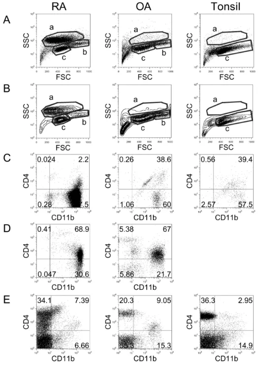
Figure 1. Flow cytometric analysis of cell populations in RA, OA, and tonsil showing by dot plots (A) and contour plots (B). X-axis shows for- ward-scattered light (FSC) and Y- axis shows side-scattered light (SSC).
The cell populations were costained with anti-CD11b and anti-CD4 and analyzed in ‘a’ area of A and B (C),
‘b’ area of A and B (D), and ‘c’ area of A and B (E). The results are representative of each RA (n=17), OA (n=3), and tonsil (n=14) group.
ELISA (R&D systems, Minneapolis, MN) according to manu- facturer’s instructions. Briefly, a 96-well microplate was coat- ed with 2μg/ml mouse anti-human CCL21 capture Ab and 100 ul of test sample or standard was added and incubate.
After washing, biotinylated goat anti-human CCL21 detection Ab, and then streptavidin conjugated to horseradish-perox-
idase solution was added. Finally, substrate solution was add- ed and optical density was measured by microplate reader (Bio Rad, Tokyo, Japan) at 450 nm.
To assay the concentration of CXCL13, a microplate coated with mouse mAb against human CXCL13 was used (R&D sys- tems). Prepared samples and standards were added into each
Figure 2. Comparison of percentage of cell populations including B cells, NK cells, NKT cells, CD4 T cells, CD8 T cells, CD4+CD11b+ monocytes, and CD4−CD11b+ macrophages in RA (n=17), OA (n=3), and tonsil (n=14). Error bar shows the standard deviation.
Figure 3. Comparison of percentage of cell populations in RA patients.
Synovial fluids which contain over 80% of CD4−CD11b+ macro- phages were grouped as macrophage high group (high, n=4), and synovial fluids which contain between 40% and 80% of macrophages were grouped as macrophage intermediate group (int, n=10). Syno- vial fluids which contain less than 40% of macrophages were grouped as macrophage low group (low, n=3).
well. After incubation with mAb against human CXCL13 con- jugated to horseradish peroxidise, substrate was added, and optical density was measured by microplate reader at 450 nm.
A standard curve was created by reducing the data using computer software generating a four parameter logistic curve-fit and used to calculate the CCL21 and CXCL13 con- centration of each sample.
RESULTS
Comparison of the diverse cell populations in RA and OA synovial fluid and tonsils
To compare the distribution of immune cells in RA synovial fluid, OA synovial fluid, and tonsils, cells were isolated and analyzed by flow cytometry (Fig. 1). Dot plot (Fig. 1A) and contour plot (Fig. 1B) analyses revealed three areas, which represented distinct cell types. The cells in ‘a’ area, which exhibited relatively high side-scattered light (SSC), were iden- tified as activated macrophages (32). RA synovial fluid showed clearly increased numbers of macrophages compared with OA synovial fluid and tonsils. Staining the cells from the ‘a’
area with anti-CD11b and anti-CD4 revealed that most of these cells were CD4−CD11b+: 97.5% in RA, 60.0% in OA, and 57.5% in tonsils (Fig. 1C). The cells in the ‘b’ area, which exhibited intermediate levels of SSC and were larger in size, were possibly monocytes (32). In tonsils, the ‘b’ area was absent, and this absence was clear in contour plot analy- sis (Fig. 1B). Co-staining of the cells in the ‘b’ area with an- ti-CD11b and anti-CD4 revealed that most of these cells were CD4+CD11b+: 68.9% in RA and 67.0% in OA (Fig. 1D). The cells in the ‘c’ area were determined to be lymphocytes, and
most of these cells were CD11b-: 86.0% in RA, 75.7% in OA, and 82.2% in tonsils (Fig. 1E).
In order to compare the proportion of each cell population in RA synovial fluid, OA synovial fluid, and tonsils, the per- centage of B cells, natural killer (NK) cells, NKT cells, CD4 T cells, CD8 T cells, CD4+CD11b+ monocytes, and CD4−CD11b+ macrophages were calculated (Fig. 2). Notably, cells that were found mainly in RA synovial fluid were macrophages (average 64.9%) and CD4+CD11b+ monocytes (∼11.2%).
In OA synovial fluid, the largest population of cells was CD4+CD11b+ monocytes (∼19.6%), while the second most common population was CD8 T cells (∼12.7%), which were increased compared with RA synovial fluid (∼5.1%). In ton- sils, B cells (∼45.4%) and CD4 T cells (22.7%) were the ma- jor populations.
RA patients divided based on the proportion of macrophages
Because each RA patient exhibits a different proportion of in- filtrated lymphocytes and activated macrophages, the RA pa- tients were categorized into three groups based on the pro- portion of macrophages found in synovial fluid (Fig. 3). The first group was the macrophage-high group, which contained more than 80% macrophages. In this group, the proportion of lymphocytes was very low (average 2.8%). The second group showed intermediate levels (between 40% and 80%) of macrophages, and the average proportion of lymphocytes was 9.6%. Ten of the 17 examined patients were assigned to this group. The third group was the macrophage-low group, which contained less than 40% macrophages and ex-
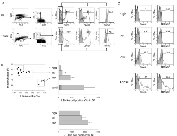
Figure 4. Identification and comparison of LTi/LTi-like cells in RA and tonsil. (A) Flow cytometric analysis of LTi-like cells. According to FSC and SSC, lymphocytes were selected and CD3-negative and CD117-positive population were gated. The expression of CD127 and RORC in CD117+CD3−CD56− cells was analyzed. Shaded histograms indicate isotype staining. (B) Correlation between the portion of macrophages and LTi-like cells in synovial fluid (SF). Percentage and absolute cell numbers of LTi-like cells in macrophage-high, intermediate (int), and low groups of RA synovial fluid were compared with tonsils. The statistical evaluation of the data was performed with Student’s t-test and one-way analysis of variance. *p<0.05, and **p<0.005 compared to macrophage- high group. (C) Expression of OX40L and TRANCE in LTi-like cells in macrophage-high, intermediate, and low groups of RA synovial fluid and tonsil. Shaded histograms indicate isotype staining.
hibited increased infiltration of lymphocytes up to 46.1% with an average lymphocyte content of 34.1%.
CD117+CD3−CD56−CD127+RORC+ LTi-like cells in RA synovial fluid
As we previously identified CD117+CD3−CD56−CD127+ RORC+OX40L+ LTi cells in human tonsils (23), we determin- ed whether LTi cells are present in RA synovial fluid. When cells were gated as CD117+CD3−CD56− cells, most of them expressed CD127 (78.2%) and RORC (80.6%) (Fig. 4A), and
these characteristics are very similar to tonsillar LTi cells.
More LTi-like cells were found in the macrophage-low group compared with the macrophage-high and -intermediate groups (Fig. 4B). Not only the proportion but also the abso- lute cell number of LTi-like cells was increased in the macro- phage-low group (p<0.05 compared to macrophage-high group).
In contrast to tonsillar LTi cells, which express high levels of OX40L and TRANCE, LTi-like cells in RA synovial tissue expressed minimal levels of OX40L and TRANCE. In fact, low
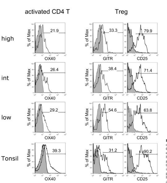
Figure 5. Flow cytometric analyses of OX40 expression on activated CD4 T cells and GITR and CD25 expression on Tregs in macrophage- high, intermediate (int), and low groups of RA synovial fluid and tonsil. Shaded histograms indicate isotype staining. The results are re- presentative of each macrophage high (n=4), intermediate (n=10), and low (n=3) group.
expression of these factors was found in the macrophage-low group, and no expression was detected in the macro- phage-high group (Fig. 4C). Thus, we categorized these cells as LTi-like cells.
Comparison of activated CD4 T cells and Tregs To analyze the activation state of CD4 T cells and CD3+Foxp3+ Tregs, the activation markers of these cells were evaluated and compared between the three macrophage groups of RA synovial fluid and tonsils (Fig. 5). All groups of CD4 T cells expressed similar levels of OX40 as tonsillar CD4 T cells. In comparison, Tregs in the macrophage-low group expressed higher levels of GITR and lower levels of CD25 compared to the other groups, suggesting that the suppressive function of Tregs in this group was downregulated (33).
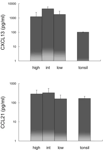
Expression of CXCL13 and CCL21 in RA synovial fluid Since CXCL13 and CCL21 recruit CXCR5- and CCR7-express- ing cells, CXCL13 and CCL21 expression was analyzed in RA synovial fluid and compared with expression in tonsils (Fig.
6). The expression of CXCL13 in RA synovial fluid was more than 12 times greater than that in tonsils, while the expression of CCL21 was comparable in all RA synovial fluid groups and tonsils.
DISCUSSION
RA, which is one of the most common autoimmune diseases, is characterized by chronic systemic inflammation with syno- vial inflammation and joint destruction. Because RA is a chronic inflammation disease, many types of immune cells in-
Figure 6. Comparison of CXCL13 and CCL21 expression in macro- phage-high, intermediate (int), and low groups of RA synovial fluid and tonsil. Error bar shows the standard deviation.
filtrate the damaged synovial tissue and in some cases, form lymphoid tissue-like structures (4,5). Although lymphoid in- filtration into the tissue is not related to clinical symptoms (34), we hypothesized that RA patients could be categorized based on the proportion of immune cells and their activation conditions in synovial fluid, and it could be related to the LTi cell numbers. In this study, we identified clear differences in the distribution of macrophages and lymphocytes in RA synovial fluid. In particular, LTi/LTi-like cells, which are crit- ical for induction of normal lymphoid tissue organogenesis (17-22), were increased in synovial fluid that contained more infiltrated lymphocytes. Compared with tonsillar LTi cells, synovial LTi/LTi-like cells in the macrophage-low, lympho- cyte-high group expressed minimal levels of OX40L, which is an important ligand for CD4 T cell memory generation (18), while the cells in the macrophage-high group did not express this ligand. In addition, the suppressive function of Tregs in
the macrophage-low, lymphocyte-high group was down- regulated, suggesting that inflammatory responses are more active in this group. Although CXCL13 and CCL21 expression was not significantly different between the RA groups, CXCL13 expression was upregulated compared with that in tonsils, and CCL21 expression was comparable in synovial fluid from all groups and in tonsils, suggesting that these che- mokine expression attracted CXCR5 and CCR7 expressing LTi/LTi-like cells (22).
In conclusion, LTi-like cells in RA synovial fluid that con- tains more infiltrated lymphocytes exhibited a more similar phenotype to tonsillar LTi cells compared to those in other RA tissues. It suggests that increased lymphocyte infiltration is correlated with an increase in LTi-like cells, and it is in- volved in the formation of normal lymphoid tissue-like struc- ture.
ACKNOWLEDGEMENTS
This research was supported by Basic Science Research Program through the National Research Foundation of Korea (NRF) funded by the Ministry of Education, Science and Technology (NRF-2011-0024205).
CONFLICTS OF INTEREST
The authors have no financial conflict of interest.
REFERENCES
1. Yanni, G., A. Whelan, C. Feighery, and B. Bresnihan. 1994.
Synovial tissue macrophages and joint erosion in rheumatoid arthritis. Ann. Rheum. Dis. 53: 39-44.
2. Kinne, R. W., R. Brauer, B. Stuhlmuller, E. Palombo-Kinne, and G. R. Burmester. 2000. Macrophages in rheumatoid arthritis. Arthritis Res. 2: 189-202.
3. Cutolo, M., A. Sulli, A. Barone, B. Seriolo, and S. Accardo.
1993. Macrophages, synovial tissue and rheumatoid arthritis.
Clin. Exp. Rheumatol. 11: 331-339.
4. Young, C. L., T. C. Adamson, 3rd, J. H. Vaughan, and R.
I. Fox. 1984. Immunohistologic characterization of synovial membrane lymphocytes in rheumatoid arthritis. Arthritis Rheum. 27: 32-39.
5. Schroder, A. E., A. Greiner, C. Seyfert, and C. Berek. 1996.
Differentiation of B cells in the nonlymphoid tissue of the synovial membrane of patients with rheumatoid arthritis.
Proc. Natl. Acad. Sci. U. S. A. 93: 221-225.
6. Takemura, S., A. Braun, C. Crowson, P. J. Kurtin, R. H.
Cofield, W. M. O'Fallon, J. J. Goronzy, and C. M. Weyand.
2001. Lymphoid neogenesis in rheumatoid synovitis. J.
Immunol. 167: 1072-1080.
7. Cupedo, T., W. Jansen, G. Kraal, and R. E. Mebius. 2004.
Induction of secondary and tertiary lymphoid structures in the skin. Immunity 21: 655-667.
8. Timmer, T. C., B. Baltus, M. Vondenhoff, T. W. Huizinga, P. P. Tak, C. L. Verweij, R. E. Mebius, and T. C. van der Pouw Kraan. 2007. Inflammation and ectopic lymphoid struc- tures in rheumatoid arthritis synovial tissues dissected by ge- nomics technology: identification of the interleukin-7 signal- ing pathway in tissues with lymphoid neogenesis. Arthritis Rheum. 56: 2492-2502.
9. Ngo, V. N., H. Korner, M. D. Gunn, K. N. Schmidt, D. S.
Riminton, M. D. Cooper, J. L. Browning, J. D. Sedgwick, and J. G. Cyster. 1999. Lymphotoxin alpha/beta and tumor ne- crosis factor are required for stromal cell expression of hom- ing chemokines in B and T cell areas of the spleen. J. Exp.
Med. 189: 403-412.
10. Wengner, A. M., U. E. Hopken, P. K. Petrow, S. Hartmann, U. Schurigt, R. Brauer, and M. Lipp. 2007. CXCR5- and CCR7-dependent lymphoid neogenesis in a murine model of chronic antigen-induced arthritis. Arthritis Rheum. 56: 3271- 3283.
11. McInnes, I. B. and G. Schett. 2007. Cytokines in the patho- genesis of rheumatoid arthritis. Nat. Rev. Immunol. 7: 429- 442.
12. Shi, K., K. Hayashida, M. Kaneko, J. Hashimoto, T. Tomita, P. E. Lipsky, H. Yoshikawa, and T. Ochi. 2001. Lymphoid chemokine B cell-attracting chemokine-1 (CXCL13) is ex- pressed in germinal center of ectopic lymphoid follicles with- in the synovium of chronic arthritis patients. J. Immunol. 166:
650-655.
13. Pickens, S. R., N. D. Chamberlain, M. V. Volin, R. M. Pope, N. E. Talarico, A. M. Mandelin, 2nd, and S. Shahrara. 2011.
Characterization of interleukin-7 and interleukin-7 receptor in the pathogenesis of rheumatoid arthritis. Arthritis Rheum. 63:
2884-2893.
14. Matsui, T., T. Akahoshi, R. Namai, A. Hashimoto, Y. Kurihara, M. Rana, A. Nishimura, H. Endo, H. Kitasato, S. Kawai, K.
Takagishi, and H. Kondo. 2001. Selective recruitment of CCR6-expressing cells by increased production of MIP-3 alpha in rheumatoid arthritis. Clin. Exp. Immunol. 125: 155-161.
15. Bruhl, H., M. Mack, M. Niedermeier, D. Lochbaum, J.
Scholmerich, and R. H. Straub. 2008. Functional expression of the chemokine receptor CCR7 on fibroblast-like synovio- cytes. Rheumatology (Oxford) 47: 1771-1774.
16. Calmon-Hamaty, F., B. Combe, M. Hahne, and J. Morel.
2011. Lymphotoxin alpha stimulates proliferation and pro-in- flammatory cytokine secretion of rheumatoid arthritis synovial fibroblasts. Cytokine 53: 207-214.
17. Mebius, R. E., P. Rennert, and I. L. Weissman. 1997. Deve- loping lymph nodes collect CD4+CD3− LTbeta+ cells that can differentiate to APC, NK cells, and follicular cells but not T or B cells. Immunity 7: 493-504.
18. Kim, M. Y., F. M. Gaspal, H. E. Wiggett, F. M. McConnell, A. Gulbranson-Judge, C. Raykundalia, L. S. Walker, M. D.
Goodall, and P. J. Lane. 2003. CD4(+)CD3(−) accessory cells costimulate primed CD4 T cells through OX40 and CD30 at sites where T cells collaborate with B cells. Immunity 18:
643-654.
19. Eberl, G., S. Marmon, M. J. Sunshine, P. D. Rennert, Y. Choi, and D. R. Littman. 2004. An essential function for the nuclear receptor RORgamma(t) in the generation of fetal lymphoid tis- sue inducer cells. Nat. Immunol. 5: 64-73.
20. Kim, M. Y., F. M. McConnell, F. M. Gaspal, A. White, S.
H. Glanville, V. Bekiaris, L. S. Walker, J. Caamano, E.
Jenkinson, G. Anderson, and P. J. Lane. 2007. Function of CD4+CD3− cells in relation to B- and T-zone stroma in spleen. Blood 109: 1602-1610.
21. Meier, D., C. Bornmann, S. Chappaz, S. Schmutz, L. A.
Otten, R. Ceredig, H. Acha-Orbea, and D. Finke. 2007.
Ectopic lymphoid-organ development occurs through inter- leukin 7-mediated enhanced survival of lymphoid-tissue-in- ducer cells. Immunity 26: 643-654.
22. Kim, M. Y., S. Rossi, D. Withers, F. McConnell, K. M.
Toellner, F. Gaspal, E. Jenkinson, G. Anderson, and P. L.
Lane. 2008. Heterogeneity of lymphoid tissue inducer cell populations present in embryonic and adult mouse lymphoid tissues. Immunology 124: 166-174.
23. Kim, S., S. Han, D. R. Withers, F. Gaspal, J. Bae, S. Baik, H. C. Shin, K. S. Kim, V. Bekiaris, G. Anderson, P. Lane, and M. Y. Kim. 2011. CD117(+) CD3(−) CD56(−) OX40Lhigh cells express IL-22 and display an LTi phenotype in human secondary lymphoid tissues. Eur. J. Immunol. 41: 1563-1572.
24. Cupedo, T., N. K. Crellin, N. Papazian, E. J. Rombouts, K.
Weijer, J. L. Grogan, W. E. Fibbe, J. J. Cornelissen, and H.
Spits. 2009. Human fetal lymphoid tissue-inducer cells are in- terleukin 17-producing precursors to RORC+ CD127+ natu- ral killer-like cells. Nat. Immunol. 10: 66-74.
25. Kim, M. Y., K. S. Kim, F. McConnell, and P. L. Lane. 2009.
Lymphoid tissue inducer cells: architects of CD4 immune re- sponses in mice and men. Clin. Exp. Immunol. 157: 20-26.
26. Withers D. R., F. M. Gaspal, V. Bekiaris, F. M. McConnell, M. Kim, G. Anderson, and P. J. Lane. 2011. OX40 and CD30 signals in CD4+ T-cell effector and memory function: a dis- tinct role for lymphoid tissue inducer cells in maintaining CD4+ T-cell memory but not effector function. Immunol.
Rev. 244: 134-148.
27. Radin, E. L., I. L. Paul, and R. M. Rose. 1972. Role of me- chanical factors in pathogenesis of primary osteoarthritis.
Lancet 1: 519-522.
28. Buckwalter, J. A. and H. J. Mankin. 1998. Articular cartilage:
degeneration and osteoarthritis, repair, regeneration, and transplantation. Instr. Course Lect. 47: 487-504.
29. Luther, S. A., K. M. Ansel, and J. G. Cyster. 2003. Overlapp- ing roles of CXCL13, interleukin 7 receptor alpha, and CCR7 ligands in lymph node development. J. Exp. Med. 197: 1191- 1198.
30. Ohl, L., G. Henning, S. Krautwald, M. Lipp, S. Hardtke, G.
Bernhardt, O. Pabst, and R. Forster. 2003. Cooperating mech- anisms of CXCR5 and CCR7 in development and organization of secondary lymphoid organs. J. Exp. Med. 197: 1199-1204.
31. Amett, F. C., S. M. Edworthy, D. A. Bloch, D. J. McShane, J. F. Fries, N. S. Cooper, L. A. Healey, S. R. Kaplan, M. H.
Liang, H. S. Luthra, T. A. Medsger, D. M. Mitchell, D. H.
Neustadt, R. S. Pinals, J. G. Schaller, J. T. Sharp, R. L. Wilder, and G. G. Hunder. 1988. The American Rheumatism
Association 1987 revised criteria for the classification of rheu- matoid arthritis. Arthritis Rheum. 31: 315-324.
32. Wells, D. A., M. Benesch, M. R. Loken, C. Vallejo, D.
Myerson, W. M. Leisenring, and H. J. Deeg. 2003. Myeloid and monocytic dyspoiesis as determined by flow cytometric scoring in myelodysplastic syndrome correlates with the IPSS and with outcome after hematopoietic stem cell transplantat- ion. Blood 102: 394-403.
33. Shimizu, J., S. Yamazaki, T. Takahashi, Y. Ishida, and S.
Sakaguchi. 2002. Stimulation of CD25(+)CD4(+) regulatory T cells through GITR breaks immunological self-tolerance.
Nat. Immunol. 3: 135-142.
34. Thurlings, R. M., C. A. Wijbrandts, R. E. Mebius, T. Cantaert, H. J. Dinant, T. C. van der Pouw-Kraan, C. L. Verweij, D.
Baeten, and P. P. Tak. 2008. Synovial lymphoid neogenesis does not define a specific clinical rheumatoid arthritis phenotype. Arthritis Rheum. 58: 1582-1589.