702 Original Article
Korean Circulation J 2005;35:702-705
ISSN 1738-5520
ⓒ 2005, The Korean Society of Circulation CASE REPORT
A Case of Acute Myocardial Infarction and Multiorgan Involvement Secondary to Rheumatoid Vasculitis
Hyuk Hwan Choi, MD, Yong Duk Jeong, MD, Hyun O Cho, MD, Sung Jin Choi, MD, Yong Suk Jeong, MD, Eun Jeong Kim, MD and Kil Hyun Cho, MD
Department of Internal Medicine, Sunlin Hospital, Handong University, Pohang, Korea ABSTRACT
Rheumatoid arthritis patients have an increased risk of subclinical cardiovascular disease, and they also have a high prevalence of carotid disease and peripheral arterial disease as a form of vasculitis. Rheumatoid arthritis patients have an increased cardiovascular mortality rate and an increased premature death rate, and they have a higher incidence of atherosclerosis. Myocardial infarction due to vasculitis is a rare complication for patients with rheu- matoid vasculitis. We report here on a case of a patient with multiorgan involvement who developed myocardial infarction, right carotid artery occlusion and left renal artery occlusion secondary to his rheumatoid vasculitis.
(Korean Circulation J 2005;35:702-705)
KEY WORDS:Rheumatoid vasculitis;Myocardial infarction;Carotid artery stenosis;Renal artery occlusion.
Introduction
Rheumatoid arthritis is a chronic multisystem disease with an unknown etiology. This disease displays many complications, including articular manifestations as well as extraarticular ones. Among its extraarticular mani- festations, vasculitis can occur and this can affect nearly any organ system. Rheumatoid vasculitis usually occurs in patients with long standing seropositive, erosive rheu- matoid arthritis. It typically involves small to medium sized vessels. Myocardial infarction secondary to rheu- matoid vasculitis has been reported, as has vasculitic involvement of the lungs, bowel, liver, spleen, pancreas, lymph nodes, testis and kidneys.
We report here on a case of a patient with multiorgan involvement who developed myocardial infarction, right carotid artery occlusion and left renal artery occlusion secondary to his rheumatoid vasculitis.
Case
A 44-year-old male visited our hospital and he presen- ted with dyspnea and anterior chest pain on exertion.
The patient had symmetric polyarticular morning stif- fness and pain of the hands and knee joints for the past 5 years. He was admitted to the neurosurgical de- partment 4 years ago complaining of headache, and he was diagnosed with right carotid artery occlusion. After that, he felt progressive polyarticular morning stiffness and pain of the hands and knee joints, but he had vi- sited the hospital since then or received any treatment.
Three years ago, he felt severe anterior chest pain and was diagnosed with acute, inferior wall myocardial in- farction. An angiography was done with the finding of right coronary artery stenosis, and so stent insertion was performed. After this treatment, he was advised to visit the rheumatology clinic, but he refused this recomen- dation; he took medicine intermittently only for his car- diac problem. For the last three months before he was admitted, he had severe and progressive hand and knee joint pain and swelling, but he had not been evaluated and treated for this problem except for taking drugstore medication intermittently for controlling the pain. He was admitted to our hospital with complains of dyspnea and anterior chest pain on exertion that had been oc- curring for 1 month.
He was a 20-pack-year smoker. On physical exami- nation, edema with tenderness on both hand and knee joints was seen, and right carotid artery pulsation was not palpated. There were no other specific findings.
For the laboratory tests, the rheumatoid factor was 4 IU/mL and C-reactive protein was 0.44 mg/dL. The antinuclear antibody test(ANA) and anti-neutrophil cy-
Received:February 2, 2005 Revision Received:May 19, 2005 Accepted:May 24, 2005
Correspondence:Kil Hyun Cho, MD,Department of Internal Medicine, Sunlin Hospital, Handong University, 69-7 Daesin-dong, Buk-gu, Pohang 791-704, Korea
Tel: 82-54-245-5124, Fax: 82-54-245-5464 E-mail: [email protected]
Hyuk Hwan Choi, et al: Rheumatoid Vasculitis·703
toplasmic antibody test(ANCA) were negative. The car- diac enzymes were within the normal range. The chest X-ray showed no specific finding. The electrocardiogram (ECG) showed Q waves on lead III and aVF(Fig. 1).
The echocardiography showed mild hypokinesia of the inferior wall and the posterolateral wall, and grade 1 mitral regurgitation. The ejection fraction(EF) was 50%.
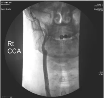
The coronary angiography(CAG) showed 90% in-stent restenosis of the proximal right coronary artery(Fig. 2), and there was a TIMI grade 1 blood supply. No stenosis findings of the left anterior descending artery and the left circumflex artery were found. The carotid angio- graphy showed total obstruction of the right internal carotid artery(Fig. 3), and the left carotid artery showed normal findings. The renal angiography showed nearly total obstruction of the left renal artery, and the right renal artery showed normal findings(Fig. 4). The ab- dominal ultrasonography demonstrated a significantly atrophied(size: 8.71 cm) left kidney and a compensa-
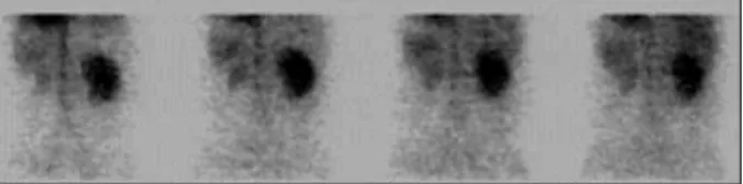
tory hypertrophied right kidney. The other organs sho- wed no specific abnormal findings. A renal scan using
99mTc-DTPA showed significantly decreased perfusion to the left kidney, but the right kidney showed normal perfusion(Fig. 5). The contribution rate of the left kid- ney to the overall glomerular filtration rate(GFR) was markedly decreased as 96:7.
Under the diagnosis of post myocardial infarction an- gina, aspirin, low molecular weight heparin(LMWH), isosorbide dinitrate, clopidogrel, and beta-blocker were administered for medical treatment, and the carotid ar- tery occlusion was referred to the neurosurgical depart- ment for treatment. Pain and swelling of the hand and
Fig. 1. The electrocardiogram showed Q waves in lead III and aVF.
aVF: augmented voltage left foot.
Fig. 2. Right coronary angiography demonstrated 90% in-stent reste- nosis.
Fig. 3. Carotid angiography showed the total obstruction of the right internal carotid artery (ICA). Rt: right, CCA: common carotid artery.
Fig. 4. Left renal angiography showed the nearly total obstruction of the left renal artery. The right renal angiography demonstrated normal per- fusion. Rt: right.
704·Korean Circulation J 2005;35:702-705
knee joints were managed according to the rheumatoid arthritis treatment guidelines.
The circulatory and neurosurgical teams are obser- ving the progress of the patient at the present time.
Discussion
Rheumatoid vasculitis is a rare complication of rheu- matoid arthritis. It typically involves the small to me- dium-sized vessels and it is associated with peripheral neuropathy, digital gangrene, nail fold infarcts and pal- pable purpura. Various clinical manifestations of rheu- matoid vasculitis can occur according to the involved vessels.
Patients with rheumatoid arthritis have a reduced life expectancy when compared with the general population.
Cardiovascular death is considered to be the leading cause of mortality in patients with rheumatoid arthritis, and it is responsible for approximately half the deaths observed in rheumatoid arthritis. The prevalence of car- diovascular comorbidity is difficult to accurately assess because cardiovascular disease has a tendency to remain silent in the rheumatoid patients.1)
Rheumatoid arthritis is a systemic disease not infre- quently involving the heart. It has been known that pericarditis, myocarditis, endocarditis, conduction dis- turbance, coronary arteritis, granulomatous arteritis are associated with rheumatoid arthritis.2)
Rheumatoid arthritis should be recognized as a mar- ker of the increased risk for myocardial infarction. Usu- ally, myocardial infarction is associated with the coronary artery disease in old people. But, it is occasionally as- sociated with coronary vasculitis in childhood and young adult.3)
The inflammatory reaction that is seen early in this disease and during disease progression is a risk factor for the progression of cardiovascular disease as well as for a shorter life span. Efficient control of the inflammation seems to be of importance not only to reduce the joint destruction, but also to alter the important mechanisms of atherothrombogenesis to a favorable direction.4)
There is growing recognition by physicians and resear- chers of an excess mortality seen in rheumatoid arthritis patients, and this is predominantly due to the increased coronary artery atherosclerosis. Atherosclerosis and its
clinical sequelae appear to be more prevalent in rheu- matoid arthritis than should be expected, and the sys- temic inflammation associated with rheumatoid arthritis may play a significant role. The effective suppression of disease activity may reduce the risk of vascular disease, and this is yet another argument for the early, aggressive, and sustained treatment of rheumatoid arthritis patients.
The relation between rheumatoid arthritis and car- diovascular disease has become a particular focus of at- tention because of the increased recognition of the inflammatory basis of atherosclerosis.5) It has long been known that T-cells play a critical role in the pathoge- nesis of rheumatoid arthritis.6) More recent data have also suggest that T-cell abnormalities may play an im- portant role in acute coronary syndrome and atheros- clerotic plaque instability.7)8) In addition, several different investigators have found that cytokines, C-reactive pro- tein and inflammatory markers, which are elevated in rheumatoid arthritis, are also elevated before and at the time of ischemic injuries.9)10)
Steroids may play a role in the increased mortality from vascular disease. Prolonged treatment with steroids accelerates the development of atherosclerosis. Abnor- mal plasma fibrinolysis in rheumatoid arthritis patients has been documented.11) Rheumatoid arthritis patients who have evidence of vascular damage in the form of vasculitis have a very marked decrease in fibrinolysis,12) and they also have increased levels of factor VIII von Willebrand factor antigen.13) This coagulation factor pro- motes thrombosis and platelet aggregation, and it may also contribute to the high cardiovascular mortality that is seen in rheumatoid arthritis patients.
The choice of disease-modifying antirheumatic drugs (DMARD) may be influenced by the vascular profile of the patient. Hydroxychloroquine appears to have fa- vorable vascular effects, while cyclosporine, methotrexate, and cyclooxygenase-2(COX-2) inhibitors may be detri- mental for patients with established atherosclerosis.14)
We report this case as a rare example of right carotid artery occlusion and left renal artery occlusion as well as acute myocardial infarction that were all secondary to the patient’s rheumatoid vasculitis.
REFERENCES
1) Goodson N. Coronary artery disease and rheumatoid arthritis.
Curr Opin Rheumatol 2002;14:115-20.
2) Jin DK, Park CG, Lee YH, et al. Asymptomatic cardiac involve- ments of rheumatoid arthritis. Korean Circ J 1997;27:884-91.
3) Baik SK, Park KS, Lee SH, Choe KH, Hwang SO. Two cases of Takayasu’s aortitis causing acute myocardial infarction. Korean Circ J 1992;22:322-9.
4) Wallberg-Jonsson S, Johansson H, Ohman ML, Rantapaa-Dahlq- vist S. Extent of inflammation predicts cardiovascular disease and overall mortality in seropositive rheummatoid arthritis: a retros- pective cohort study from disease onset. J Rheumatol 1999;26:
2562-71.
Fig. 5. Renal scan using 99mTc-DTPA revealed markedly decreased per- fusion into the left kidney and poorly defined nephrogram. There was homogeneous uptake in the right kidney and showed normal ex- cretion time to the collecting system. 99mTc-DTPA: 99mTechnetium- diethylenetriamine pentaacetic acid.
Hyuk Hwan Choi, et al: Rheumatoid Vasculitis·705
5) Ross R. Atherosclerosis: an inflammatory disease. N Engl J Med 1999;340:115-26.
6) Lee DM, Weinblatt ME. Rheumatoid arthritis. Lancet 2001;358:
903-11.
7) Weyand CM, Gornzy JJ, Luizzo G, Kopecky SL, Holmes DR Jr, Frye RL. T-cell immunity in acute coronary syndromes. Mayo Clin Proc 2001;76:1011-20.
8) Liuzzo G, Goronzy JJ, Yang H, et al. Monoclonal T-cell prolife- ration and plaque instability in acute coronary syndromes. Circu- lation 2000;101:2883-8.
9) Liuzzo G, Biasucci LM, Gallimore JR, et al. The prognostic value of C-reactive protein and serum amyloid a protein in severe unstable angina. N Engl J Med 1994;331:417-24.
10) Ridker PM, Buring JE, Shih J, Matias M, Hennekens CH. Pros- pective study of C-reactive protein and the risk of future cardio-
vascular events among apparently healthy women. Circulation 1998;98:731-3.
11) Belch JJF, McArdle B, Madhok R, et al. Decreased plasma fib- rinolysis in patients with rheumatoid arthritis. Ann Rheum Dis 1984;43:774-7.
12) Lau CS, McLaren M, Hanslip J, Kerr M, Belch JJ. Abnormal plasma fibrinolysis in patients with rheumatoid arthritis and im- paired endothelial fibrinolytic response in those complicated by vasculitis. Ann Rheum Dis 1993;52:643-9.
13) Belch JJ, Zoma A, Forbes CD, Richards IM, McLaughlin K, Strurrock RD. Vascular damage and factor-VIII-related antigen in the rheumatic disease. Rheumatol Int 1987;7:107-11.
14) van Doornum S, McColl G, Wicks IP. Accelerated atheroscle- rosis: an extraarticular feature of rheumatoid arthritis? Arthritis Rheum 2002;46:862-73.