Received:
Revised:
Accepted:
June 10, 2015 July 24, 2015 August 12, 2015
Corresponding Author: Deok-Kyu Cho, Division of Cardiology, Cardiovascular Center, Myongji Hospital, 697-24 Hwajeong-dong, Deokyang-gu, Goyang-si, Gyeonggi-do 412-270, Korea
Tel: +82-31-810-6770, Fax: +82-31-810-6778, E-mail: [email protected]
This is an Open Access article distributed under the terms of the creative Commons Attribution Non-Commercial License (http://creativecommons.org/licenses/by-nc/3.0) which permits unrestricted non-commercial use, distribution, and reproduction in any medium, provided the original work is properly cited.
pISSN 2287-2892 • eISSN 2288-2561
JLA
Missing Right Coronary Artery in a Patient with Acute Inferior ST Segment Elevation Myocardial Infarction: A Case of Extremely Rare Variation of Coronary Anatomy
Jae-Hyuk Lee1, Yongsung Suh2, In-Cheol Yoon1, Yong-Hwan Jung1, Sung-Hwa Choi1, Yun-Hyeong Cho2, Deok-Kyu Cho2
1Department of Internal Medicine, Myongji Hospital, Goyang,
2Division of Cardiology, Cardiovascular Center, Myongji hospital, Goyang, Korea
We recently encountered an interesting case of acute inferior ST segment elevation myocardial infarction (STEMI). This patient had a rare anatomic variation, single coronary artery. The right coronary artery originate from the left circumflex proper artery, not from aorta, was totally obstructed with thrombi. Though it took more time to figure out the patient’s coronary anatomy and the culprit lesion, we successfully performed primary percutaneous coronary intervention within the guideline-recommended time period. We performed left coronary angiography at the beginning. This strategy could be helpful in determining the culprit lesion and preventing unnecessary procedural delay in acute inferior STEMI.
Key Words: Coronary vessel anomalies, Myocardial infarction, Percutaneous coronary intervention
INTRODUCTION
While conducting primary percutaneous coronary inter- vention (PCI), pre-procedural localization of the culprit artery is an important step in making a procedural plan that minimizes door-to-balloon time. However, ST-segment elevation of electrocardiography (ECG) does not always correspond with the infarct related artery (IRA), especially in patients with acute inferior ST segment elevation myocardial infarction (STEMI). We recently encountered an interesting case in which the ECG-predicted IRA did not correspond with the usual one due to a rare variation in the anatomy of right coronary artery. We have conducted successful primary PCI in a patient with a single
coronary artery: right coronary artery (RCA) originates from the left circumflex (LCX) proper artery.
CASE REPORT
A 47 year-old male was admitted to the emergency room of Myongji Hospital, Goyang, Korea. He had been having squeezing epigastric pain and shortness of breath lasting more than one hour. He had taken medication for hypertension and diabetes mellitus for three years.
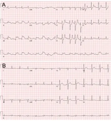
He initially had hypotension (60/40 mm Hg) and sinus tachycardia (110 beats/min). The ECG showed ST-segment elevations at II, III, and AVF leads with reciprocal ST- segment depressions at V2-V5 (Fig. 1A). ST elevation in
Fig. 1. Electrocardiography (ECG) on arrival and one day after the procedure. (A) The initial ECG showed ST segment elevations in leads II, III, and aVF, while reciprocal ST depressions were detected in leads V2-5. The ST segment in lead I is depressed, and the ST ratio II/III is less than 1.
This finding strongly suggests that the culprit lesions were in the right coronary artery (RCA) rather than in the left circumflex (LCX) artery in right dominant coronary system, (B) ST segment elevation was fully recovered one day after the procedure.
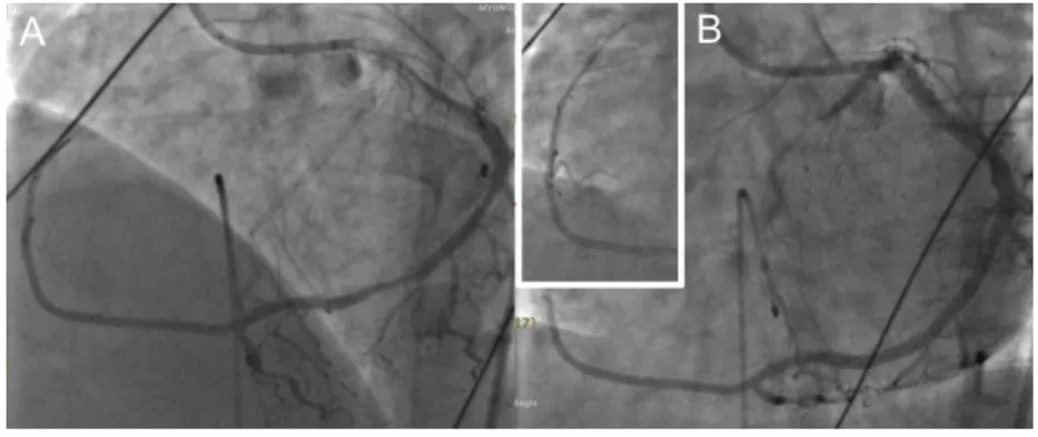
Fig. 2. Coronary angiography before revascularization. (A) Left coronary angiography showed totally occluded LCX proper artery and an intermediate lesion in the mid-segment of left anterior descending artery, (B) Aortography did not show any ostium of RCA in the aorta.
lead III was higher than in lead II, while ST segment in lead I was depressed. We assumed that hypotension of the patient would be related with RV infarction, which is frequently accompanied with inferior STEMI. Thus, we immediately started to replace intravenous (IV) volume with normal saline. At the same time, we administered him a combined dose of chewable aspirin 300 mg and clopidogrel 600 mg injecting a bolus of unfractionated heparin 5,000 IU intraveously. And then, we transferred him to the catheterization room with administering conti- nuous IV volume replacement. While transferring him to the catheterization room, the sinus rhythm changed to complete atrioventricular (AV) dissociation and the heart rate dropped to 40 rates per minute. We immediately
placed a temporary pacemaker. Because blood pressure of patient was not recovered despite of temporary pacing, we planned to insert an intra-aortic balloon pump during primary PCI as well in RCA, which is the most frequent IRA in patients with inferior ST segment elevation and AV dissociation. We first performed left coronary angio- graphy using Judkins Left 5 Fr 4.0 cm catheter which revealed a totally occluded LCX proper artery (Fig. 2A).
Since we couldn’t find an ostium of RCA despite meti- culously conducting test for several minutes with a guiding catheter (Judkins Right 7 Fr 4.0 cm), we decided to conduct aortography using a mechanical pump injector and pigtail catheter 5Fr. However, the aortography did not show any ostium of RCA in the coronary sinuses (Fig. 2B). The RCA, which was supposed to the most likely candidate of IRA, was “missing” in this emergent intervention. In this situation, we considered it would be the best option to perform revascularization for LCX proper artery first.
Angioplasty was performed using Extrabackup catheter 7 Fr 4.0 cm. After gentle advance of the guiding wire (SIONⓇ, Asahi, Japan) into the proximal stump of a totally occluded LCX proper artery, we performed balloon angioplasty at the LCX proper artery using 2.5×15 mm sized balloon catheter. The door- to-balloon time (DBT)
Fig. 4. Followed computed tomography coronary angio- graphy 3 months after procedure confirmed that the RCA originated from distal LCX proper artery.
Fig. 3. Coronary angiography after revascularization. (A) After conducting balloon angioplasty for the LCx proper artery, the missing RCA was found in the distal to LCx proper artery, (B) After stent insertion, the coronary angiography showed that the RCA originated from the LCx proper artery and extended into the right ventricular area, tapering towards the distal part.
was 45 minutes. As soon as conducting balloon angio- plasty, it was noticed the mystery of “missing RCA”, confusing us earlier, was a rare variation of the coronary anatomy. The left coronary angiography revealed that the RCA originated from the LCX proper artery and extended into the area of RCA territory, tapering towards the distal tip (Fig. 3A). Finally, we successfully implanted two overlapping everolimus-eluting stents, which were 4.0×
18 mm and 3.5×38 mm, respectively (Xience XpeditionⓇ, Abbott, Illinois, U.S.A.) (Fig. 3B). After procedure, epiga- stric pain was relieved and the ST segment elevation was resolved (Fig. 1B). Transthoracic echocardiography after revascularization showed reduced left ventricular systolic function (ejection fraction 42% by Simpson method) with akinesia of inferior LV wall. The temporary pacemaker and intra-aortic balloon pump (IABP) were successfully removed one and three days later after the procedure, respectively. Six days after conducting the procedure, the patient was successfully discharged without any significant residual symptom. We took 3 month followed computed tomography coronary angiography to evaluate the coro- nary anatomy and stent patency. In this finding, we confirmed the rare anatomic variation of single coronary artery that RCA originated from distal LCX proper artery and patent stents from distal LCX to proximal RCA. (Fig. 4).
DISCUSSION
Single coronary artery is defined as the entire coronary tree arising from the aorta through a solitary coronary ostium; it single-handedly supplies blood to the entire heart without evidence of second coronary artery.1 In patients undergoing an angiography, the incidence of single coronary artery varies between 0.024–0.066%.2 The clinical manifestations vary from benign arrhythmia to myocardial ischemia and even sudden cardiac death.3,4 Lipton et al. proposed angiographic classification of single
coronary artery, which was based on the location of coronary ostium and coronary artery course.2 According to Lipton’s classification, this case could be included in L-I group. In this case, we could safely implant stents in the segment of abnormal coronary communication since L-I group is free from the risk of external compression between the aorta and pulmonary artery.5 There were few previously reported cases, in which single coronary artery was incidentally diagnosed during evaluation for angina or as non IRA during coronary intervention.6-8 Unlike the previous report, this case is the first one in which primary PCI was successfully performed at abnor- mally originated artery from the other coronary artery in the patient with single coronary artery of L-I group of Lipton’s classification.
When the DBT is shorter, it is significantly related with the better outcomes in patients with STEMI.9 Current guidelines recommend performing revascularization within at most 90 minutes in hospitals having the primary PCI service.10,11 Differential diagnosis for IRA according to ECG analysis is an important pre-procedural step to skip unnecessary procedures during primary PCI. When ST segment elevations showed anterior STEMI pattern, IRA would be in the left coronary system with high specificity.
In contrast, ST segment elevation in inferior leads, such as II, III, and aVF does not always indicate the RCA as the IRA. In some cases, this could indicate the dominant LCX artery or rarely left anterior descending artery. In a previous study, it has been reported that when there is ST segment depression in lead I and the ST segment eleva- tion ratio of lead II/III is less than 1, it generally indicates that the RCA would be IRA.12 According to this algorithm, RCA was strongly suspected as the IRA in this case (Fig.
1A.). However, recent studies have been reporting that this algorithm has low specificity, especially in patients with left dominant coronary system or variation in anatomy.13,14 The actual IRA in our case was the LCX proper artery that branches into the RCA territory. This finding
agrees well with the findings of studies conducted lately.
In our usual clinical practice, we first take left coronary angiography during primary PCI of acute inferior STEMI, while some physicians could be performing revasculari- zation without conducting left coronary angiography in order to reduce the DBT in patients with acute inferior STEMI. To the best of our knowledge, there is no guideline that recommends you take contralateral coronary angio- graphy before conducting primary PCI. We believe that it might be a better strategy to perform left coronary angiography first in tackling cases with acute inferior STEMI, although no specific data would support this angiographic strategy. The first reason is, as mentioned above, the LCX artery could be the IRA in acute inferior STEMI. In those cases, we could spend lots of time to find the RCA ostium in which it was neither easy nor possible to engage the catheter into the RCA. Since we already knew that the LCX proper artery was totally occluded in our case, we could save some time by a swift decision to re-vascularize the LCX artery, even if some time got wasted to confirm the RCA status, such as diminutive or chronic total occlusion of RCA. We assume that we might spend longer time to find the missing RCA in the aorta without known information of LCX artery in advance in this case. The second reason is additional information, including coronary artery dominancy or disease status of contralateral coronary artery, could be helpful in determining the culprit lesion in patients with multi-vessel lesions or chronic total occlusion. On the contrary, we do not think that right coronary angiography is mandatory before revascularization in acute anterior STEMI. To confirm this hypothesis, well-designed study could be needed in the near future.
In conclusion, we report a case that a single coronary artery, a rare anatomic variation, confused us to prolong DBT in an acute inferior STEMI patient. This implicates that complete understanding of coronary anatomy including contralateral system would be very important
to make a proper decision on primary revascularization, especially in acute inferior STEMI. Additionally, if we encounter another similar situation such that highly suspected IRA is found in left coronary artery in an inferior STEMI and we could not easily engage the RCA with right coronary catheter, it could be a better option to perform revascularization first the LCA suspected IRA, rather than to spend more time and effort to find out RCA.
REFERENCES
1. Sharbaugh AH, White RS. Single coronary artery. Analysis of the anatomic variation, clinical importance, and report of five cases. JAMA 1974;230:243-246.
2. Lipton MJ, Barry WH, Obrez I, Silverman JF, Wexler L.
Isolated single coronary artery: diagnosis, angiographic classification, and clinical significance. Radiology 1979;
130:39-47.
3. Taylor AJ, Rogan KM, Virmani R. Sudden cardiac death associated with isolated congenital coronary artery anomalies. J Am Coll Cardiol 1992;20:640-647.
4. Angelini P, Velasco JA, Flamm S. Coronary anomalies:
incidence, pathophysiology, and clinical relevance. Circu- lation 2002;105:2449-2454.
5. Yamanaka O, Hobbs RE. Coronary artery anomalies in 126,595 patients undergoing coronary arteriography.
Cathet Cardiovasc Diagn 1990;21:28-40.
6. Chung SK, Lee SJ, Park SH, Lee SW, Shin WY, Jin DK.
An extremely rare variety of anomalous coronary artery:
right coronary artery originating from the distal left circumflex artery. Korean Circ J 2010;40:465-467.
7. Kim D, Jeong MH, Lee KH, Lee MG, Park KH, Sim DS, et al. Successful primary percutaneous coronary inter- vention in a patient with acute myocardial infarction and single coronary artery ostium. Korean Circ J 2012;42:
284-287.
8. Choi KL, Kwon JI, Jung WH, Kim EA, Choi SJ, Jin DK, et al. Stenting of an anormalous coronary artery in acute myocardial infarction. Korean Circ J 1998;28:1378-1381.
9. Nallamothu BK, Normand SL, Wang Y, Hofer TP, Brush JE Jr, Messenger JC, et al. Relation between door-to- balloon times and mortality after primary percutaneous coronary intervention over time: a retrospective study.
Lancet 2015;385:1114-1122.
10. Steg PG, James SK, Atar D, Badano LP, Blömstrom- Lundqvist C, Borger MA, et al. Task Force on the management of ST-segment elevation acute myocardial infarction of the European Society of Cardiology (ESC).
ESC Guidelines for the management of acute myocardial infarction in patients presenting with ST-segment elevation. Eur Heart J 2012;33:2569-2619.
11. O'Gara PT, Kushner FG, Ascheim DD, Casey DE Jr, Chung MK, de Lemos JA, et al. Society for Cardiovascular Angiography and Interventions. 2013 ACCF/AHA guide- line for the management of ST-elevation myocardial infarction: executive summary: a report of the American College of Cardiology Foundation/American Heart Association Task Force on Practice Guidelines: developed in collaboration with the American College of Emergency Physicians and Society for Cardiovascular Angiography and Interventions. Catheter Cardiovasc Interv 2013;82:
E1-E27.
12. Chia BL, Yip JW, Tan HC, Lim YT. Usefulness of ST elevation II/III ratio and ST deviation in lead I for identifying the culprit artery in inferior wall acute myocardial infarction. Am J Cardiol 2000;86:341-343.
13. Verouden NJ, Barwari K, Koch KT, Henriques JP, Baan J, van der Schaaf RJ, et al. Distinguishing the right coronary artery from the left circumflex coronary artery as the infarct-related artery in patients undergoing primary percutaneous coronary intervention for acute inferior myocardial infarction. Europace 2009;11:1517- 1521.
14. Taglieri N, Saia F, Alessi L, Cinti L, Reggiani ML, Lorenzini M, et al. Diagnostic performance of standard electro- cardiogram for prediction of infarct related artery and site of coronary occlusion in unselected STEMI patients undergoing primary percutaneous coronary intervention.
Eur Heart J Acute Cardiovasc Care 2014;3:326-339.