INTRODUCTION
For young adults, blunt chest trauma is one of the nonathe- rosclerotic mechanisms leading to acute myocardial infarction (MI). Not only a severe trauma, but also a mild trauma such as sports trauma can cause acute myocardial infarction (1-3).
Myocardial infarction after blunt chest trauma, however, is an extremely rare entity (4, 5) and 24 cases have been reported over the world in the decade with most cases received conser- vative therapy. In Korea, only one case of traumatic MI was reported, which was treated with conservative method (6). This report describes a case of MI that occurred in an adolescent after blunt chest trauma and that caused severe left ventricular dysfunction. This is the youngest case of traumatic MI ever reported, successfully treated with percutaneous coronary inter- vention (PCI).
CASE REPORT
A 16-yr-old man was transferred to the emergency room with a comatose mental state and rapid respiration rate. He ran into guardrail while riding a motorcycle. When he arrived at the hospital, four hours after the accident, he was in a co- matose mental state. He had no risk factors and family history for a cardiovascular disease. In the emergency room, his mental state was comatose, and the respiration was shallow and rapid.
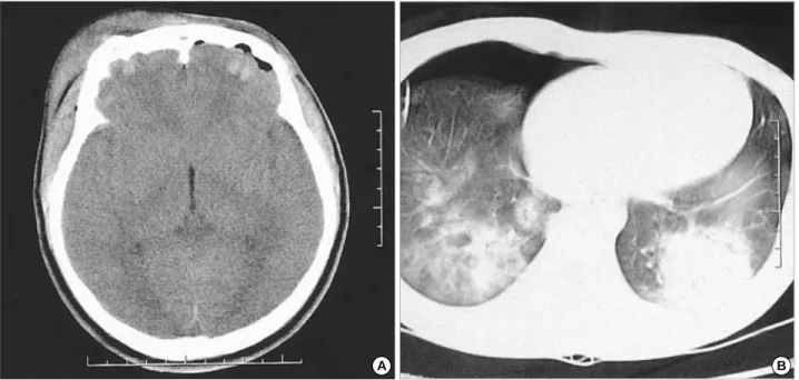
His blood pressure was 80/60 mmHg, pulse rate 130/min, respiratory rate 40/min, and body temperature 36.7℃. He seemed in an acute ill appearance, and there were bilateral orbital ecchymosis, multiple facial lacerations, and multiple ecchymosis in the chest. On an auscultation, no breathing sound was heard in the right chest and we heard crackles in the entire left chest. He had rapid heart sound without mur- mur or rubs. There was no distension, organomegaly, or ten- derness in the abdominal examination. A simple radiography showed no rib fracture, but pneumothorax was in the right chest apex, and consolidation was in the right upper lung field that appeared to have resulted from the intrapulmonary hema- toma, and interstitial markings increased in the entire lung fields (Fig. 1). An intratracheal tube and a chest tube were inserted immediately. ECG carried out routinely about eight hours after the accident, showed Q wave and 2 mm ST seg- ment elevation in the leads of all precordial leads, I and a VL (Fig. 2). The cardiac enzymes were also elevated: CK was 721 U/L, CK-MB was 300 U/L, aspartate transaminase was 94 U/L, lactate dehydrogenase was 618 U/L, and cardiac specific troponin I was 5.7 ng/mL. A brain computed tomography (CT) scan showed hemorrhagic contusions in the both frontal areas, and fractures were shown in the frontal bone, skull base, and facial bone (Fig. 3A). A chest CT scan showed hemo- pneumothorax and multiple hemorrhagic contusions (Fig. 3B).
Due to his mental state, he could not complain of any chest pain. Two-dimensional echocardiography showed anterosep-
Woo Seok Park, Myung Ho Jeong, Young Joon Hong, Ok Young Park, Joo Han Kim, Weon Kim,
Young Keun Ahn, Jeong Gwan Cho, Jong Chun Park, Byoung Hee Ahn, Sang Hyung Kim, Jung Chae Kang
The Heart Center of Chonnam National University Hospital, The Chonnam National University Research Institute of Medical Science, Gwangju, Korea
Address for correspondence Myung Ho Jeong, M.D.
The Heart Center of Chonnam National University Hospital, 8 Hak dong, Dong gu, Gwanju 501-575, Korea
Tel : +82.62-220-6243, Fax : +82.62-228-7174 E-mail : [email protected]
889
A Case of Acute Myocardial Infarction after Blunt Chest Trauma in a Young Man
Coronary artery injury rarely occurs after blunt chest trauma, but it can lead to extensive myocardial infarction and be frequently overlooked. A 16-yr-old man was presented with comatose mental state and rapid respiration rate. He ran into guard rail while riding a motorcycle. In routine examination, his electrocardiogram showed Q wave and 2 mm ST segment elevation in all precordial leads, I and aVL. The cardiac enzymes were also elevated: creatine kinase (CK)-MB was 300 U/L, and cardiac specific troponin I was 5.7 ng/mL. Two-dimensional echocardiography showed anteroseptal akinesia with severely depressed left ventricular function, ejection fraction of 28%. He could not receive any anticoagulation or thrombolytic therapy because of his brain lesion. Three weeks later, his mental state improved.
A diagnostic coronary angiogram revealed total occlusion in the proximal left ante- rior descending artery (LAD) with collaterals from the right coronary artery and left circumflex artery. We successfully performed a percutaneous coronary interven- tion for the LAD lesion, and the final angiogram showed a good coronary flow with- out residual stenosis.
Key Words : Myocardial Infarction; Angioplasty; Stents; Wounds, Nonpenetrating
Received : 19 August 2002 Accepted : 16 December 2002
tal akinesia in the left ventricle with severely depressed left ventricular function (ejection fraction=28%) (Fig. 4). We per- formed mechanical ventilation and used dopamine to elevate his blood pressure. He could receive neither primary percuta- neous coronary intervention (PCI) nor thrombolytic therapy because of his poor general condition and multiple hemorrhag- ic lesion in the chest and brain. We could wean mechanical ventilation on the 7th day of the admission and stopped the
infusion of dopamine on the 16th day of the admission. Three weeks later, his mental state became alert and we could try an invasive study. Before study, he took aspirin, clopidogrel, ACE inhibitor, and carvedilol. A diagnostic coronary angiog- ram revealed a total occlusion in the proximal left anterior descending artery with collaterals from the right coronary artery and left circumflex artery (Fig. 5A). Under the guidance of contralateral angiogram, we could successfully pass the guide wire through the lesion (Fig. 5B), but only a 1.5 mm Hayate- Pro�balloon could pass through the lesion. We dilated the balloon twice and exchanged it with a 3.0 mm balloon (Fig.
5C). Despite two times of inflating, residual stenosis still re- mained. We deployed a 3.0×20 mm Tsunami�stent in the lesion. The final angiogram showed a good coronary flow with- out residual stenosis (Fig. 5D). After PCI, he takes aspirin, clopidogrel, ACE inbibitor carvedilol and the left ventricular function was improved up to ejection fraction of 40% on follow-up echocardiogram.
DISCUSSION
Blunt chest trauma is a rare cause of cardiac damage includ- ing myocardial contusion, ventricular rupture, ventricular septal defect, valvular damage (7) and coronary artery occlu- sion with myocardial infarction (2, 5, 6, 8). However mild the trauma may be, coronary artery injury can occur (1). The most frequently injured vessel is the left anterior descending artery, followed by the right coronary and the circumflex coro- nary arteries (4). The mechanisms of injury contributing to infarction can include an intimal tear, subintimal hemorrhage, intraluminal thrombosis, and spasm (9). The repeated coro-
Fig. 1.Chest radiography shows inserted chest tube in right lung, subclavian catheter, endotracheal tube, pneumothorax in right api- cal region, and hematoma in right upper lung field and generalized increased interstitial markings.
Fig. 2.An electrocardiogram demonstrating deep Q wave and 2 mm ST segment elevation in the leads of all precordial leads, I and aVL.
I
II
III
II
aVR V1 V4
V5
V6 V2
V3 aVL
aVF
nary angiograms in several patients with coronary occlusion, due to blunt chest trauma, have shown that the natural history of such lesions is similar to intimal injuries following cardiac catheterization, frequently with complete healing of the lesion within 6 months (10).
The treatment of acute myocardial infarction caused by blunt chest trauma may be complicated by the severity of accompanying injuries, and most of the cases in the literature
have been managed conservatively (11, 12). Successful throm- bolytic treatments of coronary occlusions in patients with blunt chest trauma have been reported (13, 14). However, many trauma patients will not be candidates for thrombolytic ther- apy because of the risk of hemorrhage from coexisting injuries (15). In complicated cases, immediate surgical corrections were often performed (16). Recently, direct PCI has been performed in some patients and a few cases of stent insertion were reported (11, 15). To save myocardium from ischemic injury, the early diagnosis is the most important, and a routine ECG check should be performed in all the patients with chest trauma.
This case presents a rare complication of blunt chest trauma, which induced a complete thrombotic occlusion of the coro- nary arteries with subsequent myocardial infarction in coro- nary artery angiogram performed three weeks later. No remark- able long-term result has been reported about the prognosis of the coronary artery occlusion caused by the trauma. But the recent studies (17, 18) have reported that about 10-yr survival benefit could be expected if a successful PCI were given to chronic total occlusion caused by atherosclerotic change. Be- sides, insertion of the stent has been a better choice than percu- taneous coronary angioplasty (PTCA) in preventing restenosis in chronic total occlusion of coronary artery (19). According- ly, consider the patient’s young age, PCI with stent insertion was the choice for the patient.
REFERENCES
1. Atalar E, Acil T, Aytemir K, Ozer N, Ovunc K, Aksoyek S, Kes S, Ozmen F. Acute anterior myocardial infarction following a mild non- Fig. 3.(A) A brain CT scan shows hemorrhagic contusion in both frontal areas. (B) A chest CT scan shows hemopneumothorax and inserted chest tube in right lung field and multiple hemorrhagic contusions in both lung fields.
A B
Fig. 4.An echocardiogram reveals anteroseptal akinesia and severe left ventricular dysfunction with ejection fraction of 28%.
penetrating chest trauma- a case report. Angiology 2001; 52: 279-82.
2. Hazeleger R, Van der Wieken R, Slagboom T, Landsaat P. Coronary dissection and occlusion due to sports injury. Circulation 2001; 103:
1174-5.
3. Fang BR, Li CT. Acute myocardial infarction following blunt chest trauma. Eur Heart J 1994; 15: 705-7.
4. Ginzburg E, Dygert J, Parra-Davila E, Lynn M, Almeida J, Mayor M.
Coronary artery stenting for occlusive dissection after blunt chest trau- ma. J Trauma 1998; 45: 157-61.
5. Moosikasuwan JB, Thomas JM, Buchman TG. Myocardial infarction as a complication of injury. J Am Coll Surg 2000; 190: 665-70.
6. Jung HC, Lee SC, Gwon HC, Lee SH, Hong KP, Seo JD, Lee WR.
Left main coronary artery dissection after blunt chest trauma presented
as acute anterior myocardial infarction: assessment by intravascular ultrasound: a case report. J Korean Med Sci 1998; 13: 325-7.
7. Tengler ML. The spectrum of myocardial trauma. J Trauma 1985;
25: 620-7.
8. Marcum, JL, Booth DC, Sapin PM. Acute myocardial infarction caus- ed by blunt chest trauma: Successful treatment by direct coronary angioplasty. Am Heart J 1996; 132: 1275-7.
9. Lee DW, Garnic JD, Barlow GC. Acute anterior wall myocardial in- farction secondary to blunt chest trauma. A case report. Angiology 1990; 41: 82-4.
10. Kahn JK, Buda AJ. Long-term follow-up of coronary artery occlusion secondary to blunt chest trauma. Am Heart J 1987; 113: 207-10.
11. James ML, David BC, Peter SM. Acute myocardial infarction caused Fig. 5.(A) A left coronary angiogram reveals an abrupt total occlusion in the proximal left anterior descending coronary artery. (B) Under the guidance of contralateral right coronary angiogram, guide wire could pass the occlusive lesion of the left anterior descending artery successfully. (C) The proximal left anterior descending artery lesion is crossed with 1.5 mm Hayate-Pro�balloon successfully. (D) After 3.0 mm Tsunami stent placement, good coronary blood flow is obtained without residual stenosis.
A B
C D
300
300
300
300
by blunt chest trauma: successful treatment by direct coronary angio- plasty. Am Heart J 1996; 132: 1275-7.
12. Anto MJ, Cokinos SG, Jonas E. Acute anterior wall myocardial infarc- tion secondary to blunt chest trauma. Angiology 1984; 35: 802-4.
13. Calvo Orbe L, Garcia Gallego F, Sobrino N, Sotillo J, Lopez-Sendon JL, Oliver J, Coma I, Frutos A, Sobrino JA, Navarro JM. Acute myo- cardial infarction after blunt chest trauma in young people: need for prompt intervention. Cathet Cardiovasc Diagn 1991; 24: 182-5.
14. Ledley GS, Yazdanfar S, Friedman O, Kotler MN. Acute thrombotic coronary occlusion secondary to chest trauma treated with intracoro- nary thrombolysis. Am Heart J 1992; 123: 518-21.
15. Salmi A, Blank M, Slomski C. Left anterior descending artery occlu- sion after blunt chest trauma. J Trauma 1996; 40: 832-4.
16. Neiman RJ, Hui WKK. Posteromedial papillary muscle rupture as
a result of right coronary artery occlusion after blunt chest injury. Am Heart J 1992; 123: 1694-8.
17. Suero JA, Marso SP, Jones PG, Laster SB, Huber KC, Giorgi LV, Johnson WL, Rutherford BD. Procedural outcomes and long-term survival among patients undergoing percutaneous coronary interven- tion of a chronic total occlusion in native coronary arteries: A 20-year experience. J Am Coll Cardiol 2001; 38: 410-4.
18. Noguchi T, Miyazaki S, Morii I, Daikoku S, Goto Y, Nonogi H. Per- cutaneous transluminal coronary angioplasty of chronic total occlu- sions. Determinants of primary success and long-term clinical out- come. Catheter Cardiovasc Intervent 2000; 49: 258-64.
19. Colombo A, Stankovic G, Moses JW. Selection of coronary artery stents. J Am Coll Cardiol 2002; 40: 1021-33.