A Case of Acute Myocardial Infarction Caused by Distal Embolization of a Left Main Coronary Artery Thrombus
4
0
0
전체 글
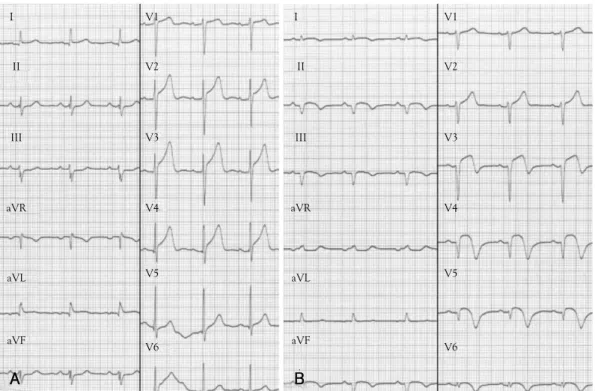
(2) Kyung-Ryun Bae, et al.·47. I. V1. I. V1. II. V2. II. V2. III. V3. III. V3. aVR. V4. aVR. V4. aVL. V5. aVL. V5. aVF. aVF. V6. A. V6. B. Fig. 1. ECG changes A: the initial ECG during typical chest pain shows no definite ST segment change. B: the follow-up ECG after coronary embolization shows ST segment elevation at lead II, III, aVF and V3-6. ECG: electrocardiogram. ECG changes.. A. B. C. D. E. F. Fig. 2. The distal embolization at coronary artery. A and B: the diagnostic coronary angiogram (CAG) shows large thrombus at left main coronary artery (LMA) (arrow head) and filling defects at distal portion of left anterior descending coronary artery (LAD) and left circumflex coronary artery (LCX) (arrow). C: after engagement of guiding catheter, there are new filling defects at the obtuse marginal branch (OM) and 1st diagonal branch (D1) and larger filling defect at LCX. D: the intravascular ultrasound reveals the large thrombus at LMA (*). E and F: even though the aspiration of thrombus at LM and multiple balloonings at LAD, LCX, D1 and OM, the most of thrombi (arrow) remain except D1. A: antero-posterior cranial view, B, C and F: right anterior oblique caudal views, D: right anterior oblique cranial view..
(3) 48·An Unusual Case of Coronary Embolization From the LMA. that time, he complained of aggravating chest pain. On CAG, the distal region of the 1st diagonal branch (D1) and the obtuse marginal branch (OM) were not visualized (Fig. 2C). We thought that this event had developed due to coronary embolization that resulted from the migration of thrombi into the LMA during CAG. Intravascular ultrasonography showed the LMA thrombus (Fig. 2D). We tried to aspirate the thrombus in the LMA using a guiding catheter and to do balloon angioplasty in the dLAD and dLCX. Balloon angioplasty was successful only for D1 (Fig. 2E). However, the dLAD, dLCX and OM had Thrombolysis In Myocardial Infarction (TIMI) grade 1 flow in spite of repeated balloon angioplasty. Applied platelet glycoprotein IIb/IIIa receptor antagonist (Abcimixab, Reopro®) and balloon angioplasty of the dLAD, OM and dLCX were done sequentially. However, filling defects in the dLAD, OM and dLCX. remained (Fig. 2F). We cared for the patient in the cardiovascular care unit. Creatine kinase-MB and cardiac troponin I levels were elevated to 349.4 ng/mL and 40.91 ng/mL, respectively. Follow up ECG showed a 3 mm ST segment elevation at leads II, III, aVF and V3-5 after angiography (Fig. 1B). We thought that the AMI developed due to distal embolization of the LMA thrombus during angiography. Laboratory profiles associated with hypercoaguability such as lupus anticoagulant, protein C activity 102% (NL 70-130%), protein S activity 74% (NL 73.7146.3%), D-dimer 0.36 μg/mL (NL 0.0-0.4 μg/mL), antithrombin III 91% (NL 80-120%) and homocysteine 12.2 μmol/L (NL 5.0-15.0 μmol/L) were in the normal range. After 3 days of intravenous unfractionated heparin (25,000 IU/day), follow-up CAG showed only slight. A. B. C. D. Fig. 3. Coronary angiography finding. A and B: after administration of glycoprotein IIb/IIIa receptor antagonist and intravenous unfractionated heparin for 3 days, the follow-up coronary angiography (CAG) shows slightly resolved thrombus at distal portion of the left anterior descending coronary artery (LAD) but the thrombi (arrow) at left circumflex coronary artery (LCX) and obtuse marginal branch (OM) remain. C and D: after triple antiplatelet agents for 1 year, the follow-up CAG shows no thrombus at all coronary arteries. A and C: right anterior oblique caudal views, B and D: right anterior oblique cranial views..
(4) Kyung-Ryun Bae, et al.·49. resolution of the filling defect and only in the dLAD, not in the OM and dLCX (Fig. 3A and B). He had no chest pain and was discharged on triple antiplatelet agent. After 1 year of this treatment, he experienced no chest pain and, on follow up CAG, had no significant stenosis or intracoronary filling defects (Fig. 3C and D).. Discussion Although rupture of atherosclerotic plaques and intracoronary thrombus formation is widely accepted as the main pathophysiological cause for the development of AMI, four to seven percent of patients with AMI do not have underlying coronary artery disease.1)3) Coronary embolism as a cause of myocardial infarction is an uncommon but important entity in terms of both etiology and treatment.1) Coronary embolism leading to myocardial infarction was first reported by Virchow in 1856.4) Previous cases of coronary emboli have been reported in association with valvular prosthesis, dilated cardiomyopathy, rheumatic valvular disease, intracardiac shunts, infective endocarditis, chronic atrial fibrillation and hypercoaguable states including pregnancy.2) In addition, primary percutaneous intervention for AMI may be complicated by distal embolization of plaque or thrombotic debris, with infarct extension.5) Henriques et al.6) reported distal embolization in patients treated with primary angioplasty is visible on the coronary angiogram in 15.2% of patients. In an unusual case, van Gaal et al.7) showed that a localized thrombus at the site of plaque rupture may embolise, causing coronary occlusion downstream in the dependent vascular territory, as in our case. Appropriate treatment of coronary embolism remains a therapeutic challenge. The strategy of intervention varies according to the acuity of the clinical presentation. If coronary embolism results in myocardial infarction associated with ST-elevation, various interventional procedures including Fogarty maneuvers, sole balloon angioplasty, stenting or thrombus aspiration have been suggested. However, in the case of non-ST-elevation myocardial infarction, there is no conclusive evidence favouring an interventional or conservative strategy.8) These techniques are particularly advocated in the setting of acute coronary syndromes.9) In particular, coronary embolism associated with an. LMA thrombus is associated with increased and extensive myocardial damage and adverse clinical outcomes such as sudden death or AMI.10) In some cases, aspiration thrombectomy has been successfully used for LMA thrombus. However, indications for this therapeutic approach are determined by coronary anatomy, clinical stability and the hemodynamic condition of the patient.10) Ahn et al.11) reported intracoronary thrombosis treated with stent and abciximab. We demonstrated that AMI developed due to coronary embolism from an LMA thrombus without any apparent cause of the systemic embolism, which progressed with angiography. Therefore, we thought that embolization of an LMA thrombus should be considered in cases where, on CAG, there are multiple coronary artery filling defects. REFERENCES 1) Kiernan TJ, Flynn AM, Kearney P. Coronary embolism causing. myocardial infarction in a patient with mechanical aortic valve prosthesis. Int J Cardiol 2006;112:e14-6. 2) Charles RG, Epstein EJ, Holt S, Coulshod N. Coronary embolism in valvular heart disease. Q J Med 1982;51:147-61. 3) Fuster V, Badimon L, Badimon JJ, Chesebro JH. The pathogenesis of coronary artery disease and the acute coronary syndromes (1). N Engl J Med 1992;326:242-50. 4) Greig LD, Leslie SJ, Denvir MA. Paradoxical coronary embolism in a young woman. Int J Cardiol 2007;115:e17-9. 5) Belli G, Pezzano A, De Biase AM, et al. Adjunctive thrombus aspiration and mechanical protection from distal embolization in primary percutaneous intervention for acute myocardial infarction. Catheter Cardiovasc Interv 2000;50:362-70. 6) Henriques JP, Zijlstra F, Ottervanger JP, et al. Incidence and clinical significance of distal embolization during primary angioplasty for acute myocardial infarction. Eur Heart J 2002;23:1112-7. 7) van Gaal WJ, West N, Banning AP. Myocardial infarction caused by distal embolisation of a ruptured left main plaque. Heart 2006; 92:1101. 8) Steinwender C, Hofmann R, Hartenthaler B, Leisch F. Resolution of a coronary embolus by intravenous application of bivalirudin. Int J Cardiol 2009;132:e115-e6. 9) Murakami T, Mizuno S, Takahashi Y, et al. Intracoronary aspiration thrombectomy for acute myocardial infarction. Am J Cardiol 1998;82:839-44. 10) Hajek P, Alan D, Vejvoda J, et al. Treatment of a large left main coronary artery thrombus by aspiration thrombectomy. J Thromb Thrombolysis 2009;27:352-4. 11) Ahn SG, Tahk SJ, Whang JC, et al. Intracoronary thrombosis treated with stent and abciximab in patient with membranous glomerulonephritis. Korean Circ J 2000;30:1307-11..
(5)
수치

관련 문서
Coronary angiogram showed severe stenosis in mid portion of left descending coronary artery (A), proximal portion of left circumflex coronary artery (B), and proximal portion of
A Case of Anomalous Right Coronary Artery Infarction Mimicking ST-Segment Elevation Myocardial Infarction at the Left Anterior Descending Artery Jong Wook Beom , MD, Joon Hyouk Choi
CAG = coronary angiography; IVUS = intravascular ultrasound; MSA = minimal stent area; ULMCA = unprotected left main coronary artery.. Left distal transradial access in the
Transbrachial peripheral angiography demonstrates (A) total occlusion of left subclavian artery (LSCA) (arrow) with patent graft from left internal mammary artery to mid left
LAD: left anterior descending coronary artery, RCA: right coronary artery. LAO view of coronary angiography showed the LCx ori- ginating from the PL branch of RCA. LAO: left
Coronary angiogram revealed total occlusion with large thrombus of distal left anterior descending artery, distal left circumflex artery and 1st diagonal artery... Fourth
Dual left anterior descending coronary artery (LAD) is a rare coronary anomaly and is incidentally detected during coronary angiography.. We report a case of a 65-year-old
1, the right coronary artery; 3, the short left anterior descending artery (LAD); 4, the long LAD; 5, the left parallel LAD; 6, the circumflex branch; 7, the septal branch from
