pISSN 2234-3180 / eISSN 2234-2591 http://dx.doi.org/10.12771/emj.2013.36.1.26
The Differences of Left Ventricular Geometry in Acute Myocardial Infarction and the Effects on Short Term Mortality
Kyung Jin Kim, In Sook Kang, Kihwan Kwon, Wook Bum Pyun, Gil Ja Shin
Division of Cardiology, Department of Internal Medicine, Ewha Womans University School of Medicine, Seoul, Korea
Objectives: This study designed to find the differences of left ventricular (LV) geometry in acute myocardial in- farction (AMI) between ST elevation myocardial infarction (STEMI) and non ST elevation myocardial infarction (NSTEMI) and the occurrences of adverse outcome according to the LV geometry.
Methods: Comprehensive echocardiographic analyses were performed in 256 patients with AMI. The left ven- tricular mass index (LVMI) and relative wall thickness (RWT) were calculated. LV geometry were classified into 4 groups based on RWT and LVMI: normal geometry (normal LVMI and normal RWT), concentric remodeling (normal LVMI and increased RWT), eccentric hypertrophy (increased LVMI and normal RWT), and concentric hypertrophy (increased LVMI and increased RWT). Cox proportional hazards models were used to evaluate the relationships among LV geometry and clinical outcomes.
Results: Patients with NSTEMI were more likely to have diabetes mellitus, hypertension, heart failure, stroke and previous myocardial infarction. By the geometric type, patients with NSTEMI were more likely to have eccen- tric hypertrophy (n=51, 34.7% vs. n=24, 22.0%, P=0.028). There was no significantly different adverse outcome between STEMI and NSTEMI patients. Fifteen patients (5.9%, 7 female [46.7%]) died and the median duration of survival was 10 days (range, 1 to 386 days). Concentric hypertrophy carried the greatest risk of all cause mortal- ity (hazard ratios, 5.83; 95% confidence interval, 1.04 to 32.7).
Conclusion: NSTEMI patients had more likely to have eccentric hypertrophy but adverse outcome after AMI was not different between STEMI and NSTEMI patients. Concentric hypertrophy had the greatest risk of short term mortality. (Ewha Med J 2013;36(1):26-34)
Key Words: Acute myocardial infarction; NSTEMI; Remodeling; STEMI; Survival
Received: February 14, 2013, Accepted: March 13, 2013
Corresponding author: Gil Ja Shin, Division of Cardiology, Depart- ment of Internal Medicine, Ewha Womans University School of Medicine, 1071 Anyangcheon-ro, Yangcheon-gu, Seoul 158-710, Korea
Tel: 82-2-2650-2843, Fax: 82-2-2655-2076 E-mail: [email protected]
Introduction
Left ventricular (LV) dysfunction begins with some
injury or stress on the myocardium and is generally a progressive process [1,2]. The principal manifestation of such progression is a change in the geometry and structure of the LV, that the chamber dilates, hyper- trophies and becomes more spherical. This process re- ferred to as cardiac remodeling [1]. Such architectural remodeling can be classified as eccentric or concentric [3]. Concentric hypertrophy is a result of systolic pres- sure overload whereas eccentric hypertrophy is a con-
sequence of volume overload [3].
The concepts of LV geometry were applied largely in clinical studies of patients with hypertension [4].
The adaptation of the LV to hypertension leads to the development of different geometric patterns and the differences of geometry are used as a risk stratification tool [5]. Hypertensive patients with concentric hyper- trophy have the highest incidence of cardiovascular events including death [6]. Subsequently LV geometry was applied to the patients after acute myocardial in- farction (AMI). In a high risk AMI, concentric hyper- trophy carries the greatest risk of adverse cardiovascular events including death [7] but uncertainty still persists with the independent prognostic value of LV geometric patterns especially after AMI. To further characterize the various geometric patterns of the left ventricle and to determine the influence of those patterns on prog- nosis in patients with AMI, and to evaluate the differ- ence of LV geometry by the type of ST change, we analyzed the patients who were diagnosed AMI with their echocardiographic data.
Methods
1. Study population
The 298 patients admitted to coronary care unit in Ewha Womans University Mokdong Hospital for AMI between January 2009 and October 2011 were included.
The patients who were suitable for clinical and echo- cardiographic data were 256 and they were constituted for the final study group. Median duration of follow-up was 212.5 days (mean, 212±174 days). Fifteen patients (5.9%, 7 female [46.7%]) died during the duration of follow-up and the median duration of survival was 10 days (range, 1 to 386 days).
2. Echocardiographic evaluation
Comprehensive echocardiography performed within 3.0 days (3.0±18.3 days) after admission. LV septal wall thickness, posterior wall thickness and cavity size were measured from the LV short-axis view by two-dimen- sionally guided M-mode echocardiography, with images of the left ventricle at the papillary muscle tip level [8]. The LV mass was calculated according to the follow-
ing formula: LV mass (g)=0.80×{1.04×[(septal wall thick- ness in diastole + LV internal diastolic diameter + posteri- or wall thickness in diastole)3 – (LV internal diastolic diameter)3]} + 0.6 g [7,9,10]. The LV mass was indexed to body surface area and LV hypertrophy was considered present when echocardiographically derived LV mass index (LVMI) was>115 g/m2 for men and >95 g/m2 for women [7,9]. The relative wall thickness (RWT) was calculated as 2×(posterior wall thickness in dia- stole)/(LV internal diastolic diameter). Increased RWT was present when this ratio was >0.42 [7,9]. The sample was divided into 4 mutually exclusive groups on the basis of LV geometry: concentric hypertrophy (LV hy- pertrophy and increased RWT), eccentric hypertrophy (LV hypertrophy and normal RWT), concentric remod- eling (normal LVMI and increased RWT), and normal geometry (normal LVMI and normal RWT) [7,9].
3. Statistical analysis
Continuous data are presented as mean±standard deviation. Baseline data were compared by means of the χ2-test for categorical variables and unpaired t test for continuous variables. Comparison between the subgroups with different patterns of LV geometry was done using ANOVA with Scheffe post hoc correction.
Multivariable cox proportional hazards models were used to determine the independent prognostic value of LV geometric patterns. Statistical analyses were per- formed using SPSS ver. 18.0 (SPSS Inc., Chicago, IL, USA). A probability value P<0.05 was considered stat- istically significant.
Results
1. Clinical and echocardiographic characteristics stratified by LV geometry
LV geometry was classified into 4 groups. Eccentric hypertrophy was present in 75 patients (29.2%), concen- tric remodeling in 15 patients (5.9%), concentric hyper- trophy in 14 patients (5.5%), and normal in 152 patients (59.4%). In total population, the mean age was 63.3±13.0 years and eighty patients were female (31.3%). Patients with eccentric hypertrophy and concentric hyper- trophy were older than normal group and patients with
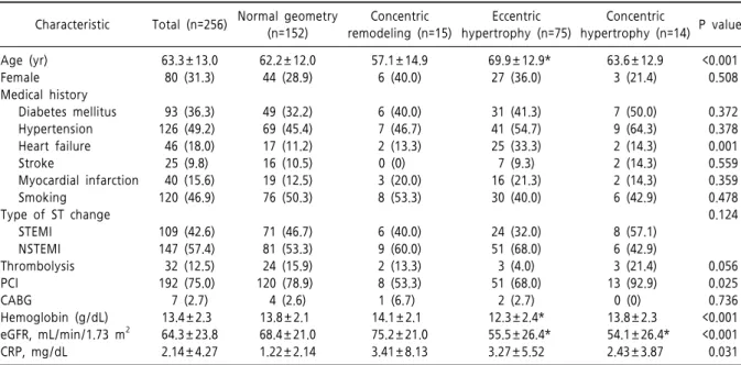
Table 1. Clinical characteristics stratified by left ventricular geometric patterns
Characteristic Total (n=256) Normal geometry (n=152)
Concentric remodeling (n=15)
Eccentric hypertrophy (n=75)
Concentric
hypertrophy (n=14) P value Age (yr)
Female Medical history Diabetes mellitus Hypertension Heart failure Stroke
Myocardial infarction Smoking
Type of ST change STEMI
NSTEMI Thrombolysis PCI
CABG
Hemoglobin (g/dL) eGFR, mL/min/1.73 m2 CRP, mg/dL
63.3±13.0 80 (31.3)
93 (36.3) 126 (49.2) 46 (18.0) 25 (9.8) 40 (15.6) 120 (46.9)
109 (42.6) 147 (57.4) 32 (12.5) 192 (75.0) 7 (2.7) 13.4±2.3 64.3±23.8 2.14±4.27
62.2±12.0 44 (28.9)
49 (32.2) 69 (45.4) 17 (11.2) 16 (10.5) 19 (12.5) 76 (50.3)
71 (46.7) 81 (53.3) 24 (15.9) 120 (78.9) 4 (2.6) 13.8±2.1 68.4±21.0 1.22±2.14
57.1±14.9 6 (40.0)
6 (40.0) 7 (46.7) 2 (13.3) 0 (0) 3 (20.0) 8 (53.3)
6 (40.0) 9 (60.0) 2 (13.3) 8 (53.3) 1 (6.7) 14.1±2.1 75.2±21.0 3.41±8.13
69.9±12.9*
27 (36.0)
31 (41.3) 41 (54.7) 25 (33.3) 7 (9.3) 16 (21.3) 30 (40.0)
24 (32.0) 51 (68.0) 3 (4.0) 51 (68.0)
2 (2.7) 12.3±2.4*
55.5±26.4*
3.27±5.52
63.6±12.9 3 (21.4)
7 (50.0) 9 (64.3) 2 (14.3) 2 (14.3) 2 (14.3) 6 (42.9)
8 (57.1) 6 (42.9) 3 (21.4) 13 (92.9)
0 (0) 13.8±2.3 54.1±26.4*
2.43±3.87
<0.001 0.508
0.372 0.378 0.001 0.559 0.359 0.478 0.124
0.056 0.025 0.736
<0.001
<0.001 0.031 Values are presented as mean±standard deviation or number (%). *P<0.01 versus patients with normal LV geometry. STEMI, ST elevation myocardial infarction; NSTEMI, non ST elevation myocardial infarction; PCI, percutaneous coronary intervention CABG, coronary artery bypass graft; eGFR, estimated glomerular filtration rate; CRP, C-reactive protein.
Table 2. Echocardiographic characteristics stratified by left ventricular geometric patterns
Total (n=256) Normal geometry (n=152)
Concentric remodeling (n=15)
Eccentric hypertrophy (n=75)
Concentric
hypertrophy (n=14) P value EDVI (mL/m2)
ESVI (mL/m2) LVEF (%) RWT LVMI (g/m2) LAVI (mL/m2) E/A
DT (ms) E/e’
MR grade (%) 0
I II III
72.2±95.3 38.8±61.8 50.9±13.3 0.34±0.08 107.7±31.3 27.8±13.5 1.0±0.5 202.6±52.6
12.7±7.0
62.9 23.0 12.3 0.4
69.1±119.0 30.8±44.1 53.1±12.9 0.32±0.06 89.3±15.6 25.0±10.6 1.0±0.5 203.3±48.9
11.3±5.8
71.8 17.4 10.7 0
42.5±13.1 15.9±3.7 64.0±8.4*
0.47±0.05*
92.2±15.1 23.2±12.5 0.9±0.3 219.7±42.3
11.3±2.1
80.0 20.0 0 0
84.0±45.4 58.2±89.1*
44.0±11.9*
0.33±0.61 142.9±24.9*
32.5±13.2*
1.0±0.6 201.7±60.1
16.0±8.7*
57.1 35.7 7.1 0
57.9±15.9 28.8±8.0 50.6±9.6 0.49±0.59*
135.4±20.8*
37.5±26.7*
0.8±0.4 175.7±57.9
13.9±7.9
45.9 33.8 18.9 1.4
0.610 0.038
<0.001
<0.001
<0.001
<0.001 0.478 0.211
<0.001 0.018
Values are presented as mean±standard deviation. EDVI, end-diastolic volume index; ESVI, end-systolic volume index; LVEF, left ventricular ejection fraction; RWT, relative wall thickness; LVMI, left ventricular mass index; LAVI, left atrial volume index; DT, deceleration time; MR, mitral regurgitation. *P<0.01 vs. patients with normal left ventricular geometry.
eccentric hypertrophy were the oldest (P<0.001 to nor- mal group). There was no significant difference of sex proportion between 4 groups. Medical history of dia- betes mellitus (DM), hypertension, heart failure (HF), stroke and myocardial infarction (MI) were included.
Prevalence of DM and hypertension were higher in patients with concentric hypertrophy with no statistical significance. Prevalence of HF was significantly differ- ent between 4 groups (P=0.001) and the patients with eccentric hypertrophy showed the highest. In addition,
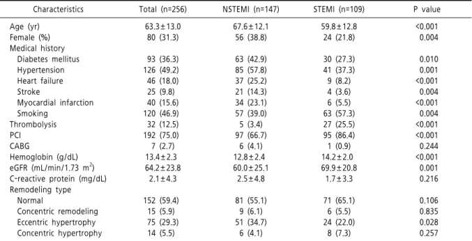
Table 3. Clinical characteristics stratified by type of ST change
Characteristics Total (n=256) NSTEMI (n=147) STEMI (n=109) P value
Age (yr) Female (%) Medical history Diabetes mellitus Hypertension Heart failure Stroke
Myocardial infarction Smoking
Thrombolysis PCI
CABG
Hemoglobin (g/dL) eGFR (mL/min/1.73 m2) C-reactive protein (mg/dL) Remodeling type
Normal
Concentric remodeling Eccentric hypertrophy Concentric hypertrophy
63.3±13.0 80 (31.3)
93 (36.3) 126 (49.2) 46 (18.0) 25 (9.8) 40 (15.6) 120 (46.9) 32 (12.5) 192 (75.0) 7 (2.7) 13.4±2.3 64.2±23.8
2.1±4.3
152 (59.4) 15 (5.9) 75 (29.3) 14 (5.5)
67.6±12.1 56 (38.8)
63 (42.9) 85 (57.8) 37 (25.2) 21 (14.3) 34 (23.1) 57 (39.0) 5 (3.4) 97 (66.7)
6 (4.1) 12.8±2.4 60.0±25.1
2.5±4.8
81 (55.1) 9 (6.1) 51 (34.7)
6 (4.1)
59.8±12.8 24 (21.8)
30 (27.3) 41 (37.3) 9 (8.2) 4 (3.6) 6 (5.5) 63 (57.3) 27 (25.5) 95 (86.4) 1 (0.9) 14.2±2.0 69.9±20.8
1.7±3.3
71 (65.1) 6 (5.5) 24 (22.0)
8 (7.3)
<0.001 0.004
0.010 0.001
<0.001 0.004
<0.001 0.004
<0.001
<0.001 0.244
<0.001 0.001 0.216
0.106 0.835 0.028 0.257 Values are presented as mean±standard deviation or number (%). NSTEMI, non ST elevation myocardial infarction; STEMI, ST elevation myocardial infarction; PCI, percutaneous coronary intervention; CABG, coronary artery bypass graft; eGFR, estimated glomerular filtration rate.
patients with LV hypertrophy had lower estimated glo- merular filtration rate and lower hemoglobin level (Table 1). The mean LVMI was 107.7±31.3 g/m2 (range, 49.9 to 253.3 g/m2) and the mean RWT was 0.34±0.08 (range, 0.13 to 0.66). The patients with LV hypertrophy had significantly lower ejection fraction, higher left atrial volume index (LAVI), higher E/e’ and severe mi- tral regurgitation (Table 2).
2. Clinical and echocardiographic characteristics stratified by type of ST change
Type of ST change divided the patients into 2 groups.
Patients with non ST elevation myocardial infarction (NSTEMI) were 147 (57.4%) and the patients with ST elevation myocardial infarction (STEMI) were 109 (42.6%). The patients with NSTEMI were older than the patients with STEMI (P<0.001). The patients with NSTEMI had higher female proportion than the patients with STEMI (P=0.004). In addition, the patients with NSTEMI had higher prevalence of DM (P=0.010), hyper- tension (P=0.001), HF P<0.001), stroke (P=0.004) and previous MI (P<0.001) significantly than the patients
with STEMI. Whereas the patients with STEMI were younger (P<0.001), more likely to be men (P=0.004) and smokers (P=0.04) than the patients with NSTEMI.
Additionally the patients with NSTEMI showed more eccentric hypertrophy significantly than the patients with STEMI (P=0.028), but the other LV geometric types were not significantly different. Besides patients with NSTEMI had lower estimated glomerular filtration rate and lower hemoglobin level (Table 3). Echocardiograph- ic characteristics showed that the patients with NSTEMI had significantly higher LVMI and LAVI than the pa- tients with STEMI (Table 4).
3. Relationship between LV geometry and clinical outcomes
Of the 256 patients, 15 patients (5.9%, 7 female [46.7%]) died, and 11 patients of them (4.3%) experi- enced a cardiovascular death. After discharged, 7 patients (2.7%) had coronary artery bypass graft, 6 patients (2.3%) were readmitted with HF, 4 patients (1.6%) had re- current MI, and 5 patients (2.0%) had stroke. The in- cidence of cardiovascular event including recurrent MI,
Table 4. Echocardiographic characteristics stratified by type of ST change
Total (n=256) NSTEMI (n=147) STEMI (n=109) P value
EDVI (mL/m2) ESVI (mL/m2) LVEF (%) RWT LVMI (g/m2) LAVI (mL/m2) E/A
DT (ms) E/e’
MR grade (%) 0
I II III
72.1±95.0 38.7±61.7 51.0±13.2 0.33±0.08 107.7±31.9 27.8±13.4 1.0±0.5 202.6±52.6
12.7±7.0
63.9 23.4 12.3 0.4
70.1±39.2 44.6±73.0 50.6±14.1 0.33±0.08 111.4±34.9 30.0±14.6 1.0±0.6 205.7±59.2
13.0±7.4
59.0 24.3 16.0 0.7
74.1±127.6 33.1±48.2 51.4±12.0 0.35±0.08 102.7±25.0 24.6±11.2 1.0±0.4 198.6±42.0
12.3±6.5
70.4 22.2 7.4 0
0.781 0.220 0.607 0.078 0.020 0.002 0.491 0.282 0.494 0.122
Values are presented as mean±standard deviation. NSTEMI, non ST elevation myocardial infarction; STEMI, ST elevation myocardial infarction; EDVI, end-diastolic volume index; ESVI, end-systolic volume index; LVEF, left ventricular ejection fraction; RWT, relative wall thickness; LVMI, left ventricular mass index; LAVI, left atrial volume index; DT, deceleration time; MR, mitral regurgitation.
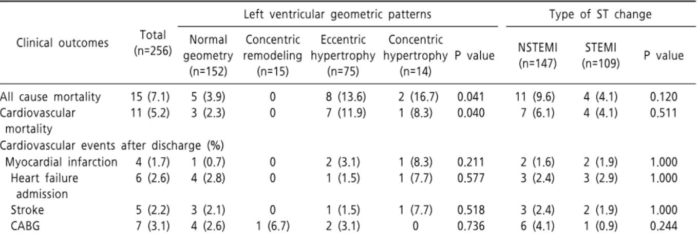
Table 5. Clinical outcomes stratified by left ventricular geometric patterns and by type of ST change
Clinical outcomes Total (n=256)
Left ventricular geometric patterns Type of ST change Normal
geometry (n=152)
Concentric remodeling
(n=15)
Eccentric hypertrophy
(n=75)
Concentric hypertrophy
(n=14)
P value NSTEMI (n=147)
STEMI
(n=109) P value
All cause mortality Cardiovascular mortality
15 (7.1) 11 (5.2)
5 (3.9) 3 (2.3)
0 0
8 (13.6) 7 (11.9)
2 (16.7) 1 (8.3)
0.041 0.040
11 (9.6) 7 (6.1)
4 (4.1) 4 (4.1)
0.120 0.511
Cardiovascular events after discharge (%) Myocardial infarction
Heart failure admission Stroke CABG
4 (1.7) 6 (2.6)
5 (2.2) 7 (3.1)
1 (0.7) 4 (2.8)
3 (2.1) 4 (2.6)
0 0
0 1 (6.7)
2 (3.1) 1 (1.5)
1 (1.5) 2 (3.1)
1 (8.3) 1 (7.7)
1 (7.7) 0
0.211 0.577
0.518 0.736
2 (1.6) 3 (2.4)
3 (2.4) 6 (4.1)
2 (1.9) 3 (2.9)
2 (1.9) 1 (0.9)
1.000 1.000
1.000 0.244 Values are presented as number (%). NSTEMI, non ST elevation myocardial infarction; STEMI, ST elevation myocardial infarction;
CABG, coronary artery bypass graft.
admission due to heart failure, cardiac death and stroke were not significantly different between LV geometric types and between STEMI and NSTEMI (Table 5).
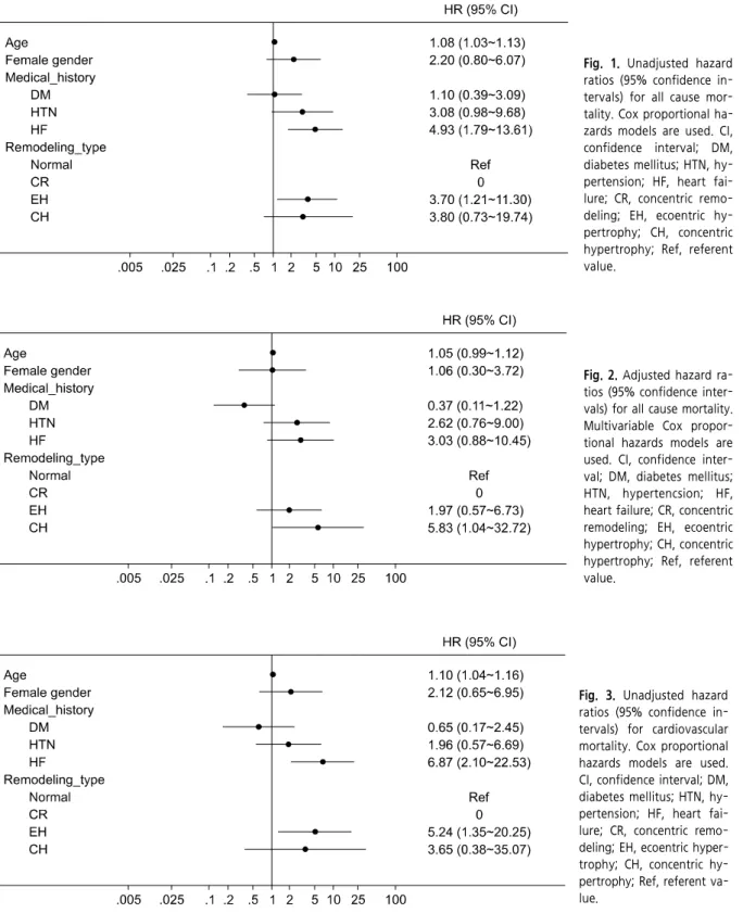
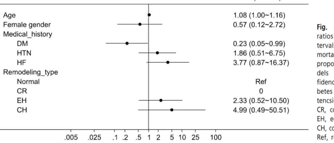
In an univariate analysis for all-cause mortality, age, history of HF and eccentric hypertrophy carried higher risk (Fig. 1) but in a multivariate analysis, only concen- tric hypertrophy carried the greatest risk of all cause mortality (hazard rations [HR], 5.83; 95% confidence interval [CI], 1.04 to 32.72) (Fig. 2).
In an univariate analysis for cardiovascular mortality, age, history of HF and eccentric hypertrophy carried
higher risk (Fig. 3) but in a multivariate analysis, age carried higher risk and history of DM carried lower risk but no specific LV geometry had significantly high- er risk for cardiovascular mortality (Fig. 4).
Discussion
Remodeling may be physiological and adaptive dur- ing normal growth or pathological due to myocardial infarction and hypertension [11]. After myocardial in- farction, myocyte necrosis and the resultant increase
Fig. 1. Unadjusted hazard ratios (95% confidence in- tervals) for all cause mor- tality. Cox proportional ha- zards models are used. CI, confidence interval; DM, diabetes mellitus; HTN, hy- pertension; HF, heart fai- lure; CR, concentric remo- deling; EH, ecoentric hy- pertrophy; CH, concentric hypertrophy; Ref, referent value.
Fig. 2. Adjusted hazard ra- tios (95% confidence inter- vals) for all cause mortality.
Multivariable Cox propor- tional hazards models are used. CI, confidence inter- val; DM, diabetes mellitus;
HTN, hypertencsion; HF, heart failure; CR, concentric remodeling; EH, ecoentric hypertrophy; CH, concentric hypertrophy; Ref, referent value.
Fig. 3. Unadjusted hazard ratios (95% confidence in- tervals) for cardiovascular mortality. Cox proportional hazards models are used.
CI, confidence interval; DM, diabetes mellitus; HTN, hy- pertension; HF, heart fai- lure; CR, concentric remo- deling; EH, ecoentric hyper- trophy; CH, concentric hy- pertrophy; Ref, referent va- lue.
in load initiates dilatation, hypertrophy, and the for- mation of a discrete collagen scar. Ventricular remodel- ing may continue until the distending forces are coun-
terbalanced by the tensile strength of the collagen scar.
This balance is determined by the size, location, and transmurality of the infarct and the patency of the in-
Fig. 4. Adjusted hazard ratios (95% confidence in- tervals) for cardiovascular mortality. Multivariable Cox proportional hazards mo- dels are used. CI, con- fidence interval; DM, dia- betes mellitus; HTN, hyper- tencsion; HF, heart failure;
CR, concentric remodeling;
EH, ecoentric hypertrophy;
CH, concentric hypertrophy;
Ref, referent value.
farct-related artery [12]. Many studies have tried to find the implication of LV geometry in AMI, but the differences in LV geometry between STEMI and NSTEMI have not been studied. Therefore this study tried to evaluate the differences of LV geometry be- tween STEMI and NSTEMI and found that the patients with NSTEMI had higher co-morbidities and higher rate of eccentric hypertrophy than the patients with STEMI significantly, but adverse outcome was not dif- ferent between STEMI and NSTEMI patients.
Changes in LV geometry and structure strongly asso- ciated with major cardiovascular events [1,13-17].
However, discerning the independent prognostic value afforded by alterations in LV shape has proved more controversial [17]. There have been many studies tried to perceive the prognostic implications of LV geometry.
Initially, the concepts of LV geometry were applied largely in clinical studies of patients with hypertension [4,18]. Koren et al. [6] were among the first to use M-mode echocardiography to study the relationship of LV geometry to clinical outcomes and the study showed that hypertensive patients with concentric hypertrophy revealed the highest incidence of cardiovascular events including death [6,7]. Subsequently, LV morphologic changes in patients after AMI and the relationship of such findings to clinical course were studied [19-23].
LV end-systolic and end-diastolic volumes are effective metrics for the severity of post-MI remodeling, and their changes are closely associated with clinical outcomes [24]. Within broader populations, LV mass is a car-
diovascular risk factor independent of blood pressure [13,16,23,24]. Recently, Verma et al. [7] related echo- cardiographic patterns of LV remodeling an average of 5 days after MI to the incidence of subsequent car- diovascular events. Patients with concentric hyper- trophy are at greatest risk for the combined end point of cardiovascular death, recurrent MI, HF, stroke, or resuscitation after cardiac arrest [7,24].
In this study, the subdivided groups stratified by LV geometry showed significantly different outcome.
Eccentric hypertrophy showed significantly higher risk for all cause mortality (HR, 3.70; 95% CI, 1.21 to 11.30) and cardiovascular mortality (HR, 5.24; 95% CI, 1.35 to 20.25) on univariate analysis. But after adjustment with age, sex, history of DM, hypertension and HF, the significance of mortality risk disappeared and only concentric hypertrophy showed the highest risk of all cause mortality (HR, 5.83; 95% CI, 1.04 to 32.72).
Postinfarction remodeling is divided into two phases.
The early phase involves expansion of the infarct zone and late remodeling involves time-dependent dilatation, the distortion of ventricular shape, and mural hyper- trophy [11]. Therefore, by 5 days, the expected struc- tural change after MI would be characterized by early dilation and eccentric hypertrophy [24]. However, in this study, the LV geometry with the greatest risk of mortality after MI was concentric hypertrophy [7,25].
Konstam [24] suspected that this could be explained by understanding the role of antecedent hypertension and its structural consequences on the clinical course
after MI. The pathologic hypertrophy, particularly con- centric hypertrophy represents a marker for the sys- temic consequences of hypertension, including vascular remodeling and results in both cerebral and myocardial ischemic events [24]. In this study [24] and the study by Verma et al. [7], the relative prevalence of hyper- tension follows the same patterns as the relative in- cidence of subsequent clinical outcomes: concentric hy- pertrophy had the greatest risk of hypertension and eccentric hypertrophy, concentric remodeling and nor- mal pattern were followed. Conclusively we can hy- pothesize that at the time of MI, antecedent structural consequences of hypertension carry the LV geometric change and also higher risk of mortality rate.
The limitation of this study is followings. First, 2-di- mensional echocardiography is limited in its accuracy for measuring LV mass because all methods assume a uniform LV thickness. Second, this result is based on the patients with AMI which limits generalization.
Third, the already known predictors of cardiovascular mortality, e.g., initial Killip class and severity of coronary vascular disease were not analyzed in this study. Finally, this study did not assess for serial changes in LV mass and its geometrical patterns and potential influence on cardiovascular risk. Therefore prospective study with serial echocardiographic analysis should be followed.
In this report, patients with NSTEMI were more likely to have eccentric hypertrophy but adverse outcome after AMI was not different between STEMI and NSTEMI patients. The baseline LV geometry represents the prognostic predictors to the patients with AMI and the concentric hypertrophy carries the greatest risk of short term mortality. Therefore routine assessment of LV mass and RWT can help us to assess the prognosis of the patients with AMI.
References
1. Hunt SA; American College of Cardiology; American Heart Association Task Force on Practice Guidelines (Writing Committee to Update the 2001 Guidelines for the Evaluation and Management of Heart Failure).
ACC/AHA 2005 guideline update for the diagnosis and management of chronic heart failure in the adult: a report of the American College of Cardiology/American
Heart Association Task Force on Practice Guidelines (Writing Committee to Update the 2001 Guidelines for the Evaluation and Management of Heart Failure). J Am Coll Cardiol 2005;46:e1-e82.
2. Kim SS, Jeon HK, Cho GM, Lee JH, Kim SJ, Park MY, et al. Evaluation of cardiac function by trans- thoracic echocardiography in subjects with st-segment elevation myocardial infarction following primary per- cutaneous coronary intervention according to valsartan dose: the valsartan one center trial. J Cardiovasc Ultrasound 2010;18:77-83.
3. Gaasch WH, Zile MR. Left ventricular structural remod- eling in health and disease: with special emphasis on volume, mass, and geometry. J Am Coll Cardiol 2011;58:
1733-1740.
4. Gaasch WH. Left ventricular radius to wall thickness ratio. Am J Cardiol 1979;43:1189-1194.
5. Castello Brescane R. The prognostic significance of left ventricular geometry: fantasy or reality? Rev Esp Cardiol 2009;62:235-238.
6. Koren MJ, Devereux RB, Casale PN, Savage DD, Laragh JH. Relation of left ventricular mass and geome- try to morbidity and mortality in uncomplicated essen- tial hypertension. Ann Intern Med 1991;114:345-352.
7. Verma A, Meris A, Skali H, Ghali JK, Arnold JM, Bourgoun M, et al. Prognostic implications of left ven- tricular mass and geometry following myocardial in- farction: the VALIANT (VALsartan In Acute myocardial iNfarcTion) Echocardiographic Study. JACC Cardiovasc Imaging 2008;1:582-591.
8. Carluccio E, Tommasi S, Bentivoglio M, Buccolieri M, Filippucci L, Prosciutti L, et al. Prognostic value of left ventricular hypertrophy and geometry in patients with a first, uncomplicated myocardial infarction. Int J Cardiol 2000;74:177-183.
9. Lang RM, Bierig M, Devereux RB, Flachskampf FA, Foster E, Pellikka PA, et al. Recommendations for cham- ber quantification: a report from the American Society of Echocardiography's Guidelines and Standards Committee and the Chamber Quantification Writing Group, developed in conjunction with the European Association of Echocardiography, a branch of the European Society of Cardiology. J Am Soc Echocardiogr 2005;18:1440-1463.
10. Devereux RB, Alonso DR, Lutas EM, Gottlieb GJ, Campo E, Sachs I, et al. Echocardiographic assessment of left ventricular hypertrophy: comparison to necropsy findings. Am J Cardiol 1986;57:450-458.
11. Sutton MG, Sharpe N. Left ventricular remodeling after myocardial infarction: pathophysiology and therapy.
Circulation 2000;101:2981-2988.
12. Pfeffer MA, Braunwald E. Ventricular remodeling after myocardial infarction. Experimental observations and clinical implications. Circulation 1990;81:1161-1172.
13. Levy D, Garrison RJ, Savage DD, Kannel WB, Castelli WP. Prognostic implications of echocardiographically determined left ventricular mass in the Framingham Heart Study. N Engl J Med 1990;322:1561-1566.
14. Ghali JK, Kadakia S, Cooper RS, Liao YL. Impact of left ventricular hypertrophy on ventricular arrhythmias in the absence of coronary artery disease. J Am Coll Cardiol 1991;17:1277-1282.
15. Levy D, Garrison RJ, Savage DD, Kannel WB, Castelli WP. Left ventricular mass and incidence of coronary heart disease in an elderly cohort. The Framingham Heart Study. Ann Intern Med 1989;110:101-107.
16. Vakili BA, Okin PM, Devereux RB. Prognostic im- plications of left ventricular hypertrophy. Am Heart J 2001;141:334-341.
17. Chahal NS, Lim TK, Jain P, Chambers JC, Kooner JS, Senior R. New insights into the relationship of left ven- tricular geometry and left ventricular mass with cardiac function: A population study of hypertensive subjects.
Eur Heart J 2010;31:588-594.
18. Hwang JW, Kang SJ, Lim HS, Choi BJ, Choi SY, Hwang GS, et al. Impact of arterial stiffness on regional my- ocardial function assessed by speckle tracking echo- cardiography in patients with hypertension. J Cardiovasc Ultrasound 2012;20:90-96.
19. Erlebacher JA, Weiss JL, Eaton LW, Kallman C, Weisfeldt ML, Bulkley BH. Late effects of acute infarct dilation on heart size: a two dimensional echocardio- graphic study. Am J Cardiol 1982;49:1120-1126.
20. Pfeffer MA, Pfeffer JM. Ventricular enlargement and reduced survival after myocardial infarction. Circulation 1987;75(5 Pt 2):IV93-IV97.
21. St John Sutton M, Pfeffer MA, Plappert T, Rouleau JL, Moye LA, Dagenais GR, et al. Quantitative two-di- mensional echocardiographic measurements are major predictors of adverse cardiovascular events after acute myocardial infarction: the protective effects of captopril.
Circulation 1994;89:68-75.
22. White HD, Norris RM, Brown MA, Brandt PW, Whitlock RM, Wild CJ. Left ventricular end-systolic volume as the major determinant of survival after recovery from myocardial infarction. Circulation 1987;76:44-51.
23. Konstam MA, Udelson JE, Anand IS, Cohn JN.
Ventricular remodeling in heart failure: a credible surro- gate endpoint. J Card Fail 2003;9:350-353.
24. Konstam MA. Patterns of ventricular remodeling after myocardial infarction: clues toward linkage between mechanism and morbidity. JACC Cardiovasc Imaging 2008;1:592-594.
25. Ghali JK, Liao Y, Cooper RS. Influence of left ventricular geometric patterns on prognosis in patients with or with- out coronary artery disease. J Am Coll Cardiol 1998;31:
635-1640.