A Case of Anomalous Right Coronary Artery Infarction Mimicking ST-Segment Elevation Myocardial Infarction at the Left Anterior Descending Artery
4
0
0
전체 글
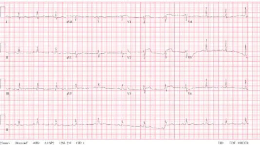
(2) An Anomalous RCA infarction Mimicking STEMI at LAD artery. Author Contributions Writing - original draft: Beom JW, Boo KY, Lee JG. Writing - review & editing: Beom JW, Kim SY, Joo SJ. Supervision: Choi JH.. Figure 2. Electrocardiogram after percutaneous coronary intervention to the mid-LAD artery lesion showed improvement in ST-segment elevations in the precordial leads. LAD = left anterior descending.. Pre. Pre Mid LAD. RCA. Mid LAD. RCA. A Post. B Post. Mid LAD. RCA Mid LAD. RCA. C. D. Figure 3. Left coronary angiograms showing an anomalous origin of the RCA from the mid-LAD artery with a significant stenosis of the RCA ostium (A, B). Left coronary angiograms showing successful revascularization at the bifurcation site between the LAD artery and the RCA (C, D). LAD = left anterior descending; RCA = right coronary artery.. A single coronary artery is an extremely rare congenital anomaly of the coronary system in structurally normal heart.1-3) To our knowledge, only one patient has been reported to undergo PCI for a true LAD artery/RCA bifurcation lesion treated using two-stent technique.4) We emphasize the importance of clinical effort to evaluate all coronary arteries in patients with coronary artery anomalies.. https://e-kcj.org. https://doi.org/10.4070/kcj.2020.0115. 952.
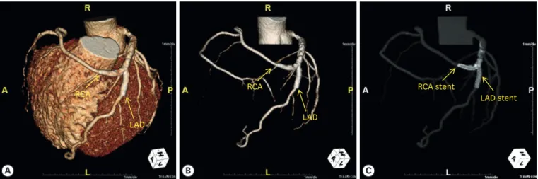
(3) An Anomalous RCA infarction Mimicking STEMI at LAD artery. RCA. RCA. LAD stent. LAD. LAD. A. RCA stent. B. C. Figure 4. Three-dimensional computed tomography coronary angiographic images revealing an anomalous origin of the RCA from the LAD artery (A, B), and showing patent stents at the bifurcation site between the LAD artery and the RCA (C). LAD = left anterior descending; RCA = right coronary artery.. SUPPLEMENTARY MATERIALS Supplementary Video 1 Left coronary angiogram showing approximately 80% segmental stenosis of the mid-LAD artery and total occlusion of the septal branch of the LAD artery. LAD = left anterior descending. Click here to view. Supplementary Video 2 Left coronary angiogram showing successful revascularization of the LAD artery. Click here to view. Supplementary Video 3 Coronary angiogram via a pigtail catheter. The right coronary artery was not visualized. Click here to view. Supplementary Video 4 A diastolic D-shape of left ventricle was observed in the parasternal short axis view of echocardiography, suggesting right ventricular infarction. Click here to view. REFERENCES 1. Topaz O, DeMarchena EJ, Perin E, Sommer LS, Mallon SM, Chahine RA. Anomalous coronary arteries: angiographic findings in 80 patients. Int J Cardiol 1992;34:129-38. PUBMED | CROSSREF. 2. Barriales Villa R, Morís C, López Muñiz A, et al. Adult congenital anomalies of the coronary arteries described over 31 years of angiographic studies in the Asturias Principality: main angiographic and clinical characteristics. Rev Esp Cardiol 2001;54:269-81. PUBMED | CROSSREF. https://e-kcj.org. https://doi.org/10.4070/kcj.2020.0115. 953.
(4) An Anomalous RCA infarction Mimicking STEMI at LAD artery. 3. Yamanaka O, Hobbs RE. Coronary artery anomalies in 126,595 patients undergoing coronary arteriography. Cathet Cardiovasc Diagn 1990;21:28-40. PUBMED | CROSSREF. 4. Khan UA, Sharma D, McGlinchey P, Peace A. Percutaneous coronary intervention to left anterior descending artery/right coronary artery bifurcation: this is not a typo! A case report. Eur Heart J Case Rep 2019;3:ytz137. PUBMED | CROSSREF. https://e-kcj.org. https://doi.org/10.4070/kcj.2020.0115. 954.
(5)
수치

관련 문서
ST-segment elevation myocardial infarction (STEMI) is one of the most fatal cardiac diseases caused by acute rupture of atherosclerotic plaque and thrombotic total occlusion
Given the clin- ical suspicion of coronary artery disease (CAD), he underwent coronary angiography, which revealed a total occlusion of the proximal left anterior descending
Percutaneous coronary intervention-related time delay, patient’ s risk profile, and survival benefits of primary angioplasty vs lytic therapy in ST- segment elevation
Large Chronic Pseudoaneurysm of Left Ventricle Complicating Anterior Myocardial Infarction Won Kyoun Park , MD, Dae Hyun Kim , MD, PhD, and Sang-Ho Cho , MD, PhD Department of
B: LAO caudal protection marking the site of LAD stump (arrow). C: AP caudal view showing the separate LAD ostium di- rectly originating from the left coronary aortic sinus. D:
Hypothesis: We analyzed data from the Korean Acute Myocardial Infarction Registry (KAMIR) to assess gender differences in in-hospital outcomes post ST-segment
We report a case of rescue thrombolysis followed by salvage percutaneous coronary intervention (PCI) for the treatment of inferior ST elevation myocardial infarction (STEMI)
Clinical Benefit of Low Molecular Weight Heparin for ST-segment Elevation Myocardial Infarction Patients Undergoing Primary Percutaneous Coronary Intervention with
