https://doi.org/10.14734/PN.2018.29.4.147 pISSN 2508-4887•eISSN 2508-4895
Yae Heun Lee, MD1, Yoo Jung Lee, MD1, Sun Young Jung, MD1, Suk Young Kim, MD1, Dong Woo Son, MD2, Il Hye Seo, MD3
Departments of 1Obstetric Gynecology, 2Pediatrics, 3Clinical Pathology, Gachon University College of Medicine, Incheon, Korea
Objective: This study examines whether maternal group B Streptococcus (Streptococcus agalactiae, GBS) infection was associated with preterm births and premature neonatal outcomes.
Methods: Maternal and neonatal outcomes were examined among singleton pregnant women with preterm birth (from 24+0 weeks to 36+6 weeks) who were tested for GBS (n=203) during the pregnancy. Data were collected retrospectively from the medical records of women who delivered at our hospital from January 2015 to February 2017. We compared obstetrical factors (causes of preterm birth) and neonatal (gestational age at delivery, birth weight, Apgar score 1 min/5 min, hospitaliza
tion period, duration of mechanical ventilation, neonatal Creactive protein within three days, and other complication [respiratory distress syndrome, neonatal deaths]) outcomes between GBSinfect
ed and noninfected pregnant women.
Results: There were 203 singleton pregnant women included in the study, 25 of whom were con
firmed to have a GBS infection during the pregnancy. There was no difference in neonatal outcomes by GBS status. Preterm premature rupture of membranes (pPROM), as an obstetric factor, was asso
ciated with GBS infection (P=0.022). GBS infection raised the risk of pPROM by 3.6 times (odds ratio 3.648, 95% confidence interval 1.4769.016, P=0.005).
Conclusion: GBS infection in preterm birth was associated with pPROM but did not result in adverse neonatal outcomes. Continuous attention and evaluation of GBS infection, a major cause of neonatal sepsis and pneumonia, are needed.
Key Words: Streptococcus agalactiae, Premature birth, Fetal membranes, Premature rupture
Introduction
Group B Streptococcus (Streptococcus agalactiae, GBS) is a Gram-positive coccus that frequently colonizes the human genital and gastrointestinal tracts, as well as the upper respiratory tract of young infants.1-3 GBS infections in pregnant women include urinary tract infection, upper genital tract infection, intra-amniotic infection, endometritis, and bac- teremia.4,5 Neonatal sepsis occurs in 80-85% of cases of early-onset GBS disease.6 The mortality rate is 4-6%, and in the case of preterm birth, up to 30%. GBS can be acquired during labor or in utero by transmission from maternal-vaginal or anorectal-colonized mucosa.7-9
In 2002, the US Centers for Disease Control and Prevention (CDC) recommended univer- sal antenatal screening at 35 to 37 weeks of pregnancy and intrapartum chemoprophylaxis for women with GBS colonization.10 An additional reduction in invasive GBS-related dis- ease was reported after publication of this guideline.6,11 Reported maternal colonization rates are quite variable but generally range from 20% to 30%.10,12 The differences in colo- Received: 20 March 2018
Revised: 25 April 2018 Accepted: 8 August 2018 Correspondence to Suk Young Kim, MD
Department of Obstetric Gynecology, Gachon University Gil Medical Center, Gachon University College of Medicine, 21 Namdongdaero 774beongil, Namdonggu, Incheon 21565, Korea
Tel: +82324603254 Fax: +82324603290 E-mail: [email protected] Copyright© 2018 by The Korean Society of Perinatology
This is an Open Access article distributed under the terms of the Creative Com
mons Attribution NonCommercial License (http://creativecommons.org/
license/bync/4.0/), which permits unrestricted noncommercial use, distribution, and reproduction in any
Pregnancy and Neonatal Outcomes of Group B Streptococcus Infection in Pre
term Births
identify GBS.
Korea has not established a guideline for GBS screening during pregnancy, and there are few reports indicating a Korean interest in perinatal complications related to GBS infection in pregnant women. This lack of concern is probably due to the low GBS infection rate in Korea. Before 2010, according to national reports, the rate of GBS infection in Korean pregnant women was 0.3-5.9%. However, two recent studies reported a 2010 GBS infection rate of 8.3% or 10.0%.13,14
Despite major advances in perinatal care, preterm birth is still the predominant cause of perinatal mortality and a major cause of neurological morbidity in surviving infants. Although the causes of preterm birth are uncertain, evidence suggests that maternal genital tract colonization by specific organisms increases the risk of preterm rupture of membranes and preterm birth.15
The aim of this study was to examine the relationship between maternal GBS infection and preterm birth outcomes.
Methods
1. Patient population
The study was approved by the institutional review board of Gachon University Gil Medical Center. The medical records of the 1,201 women admitted to our hospital for delivery between January 2015 and February 2017 were reviewed. Of the 1,201 patients, only 395 patients underwent GBS culture. Two hun- dred three singleton pregnant women with preterm birth (from 24+0 weeks to 36+6 weeks) were enrolled. One hundred ninety- two were excluded based on exclusion criteria. The exclusion criteria were multiple pregnancies, term birth, and intrauterine fetal death of unknown cause. Cases in which the baby was transferred to another hospital were also excluded.
2. Data collection
Vaginal cultures were performed for all women who were admitted due to the risk of preterm birth. Indications at such admissions included spontaneous preterm labor (SPTL), pre- term premature rupture of membrane (pPROM), incom petent internal os of cervix (IIOC), pregnancy-induced hypertension (PIH), preeclampsia, placenta previa with bleeding, placental
abruption, uterine rupture, sign of fetal distress, fetal growth restriction (FGR), and fetal anomaly. IIOC was diagnosed when cervical length shortening was observed on ultrasound imaging in the second trimester without contractions or labor and in the absence of other clear pathology. Women with pPROM received antibiotic treatment (intravenous cephalosporin 2 g injection) until the culture results were available. If the culture was confirmed positive for GBS before delivery, penicillin or cephalosporin treatment was administered. Women without evidence of pPROM were not treated with antibiotics until culture results were confirmed.
The culture method was changed during the study period.
Between January 2015 and February 2016, we obtained speci- mens from the vagina and anus separately. From March 2016, we used a rectovaginal swab method in which a specimen was obtained from the vaginal entrance and anus sequentially. In the vagina/rectal separate method, if either the vagina or the rectal was positive in culture, it was defined as GBS infection.
Included in our assessments were subgroup analyses to detect differences between the two culture methods. Subgroup analy- sis was performed on all women who underwent GBS analysis (n=395) to examine the effect on GBS detection rate according to the culture method used.
3. Outcome definitions
Maternal and neonatal medical records were analyzed to determine whether the following factors were associated with GBS infection: 1) Obstetric factors (causes of preterm birth) were categorized into five types: SPTL, pPROM, IIOC, maternal complications, and fetal complications. PIH, preeclampsia, pla- centa previa, placental abruption, and uterine rupture were included as maternal complications, while fetal distress, FGR, and fetal anomaly were included as fetal complications. And 2) Neo natal outcomes such as gestational age at delivery, birth weight, Apgar score at 1 min/5 min, hospitalization period, du- ration of mechanical ventilation, neonatal C-reactive protein (CRP) within three days, and other complications (respiratory distress syndrome [RDS] and neonatal death) were included.
4. Statistical analysis
To determine whether the presence of GBS statistically differed by pregnancy and neonatal outcome variables chi-
GBS-positive group than the GBS-negative group (P=0.022).
Among the study subjects, pPROM was the most common cause of preterm birth (n=68, 56.0%). However, the incidence of SPTL was significantly higher in the GBS-negative group (P=0.005). IIOC and other causes of preterm birth were not associated with maternal GBS colonization (P>0.05) (Table 2).
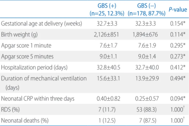
Neonatal outcomes such as gestational age at delivery, birth weight, Apgar score 1 min/5 min, hospitalization period, dura- tion of mechanical ventilation, neonatal CRP within 3 days, and other complications did not differ by GBS status (P>0.05). Two squared test was used for categorical variables and the Mann-
Whitney U test was used for continuous variables. Univariate analysis and multivariate logistic regression analysis were performed to detect associations with factors and to calculate an odds ratio (OR). Statistical analyses were performed by using SPSS (released 2009, PASW Statistics for Windows, version 18.0; SPSS Inc., Chicago, IL, USA). Null hypotheses of no difference were rejected if P values were less than 0.05.
Results
During the study period, 1,201 deliveries had occurred.
There were 732 term births and 469 preterm births. Two hundred three cases met the inclusion criteria of being a GBS carrier with a preterm birth (24+0 weeks to 36+6 weeks). Of the 203 women that underwent GBS culture analysis, 25 (12.3%) had a positive GBS culture result. Interestingly, the GBS detec- tion rate from samples obtained via the separate vagina/rectal swab method was 6.3%, whereas that from the rectovaginal swab method was 13.2%.
Characteristics of GBS-infected and non-infected subjects were not statistically different. Neither were sexually trans- mitted disease (STD) infection status nor chorioamnionitis status, which was diagnosed according to pathologic observa- tions, associated with the presence of GBS infection (P>0.05).
Moreover, there were no differences in the results of blood tests (maternal white blood cell count [WBC] and CRP) that could indicate signs of infection (P>0.05) (Table1).
The incidence of pPROM was significantly higher in the
Table 1. Study Subject Characteristics (n=203) GBS (+)
(n=25, 12.3%) GBS (−)
(n=178, 87.7%) P-value
Maternal age (years) 32.6±4.6 33.0±4.9 0.300*
STD infection (%) 4 (9.5) 38 (90.5) 0.610†
Chorioamnionitis (%) 8 (15.1) 45 (84.9) 0.675†
Maternal CRP 1.21±2.65 1.31±3.09 0.389*
Maternal WBC 11,125±3,581 10,923±3,581 0.445*
Values are presented as mean±standard deviation or number (%).
Abbreviations: GBS, group B Streptococcus agalactiae; STD, sexually transmitted disease; CRP, Creactive protein; WBC, white blood cell.
*MannWhitney test.
†Chisquared test.
Table 2. Obstetric Factors between GBS (+) and GBS (-) in Preterm Birth
GBS (+)
(n=25, 12.3%) GBS (−)
(n=178, 87.7%) P-value*
Causes of preterm birth (%)
SPTL 2 (8.0) 64 (36.0) 0.005
pPROM 14 (56.0) 54 (30.3) 0.022
IIOC 1 (4.0) 6 (3.4) 1.000
Maternal complications† 7 (28.0) 44 (24.7) 0.806
Fetal complications‡ 1 (4.0) 10 (5.6) 1.000
Values are presented as mean±standard deviation or number (%).
Abbreviations: GBS, group B Streptococcus agalactiae; SPTL, spontaneous pre
term labor; pPROM, preterm premature rupture of membrane; IIOC, incompetent internal os of cervix.
*Chisquared test.
†Maternal complications: pregnancyinduced hypertension, preeclampsia, placenta previa, placental abruption, uterine rupture.
‡Fetal complications: fetal distress, fetal growth restriction, fetal anomaly.
Table 3. Neonatal Outcomes between GBS (+) and GBS (−) in Preterm Birth
GBS (+)
(n=25, 12.3%) GBS (−)
(n=178, 87.7%) P-value Gestational age at delivery (weeks) 32.7±3.3 32.3±3.3 0.154*
Birth weight (g) 2,126±851 1,894±676 0.114*
Apgar score 1 minute 7.6±1.7 7.6±1.9 0.295*
Apgar score 5 minutes 9.0±1.1 9.0±1.4 0.273*
Hospitalization period (days) 32.8±40.5 32.7±40.0 0.412*
Duration of mechanical ventilation (days)
15.6±33.1 13.9±29.9 0.494*
Neonatal CRP within three days 0.40±0.82 0.25±0.57 0.094*
RDS (%) 7 (11.7) 53 (88.3) 1.000†
Neonatal deaths (%) 1 (12.5) 7 (87.5) 1.000†
Values are presented as mean±standard deviation or number (%).
Abbreviations: GBS, group B Streptococcus agalactiae; CRP, Creactive protein;
RDS, respiratory distress syndrome.
*MannWhitney test.
†Chisquared test.
neonates from GBS-infected women experienced GBS sepsis, and one neonate died (Table 3).
Based on univariate logistic analysis, GBS infection (OR 2.923, 95% confidence interval [CI] 1.247-6.851, P=0.014) was associated with pPROM. Among the maternal factors (ma- ternal age, STD infection, chorioamnionitis, maternal CRP, maternal WBC) and neonatal outcomes (gestational age at de- livery, birth weight, Apgar score 1 min/5 min, hospitalization period, duration of mechanical ventilation, neonatal CRP within three days, RDS, neonatal deaths), only gestational age at delivery (OR 1.020, 95% CI 1.005-1.034, P=0.007) and birth weight (OR 1.001, 95% CI 1.000-1.001, P<0.001) were asso- ciated with pPROM, based on the univariate logistic analysis results. After adjustment for gestational age at delivery and birth weight, GBS infection increased the risk of pPROM by 3.6 times (OR 3.648, 95% CI 1.476-9.016, P=0.005) (Table 4).
Discussion
Since the rate of GBS infection in Korea is considered low, there has been little apparent interest in perinatal complications associated with GBS infection in pregnant women. For this reason, Korea has not established guidelines for performing GBS screening during pregnancy. We suggest that, if multi- center medical facilities increase their level of interest in GBS infection and routinely conduct GBS analyses, the GBS dia- gnosis rate in Korea may increase.
In our review, 395 of 1,201 patients underwent GBS infection assessment. Thirty-eight (38/395, 9.6%) of the swab culture- assessed patients were positive for GBS. The percentage of GBS-positive patients with preterm births in the study sample was 12.3% (25/203). This incidence is higher than the infection rate previously reported in Korea but lower than that of other countries.10,12-14,16
GBS prevalence and GBS serotype distribution differ by study region and country. Early-onset neonatal GBS disease has a different distribution among pregnant women in Korea than among those in other countries.6,17,18 The apparent incre- ase in GBS infection may be due to a change in serotype follow- ing changes in dietary habits and globalization. Further epide- miological studies that include large sample sizes are needed in Korea.
A CDC guideline recommends the use of rectovaginal sam- pling for GBS detection.3 Based on our swab culture analysis results, after changing the GBS culture method from the use of separate vagina/rectum swabs to using a combined rectova- ginal swab, the GBS detection rate increased. Previous studies have also shown that rectovaginal sampling results in an in- creased GBS-positive rate.19 However, our study results have a limitation in that no comparisons were made between the two swabbing methods in the same patients.
In our study, the most common cause of preterm birth was pPROM. All patients diagnosed with pPROM were treated with antibiotics, and patients without evidence of pPROM were not treated with antibiotics. Until the vaginal culture results were available, intravenous cephalosporin injections were given to treat a broad spectrum of bacteria in pPROM patients. If the presence of GBS before delivery was confirmed, penicillin or cephalosporin treatment was applied. Treatment for GBS involves administration of penicillin intravenously every 4 hours until delivery, while cephalosporin treatment can be used for patients with a penicillin allergy. For most of our study subjects, delivery was complete before culture results were confirmed, and most patients with pPROM were treated with cephalosporin, which is not the optimal treatment for GBS in- fection. Failure to apply the most appropriate antibiotic for GBS is a weakness of the treatments reported in this study.
In this study, SPTL appeared to be related to GBS presence.
However, the univariate logistic analysis calculated OR was Table 4. Logistic Regression-Derived ORs and 95% CIs for the Association between GBS and pPROM
Univariate Multivariate
OR (95% CI) P-value OR (95% CI) P-value
Gestational age at delivery 1.020 (1.0051.034) 0.007 0.987 (0.9621.014) 0.353
Birth weight 1.001 (1.0001.001) <0.001 1.001 (1.0001.002) 0.002
GBS infection 2.923 (1.2476.851) 0.014 3.648 (1.4769.016) 0.005
Abbreviations: GBS, group B Streptococcus agalactiae; pPROM, preterm premature rupture of membrane; OR, odds ratio; Cl, confidence interval.
0.154 (95% CI 0.035-0.672, P=0.13). The causes of SPTL are various, and sometimes it is caused by an unknown factor. Our data suggest that maternal GBS infection is not a major cause of SPTL. However, the frequency of SPTL among GBS-infected women was too small (8.0%) to produce accurate results.
In our study, GBS infection was a risk factor for pPROM. We assume that when a maternal GBS infection occurs, it advances through an increasing abundance of GBS, eventually leading to amniotic membrane rupture.
Each of the maternal and fetal complication factors in this study was grouped and classified into two broad categories (maternal or fetal) because each of the individual factors had a small sample size and produced no statistically significant difference related to GBS infection status.
During the study period, there were 68 cases of pPROM in women with preterm births. According to the hospital protocol, all neonates born with pPROM are treated with antibiotics, usually amoxicillin. In this study, all of the neonates whose mothers were identified as GBS infected were observed for at least 24 hours for signs of sepsis and were treated with amo- xicillin prophylactically. This protocol has been previously reported to prevent GBS infection and improve neonatal out- comes.20 Perhaps this protocol was responsible for our results that GBS infection did not affect factors such as Apgar scores, hospitalization periods, duration of mechanical ventilation, RDS, and infant mortality rates in our study.
In the case of neonatal death in this study, the GBS-infected mother had a history of gestational diabetes mellitus and SPTL. She had been treated with tocolytics at 31+5 weeks for 1 week and delivered at 34+5 weeks. She did not receive anti- biotic treatment because her GBS status was not confirmed before delivery. The newborn expired within 16 hours of birth.
Blood culture after birth revealed the presence of GBS and the neonate was diagnosed as GBS sepsis.
Ganor-Paz et al.21 concluded that GBS carriers with pPROM do not have adverse outcomes. In Ganor-Paz’s study21, all patients with pPROM were treated with appropriate antibiotics according to a hospital protocol. Those results suggest that complications related to GBS infection such as neonatal sepsis, neonatal infection, and a low 5 minutes Apgar score can be prevented. Based on the results of this and previous studies, further research examining pPROM in GBS carrier patients in
our medical center is needed.
In conclusion maternal GBS infection in preterm birth was associated with pPROM, but it did not affect neonatal outcome.
Considering the prognosis of GBS, a major cause of neonatal sepsis and pneumonia, national GBS-related protocols are needed. Along with a GBS screening protocol, accurate screen- ing methods and intrapartum prophylactic antibiotic admini- stration are needed to prevent adverse neonatal outcomes in GBS-infected mothers.
References
1) Edwards MS, Nizet V, Baker CJ. Group B Streptococcal Infections. In:
Remington JS, Klein JO, Wilson CB, et al. editors. Infectious Diseases of the Fetus and Newborn Infant. 7th ed, Philadelphia, Elsevier Saunders, 2011, p419-69.
2) Eichenwald EC. Perinatally transmitted neonatal bacterial infections.
Infect Dis Clin North Am 1997;11:223-39.
3) Verani JR, McGee L, Schrag SJ; Division of Bacterial Diseases, National Center for Immunization and Respiratory Diseases, Centers for Disease Control and Prevention (CDC). Prevention of perinatal group B strepto- coccal disease--revised guidelines from CDC, 2010. MMWR Recomm Rep 2010;59(RR-10):1-36.
4) Regan JA, Klebanoff MA, Nugent RP, Eschenbach DA, Blackwelder WC, Lou Y, et al. Colonization with group B streptococci in pregnancy and adverse outcome. VIP Study Group. Am J Obstet Gynecol 1996;174:
1354-60.
5) Krohn MA, Hillier SL, Baker CJ. Maternal peripartum complications asso- ciated with vaginal group B streptococci colonization. J Infect Dis 1999;
179:1410-5.
6) Phares CR, Lynfield R, Farley MM, Mohle-Boetani J, Harrison LH, Petit S, et al. Epidemiology of invasive group B streptococcal disease in the United States, 1999-2005. JAMA 2008;299:2056-65.
7) Koenig JM, Keenan WJ. Group B streptococcus and early-onset sepsis in the era of maternal prophylaxis. Pediatr Clin North Am 2009;56:689- 708, Table of Contents.
8) Gerdes JS. Diagnosis and management of bacterial infections in the neonate. Pediatr Clin North Am 2004;51:939-59, viii-ix.
9) Heath PT, Balfour G, Weisner AM, Efstratiou A, Lamagni TL, Tighe H, et al.
Group B streptococcal disease in UK and Irish infants younger than 90 days. Lancet 2004;363:292-4.
10) Schrag S, Gorwitz R, Fultz-Butts K, Schuchat A. Prevention of perinatal group B streptococcal disease. Revised guidelines from CDC. MMWR Recomm Rep 2002;51(RR-11):1-22.
11) Van Dyke MK, Phares CR, Lynfield R, Thomas AR, Arnold KE, Craig AS, et al. Evaluation of universal antenatal screening for group B streptoco- ccus. N Engl J Med 2009;360:2626-36.
12) Benitz WE, Gould JB, Druzin ML. Risk factors for early-onset group B streptococcal sepsis: estimation of odds ratios by critical literature re- view. Pediatrics 1999;103:e77.
13) Hong JS, Choi CW, Park KU, Kim SN, Lee HJ, Lee HR, et al. Genital group B Streptococcus carrier rate and serotype distribution in Korean preg- nant women: implications for group B streptococcal disease in Korean neonates. J Perinat Med 2010;38:373-7.
14) Lee BK, Song YR, Kim MY, Yang JH, Shin JH, Seo YS, et al. Epidemiology of group B streptococcus in Korean pregnant women. Epidemiol Infect 2010;138:292-8.
15) Zhang LX, Sun Y, Zhao H, Zhu N, Sun XD, Jin X, et al. A bayesian stepwise discriminant model for predicting risk factors of preterm premature rupture of membranes: a case-control study. Chin Med J (Engl) 2017;
130:2416-22.
16) Yoon IA, Jo DS, Cho EY, Choi EH, Lee HJ, Lee H. Clinical significance of serotype V among infants with invasive group B streptococcal infec- tions in South Korea. Int J Infect Dis 2015;38:136-40.
17) Persson E, Berg S, Trollfors B, Larsson P, Ek E, Backhaus E, et al. Serotypes and clinical manifestations of invasive group B streptococcal infections in western Sweden 1998-2001. Clin Microbiol Infect 2004;10:791-6.
18) Lachenauer CS, Kasper DL, Shimada J, Ichiman Y, Ohtsuka H, Kaku M, et al. Serotypes VI and VIII predominate among group B streptococci isolated from pregnant Japanese women. J Infect Dis 1999;179:1030-3.
19) El Aila NA, Tency I, Claeys G, Saerens B, Cools P, Verstraelen H, et al.
Comparison of different sampling techniques and of different culture methods for detection of group B streptococcus carriage in pregnant women. BMC Infect Dis 2010;10:285.
20) Benitz WE, Gould JB, Druzin ML. Antimicrobial prevention of early-onset group B streptococcal sepsis: estimates of risk reduction based on a critical literature review. Pediatrics 1999;103:e78.
21) Ganor-Paz Y, Kailer D, Shechter-Maor G, Regev R, Fejgin MD, Biron- Shental T. Obstetric and neonatal outcomes after preterm premature rupture of membranes among women carrying group B streptococcus.
Int J Gynaecol Obstet 2015;129:13-6.