https://doi.org/10.14734/PN.2017.28.4.119 pISSN 2508-4887•eISSN 2508-4895
Jeong Min Shin, MD1,2, Jin A Lee, MD, PhD1,3, Hye Ri Yun, MD1,2, Sohee Oh, PhD4, Jin A Sohn, MD, PhD1,3, Seung Han Shin, MD, PhD1,2, Chang Won Choi, MD, PhD1,5, Ee-Kyung Kim, MD, PhD1,2, Han-Suk Kim, MD, PhD1,2, Byeong-Il Kim, MD, PhD1,5
1Department of Pediatrics, Seoul National University College of Medicine, 2Department of Pediatrics, Seoul National University Hospital,
3Department of Pediatrics, 4Depart
ment of Biostatistics, Seoul National University Boramae Medical Center,
5Department of Pediatrics, Seoul National University Bundang Hospi tal, Seoul, Korea
Objective: We assessed the influence of small for gestational age (GA) with placental disorders (SGAP) and histologic chorioamnionitis (HCA) on the inhospital outcomes of preterm infants.
Methods: Preterm infants with a GA <32 weeks born at Seoul National University Hospital between 2007 and 2014 were included and divided into 4 groups according to the presence of SGAP and HCA: group 1, SGAP only; group 2, HCA only; group 3, both SGAP and HCA; and group 4, no SGAP or HCA. Multivariate logistic regression was done to compare neonatal outcomes including death, moderate to severe bronchopulmonary dysplasia (BPD) or death, patent ductus arteriosus with treat
ment, sepsis, necrotizing enterocolitis ≥stage 2b, and intraventricular hemorrhage ≥grade 3.
Results: A total of 572 infants were included. There were 77 patients (13.5%) in group 1, 226 patients (39.5%) in group 2, and 24 patients (4.2%) in group 3. After adjusting for GA, cesarean section, 5 minute Apgar score, multiple pregnancy, premature rupture of membrane before 18 hours prior to delivery, and preeclampsia, group 1 showed higher risks of mortality (adjusted odds ratio [aOR] 3.15, 95% confidence interval [CI] 1.138.80), moderate to severe BPD or death (aOR 9.12, 95% CI 3.98
20.90), sepsis (aOR 2.12, CI 1.014.46), and pulmonary hypertension (aOR 3.26, 95% CI 1.159.22) compared with group 4. There were no significant differences in mortality and inhospital outcomes between groups 2 and 4 or between groups 3 and 4.
Conclusion: Close monitoring and early intervention are suggested in SGAP infants.
Key Words: Preterm infants, Placenta disorders, Fetal growth retardation, Chorioamnionitis, Patient outcome assessment
Introduction
Preterm infants have a high mortality and morbidities throughout their lives. Previous reports have suggested that pregnancy disorders causing preterm delivery also affect the neonatal outcomes of preterm infants.1 Recently, many epidemiologists have been trying to group pregnancy disorders associated with preterm delivery to find common therapeutic interventions. McElrath et al.2 and Klebanoff et al.3 classified disorders leading to preterm delivery into 2 groups: disorders with intrauterine inflammation and those associated with primary aberrations of placentation. Histologic chorioamnionitis (HCA) is a representative disorder of intrauterine inflammation, and hypertensive disorders of pregnancy and intrau
terine growth restriction (IUGR) are representative disorders of aberrant placentation.
IUGR is defined as inutero growth retardation, and small for gestational age (SGA) is Received: 16 January 2017
Revised: 5 July 2017 Accepted: 5 September 2017 Correspondence to Jin A Lee, MD, PhD
Department of Pediatrics, Seoul Naitonal University Boramae Medical Center, 20 Boramaero 5gil, Dongjakgu, Seoul 07061, Korea Tel: +8228702364
Fax: +8228312826 E-mail: [email protected]
Copyright© 2017 by The Korean Society of Perinatology
This is an Open Access article distributed under the terms of the Creative Com
mons Attribution NonCommercial License (http://creativecommons.org/
license/bync/4.0/), which permits
The Influence of Pregnancy Disorders
Caus ing Preterm Delivery on In-Hospital
Outcomes in Preterm Infants at Less than
32 Weeks of Gestation
ably, and only 12% were not SGA among IUGR neonates. How
ever, some neonatologist reported that about 1/3 of SGA cases were not fetal growth restriction.4 Among maternal, environ
mental and fetal causes of IUGR or SGA, placental problems such as placental insufficiency, preeclampsia, and placental abruption leads to a preterm birth. There have been studies that have reported that SGA is a significant risk factor of mor
tality, bronchopulmonary dysplasia (BPD), or sepsis.5 Addi
tionally, others have reported that such associations occurred regardless of whether IUGR was suspected antenatally.4
For the association between HCA and neonatal outcomes of preterm infants, some studies have reported that HCA decre
ases the frequency of respiratory distress syndrome (RDS) in preterm infants.6 However, it is still controversial whether HCA increases the risk of BPD or a poor neurodevelopmental outcome.79 Such reports have classified their study population according to the presence of HCA only; thus, the control group could contain other significant pregnancy disorders, which could affect neonatal outcomes, and do not represent a ‘normal’
con trol group. Additionally, both SGA and HCA are stressful conditions, and mortality and adverse neonatal outcomes can increase due to the combined effects of SGA and HCA; how
ever, there are few reports on the proportion of infants with both SGA and HCA as well as on comparing the combined effects of SGA and HCA on neonatal outcomes.
Thus, this study assessed whether there are any differences in major neonatal morbidities in preterm infants with a gesta
tional age (GA) <32 weeks according to the presence of SGA with placental disorders (SGAP) and HCA, which are repre
sentative disorders for the 2 major types of disorders causing preterm delivery.
Methods
1. Study design
A retrospective study was done for 704 infants who were born and admitted to the neonatal intensive care unit of Seoul National University Children’s hospital between 2007 and 2014. Clinical and demographic data were collected from the reviewed medical records of the enrolled patients. Thirty
seven infants who had major congenital anomalies, 47 twin to
twin transfusion syndrome patients, 1 paroxysmal supraven
tricular tachycardia, 5 fetal hydrops and 2 maternal systemic lupus erythematosus were excluded. Twentythree patients with inadequate medical records and 1 patient admitted for more than 1 year were also excluded. Sixteen SGA patients without placental disorders (such as SGA due to maternal mal
nutrition or no specific cause) were excluded. Finally, a total of 572 preterm infants were included in the analysis (Fig. 1).
We categorized the entire study population into four groups according to the presence of SGAP or HCA. Group 1 consisted of patients with SGAP only, group 2 with HCA only, group 3 with both SGAP and HCA, and group 4 with no SGAP or HCA.
Then, we compared the baseline demographic characteristics including the GA at birth, birthweight, gender, cesarean sec
tion, multiple pregnancy, antenatal steroid use started before 12 hours from birth, preeclampsia, premature rupture of mem
brane (PROM) before 18 hours from birth, maternal gestational diabetes, cord pH, and Apgar score at 1 and 5 minutes between the two groups. We also examined the difference in the mor
tality and inhospital outcomes such as surfactant use, patent ductus arteriosus (PDA) requiring treatment, inhaled nitric oxide treatment before 14 postnatal days, pulmonary hyperten
sion, bronchopulmonary dysplasia (BPD), sepsis, necroti zing enterocolitis (NEC) ≥stage 2b, intraventricular hemorrhage (IVH) ≥grade 3, retinopathy of prematurity (ROP) requiring Fig. 1. Flow chart of the study population. A total of 572 patients with
<32 weeks of gestation born between 2007 and 2014 were included in the analysis. 77 patients (13.5%) were in group 1, 226 patients (39.5%) were in group 2, and 24 patients (4.2%) were in group 3. GA, gestational age; SNUCH, Seoul National University Children’s Hospital; TTTS, twin to twin transfusion syndrome; PSVT, paroxysmal supraventricular tachycardia; SLE, systemic lupus erythematosus; SGA-P; small for gestational age with placental disorders; HCA, histologic chorio- amnionitis.
difference in the duration of hospital stay when compared with group 4. The statistical analysis was done with IBM SPSS Sta
tistics version 20 (IBM Corp., Armonk, NY, USA) and R version 3.3.2. (The R Foundation, Vienna, Austria). P values less than 0.05 were considered statistically significant.
Results
A total of 572 patients were included in the final analysis.
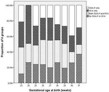
Seventyseven patients (13.5%) were in group 1, 226 patients (39.5%) in group 2, 24 patients (4.2%) in group 3, and 245 pati
ents (42.8%) in group 4 (Fig. 1). The proportion of the group 2 decreased and the proportion of the group 1 increased as the GA increased (P=0.045, Fig. 2). In group 1, causes of SGA were as follows: preeclampsia in 12 infants (15.6%), absent or re
versed umbilical artery enddiastolic flow in 26 infants (33.8
%), both preeclampsia and absent or reversed umbilical artery enddiastolic flow in 27 infants (35.1%), fetal growth restriction with fetal distress due to placental insufficiency in 11 infants (14.3%), and placental abruptio in 1 infants (1.3%). In group 3, causes of SGA were preeclampsia in 4 infants (16.7%), absent surgery or vascular endothelial growth factor (VEGF) treat
ment, discharge with respiratory support, and the duration of hospital stay between the two groups.
2. Definitions
Preeclampsia was defined as any maternal diagnoses of preeclampsia, eclampsia or hemolysis, elevated liver enzymes, and low platelet count syndrome. SGA was defined according to the definitions published by Olsen et al.10 SGAP was defined when causes of SGA were as follows: preeclampsia, absent or reversed umbilical artery enddiastolic flow, fetal growth re
striction with fetal distress due to placental insufficiency, and placenta abruptio. HCA was defined according to the definition of Salafia et al.11: the presence of acute inflammation and infil
tration of polymorphonuclear leukocytes in the amnion, chorio
nic decidua, umbilical cord, or the chorionic plate reported by pathologists in our hospital. Sepsis was defined as the pre sence of clinical symptoms and signs with proven causative organisms documented from blood cultures. If the organisms were iden
tified within 7 postnatal days, it was defined as early sepsis. If the organisms were identified after 8 or more post natal days, it was defined as late sepsis. Pulmonary hyperten sion was defin
ed according to the echocardiographic findings previously published.12 Discharge with respiratory support was defined as discharge with oxygen supply or home ventila tor support.
3. Statistical analysis
All continuous variables are expressed as the median (inter
quartile range), and all categorical variables are express ed as numbers and proportions. For the univariate analysis, continu
ous variables were compared with the KruskalWallis test, and categorical variables were compared with the chisquare test or Fisher’s exact test. To assess the independent association between the presence of SGAP or HCA and neonatal out
comes including mortality, moderate to severe BPD or death, severe BPD or death, PDA with treatment, sepsis, IVH ≥grade 3 of Papile’s classification,13 NEC ≥stage 2 of Bell’s criteria,14 ROP requiring surgery or VEGF treatment, and pulmonary hy
pertension, binary logistic regression analysis was done adju
sting for GA, cesarean section, 5 minute Apgar score, multiple pregnancy, PROM, and preeclampsia. We also performed mul
tiple linear regression analysis to assess whether there is any
Fig. 2. The proportion of 4 groups according to the gestational age at birth. The percentage of histologic chorioamnionitis group was de- creased and the percentage of small for gestational age with placental disorders was increased as the gestational age gets older (P=0.045).
SGA-P, small for gestational age with placental disorders; HCA, histolo- gic chorioamnionitis.
or reversed umbilical artery enddiastolic flow in 6 infants (25.0%), both preeclampsia and absent or reversed umbilical artery enddiastolic flow in 11 infants (45.8%), and fetal growth restriction with fetal distress due to placental insufficiency in 3 infants (12.5%).
1. Baseline and demographic characteristics
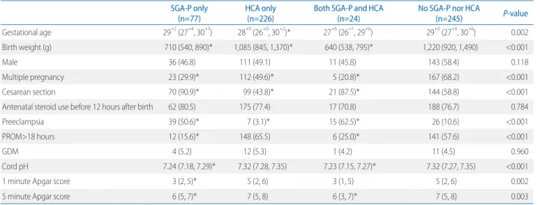
There were significant differences in the GA, birthweight, multiple pregnancy, cesarean section, cord pH, and the 1 and 5 minute Apgar scores between the four groups. When compared with group 4, group 2 was younger, and the birthweights of groups 1, 2, and 3 were lower. The proportion of infants deli
vered from mothers with preeclampsia was higher in group 1 and 3, and lower in group 2 when compared with group 4. Cord pH and 5 minute Apgar score were lower in groups 1 and 3 when compared with group 4 (Table 1).
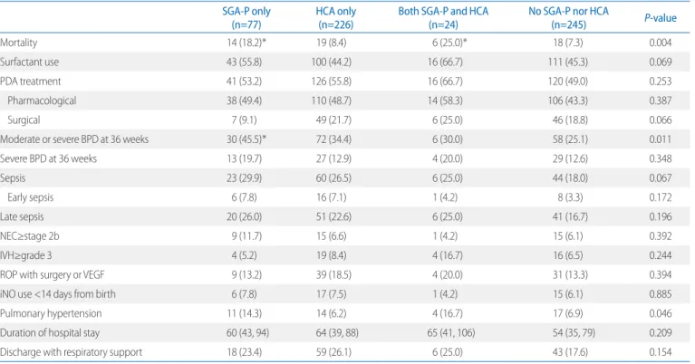
2. Mortality and in-hospital outcomes
In the univariable analysis, there were significant differences in the mortality, moderate to severe BPD at 36 weeks of cor
rected age, and pulmonary hypertension between the 4 groups.
When compared with group 4, the mortality was higher in groups 1 and 3 (Table 2). The proportion of infants with mod
erate to severe BPD was higher in group 1 when compared
with group 4.
After the multivariable analysis adjusting for GA, cesarean section, 5 minute Apgar score, multiple pregnancy, PROM be fore 18 hours prior to delivery, and preeclampsia, group 1 showed higher risks for death, moderate to severe BPD or death, sepsis, and pulmonary hypertension when compared with group 4 (Table 3). When compared with group 4, there were no signi
ficant differences of neonatal outcomes in group 2 and 3.
After multiple linear regression analysis adjusting for GA, cesarean section, 5 minute Apgar score, multiple pregnancy, PROM before 18 hours prior to delivery, and preeclampsia, there was no significant difference in the duration of hospital stay when compared with group 4 (group 1: β 10.39, 95% CI –1.8927.53, P-value=0.151; group 2: β –2.71, 95% CI –9.97- 3.06, P-value=0.405; group 3: β –0.46, 95% CI –20.18-13.99, Pvalue=0.957).
Discussion
In our study, SGAP was a major risk factor of mortality and adverse inhospital outcomes in preterm infants. However, HCA was not a significant risk factor of mortality or any neo
natal morbidities, and in infants with both SGAP and HCA,
Table 1. Baseline Demographic Characteristics of the 4 Groups According to the Presence of SGA-P and HCA SGA-P only
(n=77) HCA only
(n=226) Both SGA-P and HCA
(n=24) No SGA-P nor HCA
(n=245) P-value
Gestational age 29+2 (27+4, 30+3) 28+0 (26+0, 30+2)* 27+6 (26+1, 29+6) 29+0 (27+0, 30+6) 0.002
Birth weight (g) 710 (540, 890)* 1,085 (845, 1,370)* 640 (538, 795)* 1,220 (920, 1,490) <0.001
Male 36 (46.8) 111 (49.1) 11 (45.8) 143 (58.4) 0.118
Multiple pregnancy 23 (29.9)* 112 (49.6)* 5 (20.8)* 167 (68.2) <0.001
Cesarean section 70 (90.9)* 99 (43.8)* 21 (87.5)* 144 (58.8) <0.001
Antenatal steroid use before 12 hours after birth 62 (80.5) 175 (77.4) 17 (70.8) 188 (76.7) 0.784
Preeclampsia 39 (50.6)* 7 (3.1)* 15 (62.5)* 26 (10.6) <0.001
PROM>18 hours 12 (15.6)* 148 (65.5) 6 (25.0)* 141 (57.6) <0.001
GDM 4 (5.2) 12 (5.3) 1 (4.2) 11 (4.5) 0.960
Cord pH 7.24 (7.18, 7.29)* 7.32 (7.28, 7.35) 7.23 (7.15, 7.27)* 7.32 (7.27, 7.35) <0.001
1 minute Apgar score 3 (2, 5)* 5 (2, 6) 3 (1, 5) 5 (2, 6) 0.002
5 minute Apgar score 6 (5, 7)* 7 (5, 8) 6 (3, 7)* 7 (5, 8) 0.003
Values are presented as median (interquartile range) or number (%).
Abbreviations: SGAP, small for gestational age with placental disorders; HCA, histologic chorioamnionitis; PROM, premature rupture of membranes; GDM, gestational diabetes mellitus.
*P<0.05 versus no SGAP nor HCA.
there were no significant differences in mortality and major neonatal outcomes when compared with the no SGAP or HCA
group.
Many pregnancy disorders such as spontaneous preterm
Table 2. In-hospital Outcomes of the 4 Groups According to the Presence of SGA-P and HCA SGA-P only
(n=77) HCA only
(n=226) Both SGA-P and HCA
(n=24) No SGA-P nor HCA
(n=245) P-value
Mortality 14 (18.2)* 19 (8.4) 6 (25.0)* 18 (7.3) 0.004
Surfactant use 43 (55.8) 100 (44.2) 16 (66.7) 111 (45.3) 0.069
PDA treatment 41 (53.2) 126 (55.8) 16 (66.7) 120 (49.0) 0.253
Pharmacological 38 (49.4) 110 (48.7) 14 (58.3) 106 (43.3) 0.387
Surgical 7 (9.1) 49 (21.7) 6 (25.0) 46 (18.8) 0.066
Moderate or severe BPD at 36 weeks 30 (45.5)* 72 (34.4) 6 (30.0) 58 (25.1) 0.011
Severe BPD at 36 weeks 13 (19.7) 27 (12.9) 4 (20.0) 29 (12.6) 0.348
Sepsis 23 (29.9) 60 (26.5) 6 (25.0) 44 (18.0) 0.067
Early sepsis 6 (7.8) 16 (7.1) 1 (4.2) 8 (3.3) 0.172
Late sepsis 20 (26.0) 51 (22.6) 6 (25.0) 41 (16.7) 0.196
NEC≥stage 2b 9 (11.7) 15 (6.6) 1 (4.2) 15 (6.1) 0.392
IVH≥grade 3 4 (5.2) 19 (8.4) 4 (16.7) 16 (6.5) 0.244
ROP with surgery or VEGF 9 (13.2) 39 (18.5) 4 (20.0) 31 (13.3) 0.394
iNO use <14 days from birth 6 (7.8) 17 (7.5) 1 (4.2) 15 (6.1) 0.885
Pulmonary hypertension 11 (14.3) 14 (6.2) 4 (16.7) 17 (6.9) 0.046
Duration of hospital stay 60 (43, 94) 64 (39, 88) 65 (41, 106) 54 (35, 79) 0.209
Discharge with respiratory support 18 (23.4) 59 (26.1) 6 (25.0) 43 (17.6) 0.154
Values are presented as number (%) or median (interquartile range).
Abbreviations: SGAP, small for gestational age with placental disorders; HCA, histologic chorioamnionitis; PDA, patent ductus arteriosus; BPD, bronchopulmonary dysplasia;
NEC, necrotizing enterocolitis; IVH, intraventricular hemorrhage; ROP, retinopathy of prematurity; VEGF, vasculoendothelial growth factor; iNO, inhaled nitric oxide.
*P<0.05 versus no SGAP nor HCA.
Table 3. Multivariable Analysis of Mortality and In-hospital Outcomes according to the Presence of SGA-P or HCA (Reference: No SGA-P nor HCA)
SGA-P only HCA only Both SGA-P and HCA
aOR (95% CI) P-value aOR (95% CI) P-value aOR (95% CI) P-value
Death 3.15 (1.13, 8.80) 0.029 0.88 (0.42, 1.87) 0.741 2.76 (0.73, 10.41) 0.133
Moderate to severe BPD or death 9.12 (3.98, 20.90) <0.001 1.05 (0.61, 1.81) 0.853 1.53 (0.44, 5.32) 0.509
Severe BPD or death 3.73 (1.62, 8.58) 0.002 0.68 (0.39, 1.21) 0.191 1.82 (0.55, 6.03) 0.325
PDA treatment 0.98 (0.51, 1.90) 0.961 1.14 (0.74, 1.78) 0.553 1.14 (0.40, 3.28) 0.806
Sepsis 2.12 (1.01, 4.46) 0.046 1.38 (0.84, 2.26) 0.207 1.07 (0.35, 3.27) 0.913
Early sepsis 3.79 (1.04, 13.81) 0.044 2.06 (0.82, 5.15) 0.122 1.70 (0.17, 16.95) 0.651
Late sepsis 1.65 (0.77, 3.54) 0.200 1.19 (0.71, 2.00) 0.499 1.00 (0.33, 3.08) 0.998
IVH≥grade 3 0.48 (0.13, 1.81) 0.277 1.00 (0.47, 2.12) 0.999 1.34 (0.34, 5.36) 0.675
NEC≥stage 2b 2.42 (0.79, 7.43) 0.123 0.79 (0.35, 1.75) 0.554 0.47 (0.05, 4.28) 0.499
Pulmonary hypertension 3.26 (1.15, 9.22) 0.026 0.71 (0.33, 1.54) 0.383 2.37 (0.56, 9.97) 0.240
ROP with surgery or VEGF 1.64 (0.51, 5.24) 0.404 1.23 (0.65, 2.33) 0.535 1.65 (0.34, 8.04) 0.536
Adjusted for Gestational age at birth (weeks), cesarean section, 5 minute Apgar score, multiple pregnancy, premature rupture of membranes before 18 hours prior to delivery, and preeclampsia.
Abbreviations: SGAP, small for gestational age with placental disorders; HCA, histologic chorioamnionitis; aOR, adjusted odds ratio; CI, confidence interval; BPD, bronchopulmonary dysplasia; PDA, patent ductus arteriosus; IVH, intraventricular hemorrhage; NEC, necrotizing enterocolitis; ROP, retinopathy of prematurity; VEGF, vasculoendothelial growth factor.
labor, hypertensive disorders of pregnancy, chorioamnionitis, IUGR, and maternal hemorrhage lead to preterm birth. Some epidemiologists group the pregnancy complications ending in preterm birth according to a common etiology and relationship to outcomes: a placental histologic inflammation and poor pla
centation such as infarcts and an abundance of syncytial knots.2 In our study, because we performed a placental biopsy in all the mothers who delivered preterm infants, we collected accurate information about histologic chorioamnionitis. We included SGA infants only associated with placentation disorders because many other factors such as genetic, chromosomal, and maternal nutrition factors can also cause SGA but usually are not accom
panied with preterm birth. Most of our SGAP group repre
sented IUGR; however, because this study was not a prospec
tive study and we could not get accurate information on in
trauterine growth, we used the term SGAP instead of IUGR.
SGA is known to be a significant risk factor of neonatal death, BPD, and nosocomial infection; however, the results are still controversial.5,1517 In our study, the risk of neonatal death, BPD, sepsis, and pulmonary hypertension was higher in infants with SGAP only; however, the risk of such neonatal outcomes was not increased in infants with both SGAP and HCA. We could not examine the longterm neurodevelopmental out
come; thus, further study will be needed.
In our study, only 4.2% of infants had both SGAP and HCA.
Recently, the proportion of microbial invasion of the amniotic cavity in pregnancies with SGA detected by the 16S ribosomal DNA polymerase chain reaction method was also 6%, which is smaller than that of other conditions (13% of women with pre
term labor, 32% of preterm PROM, and about 50% of cervical insufficiency).18 Although the proportion of infants with both SGAP and HCA was low, HCA can have a protective role in SGA patients for mortality and BPD, and there were no signifi
cant differences in neonatal outcomes when compared with the control groups.
In our study, we classified our study population into 4 groups according to the presence of SGAP and HCA. Actually, hy
pertensive disorders of pregnancy including preeclampsia are also known to be due to abnormal placentation. SGA can be an indicator of the severity of preeclampsia and has been attri
buted to a restricted arteriolar supply of the uterineplacental interface.19 When we also assessed the difference in neonatal
outcomes between infants with preeclampsia or SGAP only, HCA only, both preeclampsia/SGAP and HCA, and no pre
eclampsia/SGAP or HCA, the risks of moderate to severe BPD or death and sepsis were higher only in infants with pre
eclampsia or SGAP when compared with the no preeclampsia/
SGA or HCA group, which was similar to our results (data not shown). However, when we classified our study population into infants with preeclampsia only, HCA only, both preeclampsia and HCA, and no preeclampsia or HCA, there were no signi
ficant associations between neonatal outcomes and pre
eclampsia only, HCA only or both preeclampsia and HCA (data not shown). We suggest that SGAP is a potent risk factor of mortality and major adverse outcomes in preterm infants.
According to the annual report of the Korean neonatal net
work (KNN) released on 2015, HCA is a more prevalent an
tenatal condition in extreme premature infants. HCA accounts for 46.5% of the causes of preterm birth among preterm infants born less than GA 24 weeks.20 Our results also show the same distribution of pregnancy disorders according to GA, and when there were both HCA and SGAP, the GA and birth weight were the lowest between the 4 groups. Although we wanted to match the GA and birthweight in the 4 groups, because of the small number of study subjects, we could not match our cohort, and multiple logistic regression analysis was done instead of matching.
In our cohort, HCA had no association with any neonatal morbidities, which somewhat contrasted other previous re
sults. Previous studies have reported that HCA can increase the incidence of mortality, BPD, early sepsis, and longterm adverse neurodevelopmental outcomes.8,2123 Some evidence supports that the incidence of RDS was decreased in infants with maternal HCA, and in animal studies, intraamniotic injec
tion of Escherichia coli increased surfactant protein synthesis.24 Such reports classified their study population according to the presence of HCA only; thus, the control group patients could be mixed up with other significant pregnancy disorders which did not represent a ‘normal’ control group. Our control group did not have any infants with maternal SGAP or HCA, which could give such different results for the influence on neonatal outcomes.
In many reports, neonatal mortality was higher in pregnancy disorders associated with placental dysfunction than in those
disorders with intrauterine inflammation.2 When Gagliardi et al.1 compared the disorders of placentation including hyperten
sive disorders of the pregnancy and the IUGR group to the presumed infection/inflammation group, the risk of mortality, BPD and ROP was higher especially in growthrestricted infants, and the risks of IVH and periventricular leukomalacia were lower in the disorders of placentation group.
In our study, the frequency of sepsis was higher in infants with SGAP when compared with infants with no diagnosis of SGAP or HCA. Tröger et al.25 reported that SGA increased the incidence of late onset culture proven sepsis in VLBW in
fants. Thymic atrophy and lymphopenia and deficiencies in humoral responses in SGA infants were reported.26,27 Addi
tionally, in the first week from birth, white blood cell counts are usually decreased in infants with SGA or preeclampsia which is common.
Pulmonary hypertension is known to be associated with BPD.28 Recently, there have been some reports that placental pathologic changes of maternal vascular underperfusion in BPD are associated with increased risk of pulmonary hyper
tension,29 and after a systematic review and metaanalysis, oligohydramnios and SGA were shown as significant risk fac
tors of pulmonary hypertension in infants with BPD.30 Addi
tionally, in our study, SGAP was a significant risk factor of pulmonary hypertension.
Our study has some limitations. First, this is a retrospective study with a small number of patients in a single center. Second, when compared to the prospective EPIPAGE2 study, we could not include all the antenatal factors causing preterm de
livery in the classification of the groups because of incom plete medical records due to the retrospective study design. Thus, we decided not to differentiate our whole study population according to the classification guided by antenatal pregnancy complications such as in the EPIPAGE study, and we only used SGAP and HCA as a standard to classify our groups. A pro
spective study with a more sophisticated grouping accord ing to the antenatal pregnancy disorders should be done in the future.
In our study, the proportion of infants with both SGAP and HCA was very small in preterm infants, and there were no significant differences in neonatal outcomes when compared to the control group. SGAP was a significant risk factor of mor
tality and moderate to severe BPD or death, sepsis, and pul
monary hypertension in preterm infants. We should give atten
tion to the close monitoring and prevention of sepsis, BPD, and pulmonary hypertension in the care of preterm infants with SGAP.
Acknowledgements
This study was approved by the institutional review board (IRB No. 262013126). The need for informed consent was exempted because of the retrospective nature of this study.
References
1) Gagliardi L, Rusconi F, Da Frè M, Mello G, Carnielli V, Di Lallo D, et al.
Pregnancy disorders leading to very preterm birth influence neonatal outcomes: results of the population-based ACTION cohort study.
Pediatr Res 2013;73:794-801.
2) McElrath TF, Hecht JL, Dammann O, Boggess K, Onderdonk A, Marken- son G, et al. Pregnancy disorders that lead to delivery before the 28th week of gestation: an epidemiologic approach to classification. Am J Epidemiol 2008;168:980-9.
3) Klebanoff MA, Shiono PH. Top down, bottom up and inside out:
reflections on preterm birth. Paediatr Perinat Epidemiol 1995;9:125-9.
4) Monier I, Ancel PY, Ego A, Jarreau PH, Lebeaux C, Kaminski M, et al. Fetal and neonatal outcomes of preterm infants born before 32 weeks of gestation according to antenatal vs postnatal assessments of restricted growth. Am J Obstet Gynecol 2017;216:516.e1-516.e10.
5) Giapros V, Drougia A, Krallis N, Theocharis P, Andronikou S. Morbidity and mortality patterns in small-for-gestational age infants born pre- term. J Matern Fetal Neonatal Med 2012;25:153-7.
6) Lee HJ, Kim EK, Kim HS, Choi CW, Kim BI, Choi JH. Chorioamnionitis, respiratory distress syndrome and bronchopulmonary dysplasia in extremely low birth weight infants. J Perinatol 2011;31:166-70.
7) Hartling L, Liang Y, Lacaze-Masmonteil T. Chorioamnionitis as a risk factor for bronchopulmonary dysplasia: a systematic review and meta- analysis. Arch Dis Child Fetal Neonatal Ed 2012;97:F8-17.
8) Kim SY, Choi CW, Jung E, Lee J, Lee JA, Kim H, et al. Neonatal morbidities associated with histologic chorioamnionitis defined based on the site and extent of inflammation in very low birth weight infants. J Korean Med Sci 2015;30:1476-82.
9) Hendson L, Russell L, Robertson CM, Liang Y, Chen Y, Abdalla A, et al.
Neonatal and neurodevelopmental outcomes of very low birth weight infants with histologic chorioamnionitis. J Pediatr 2011;158:397-402.
10) Olsen IE, Groveman SA, Lawson ML, Clark RH, Zemel BS. New intrau- terine growth curves based on United States data. Pediatrics 2010;125:
e214-24.
11) Salafia CM, Weigl C, Silberman L. The prevalence and distribution of acute placental inflammation in uncomplicated term pregnancies.
Obstet Gynecol 1989;73(3 Pt 1):383-9.
12) Kim SY, Shin SH, Kim HS, Jung YH, Kim EK, Choi JH. Pulmonary arterial hypertension after ibuprofen treatment for patent ductus arteriosus in very low birth weight infants. J Pediatr 2016;179:49-53.e1.
13) Papile LA, Burstein J, Burstein R, Koffler H. Incidence and evolution of subependymal and intraventricular hemorrhage: a study of infants with birth weights less than 1,500 gm. J Pediatr 1978;92:529-34.
14) Bell MJ, Ternberg JL, Feigin RD, Keating JP, Marshall R, Barton L, et al.
Neonatal necrotizing enterocolitis. Therapeutic decisions based upon clinical staging. Ann Surg 1978;187:1-7.
15) Nobile S, Marchionni P, Carnielli VP. Neonatal outcome of small for gestational age preterm infants. Eur J Pediatr 2017;176:1083-8.
16) Vayssière C, Sentilhes L, Ego A, Bernard C, Cambourieu D, Flamant C, et al. Fetal growth restriction and intra-uterine growth restriction: guide- lines for clinical practice from the French college of gynaecolo gists and obstetricians. Eur J Obstet Gynecol Reprod Biol 2015;193:10-8.
17) Eriksson L, Haglund B, Odlind V, Altman M, Ewald U, Kieler H. Perinatal conditions related to growth restriction and inflammation are associat- ed with an increased risk of bronchopulmonary dysplasia. Acta Paediatr 2015;104:259-63.
18) DiGiulio DB, Gervasi MT, Romero R, Vaisbuch E, Mazaki-Tovi S, Kusanovic JP, et al. Microbial invasion of the amniotic cavity in pregnancies with small-for-gestational-age fetuses. J Perinat Med 2010;38:495-502.
19) De Wolf F, Brosens I, Renaer M. Fetal growth retardation and the ma- ternal arterial supply of the human placenta in the absence of sus- tained hypertension. Br J Obstet Gynaecol 1980;87:678-85.
20) The Executive Committee of Korean Neonatal Network. 2015 Korean Neonatal Network Annual Report. Cheongwon: Korean Centers for Disease Control and Prevention; 2016.
21) Pugni L, Pietrasanta C, Acaia B, Merlo D, Ronchi A, Ossola MW, et al.
Chorioamnionitis and neonatal outcome in preterm infants: a clinical overview. J Matern Fetal Neonatal Med 2016;29:1525-9.
22) Lee SY, Leung CW. Histological chorioamnionitis-implication for bac- terial colonization, laboratory markers of infection, and early onset sep- sis in very-low-birth-weight neonates. J Matern Fetal Neonatal Med 2012; 25:364-8.
23) Ylijoki M, Lehtonen L, Lind A, Ekholm E, Lapinleimu H, Kujari H, et al.
Chorioamnionitis and five-year neurodevelopmental outcome in pre- term infants. Neonatology 2016;110:286-95.
24) Kramer BW, Kallapur S, Newnham J, Jobe AH. Prenatal inflammation and lung development. Semin Fetal Neonatal Med 2009;14:2-7.
25) Tröger B, Göpel W, Faust K, Müller T, Jorch G, Felderhoff-Müser U, et al.
Risk for late-onset blood-culture proven sepsis in very-low-birth weight infants born small for gestational age: a large multicenter study from the German Neonatal Network. Pediatr Infect Dis J 2014;33:238-43.
26) Xanthou M. Immunologic deficiencies in small-for-dates neonates.
Acta Paediatr Scand Suppl 1985;319:143-9.
27) Tröger B, Müller T, Faust K, Bendiks M, Bohlmann MK, Thonnissen S, et al.
Intrauterine growth restriction and the innate immune system in preterm infants of </=32 weeks gestation. Neonatology 2013;103:199- 204.
28) Mirza H, Ziegler J, Ford S, Padbury J, Tucker R, Laptook A. Pulmonary hypertension in preterm infants: prevalence and association with bron- chopulmonary dysplasia. J Pediatr 2014;165:909-14.e1.
29) Mestan KK, Check J, Minturn L, Yallapragada S, Farrow KN, Liu X, et al.
Placental pathologic changes of maternal vascular underperfusion in bronchopulmonary dysplasia and pulmonary hypertension. Placenta 2014;35:570-4.
30) Nagiub M, Kanaan U, Simon D, Guglani L. Risk factors for development of pulmonary hypertension in infants with bronchopulmonary dyspla- sia: systematic review and meta-analysis. Paediatr Respir Rev 2017;23:
27-32.