대한소화기학회지 2004;44:147~152
서 론
1)암유전자 HER-2/neu는 tyrosine kinase 활성도를 나타내 는 185 kDa의 세포표면 수용체인 HER-2/neu 단백을 coding 하며 c-erbB2라고도 알려져 있다.12)HER-2/neu 단백은 상피
접수: 2004년 4월 20일, 승인: 2004년 7월 25일 연락처: 손진희, 110-746, 서울시 종로구 평동 108
강북삼성병원 병리과
Tel: (02) 2001-2391, Fax: (02) 2001-2398 E-mail: [email protected]
성장인자 수용체와 구조가 거의 일치하기 때문에 정상 세포 의 성장과 증식 과정에 관여한다.2 HER-2/neu 암유전자를 인간 유방 세포주에 이입(transfection) 시켰을 때 HER-2/neu 단백의 생성이 증가되면서 세포의 악성 변화가 촉진된다.3 HER-2/neu 유전자에 대한 임상 연구가 활발히 진행되어 유
Correspondence to: Jin Hee Sohn, M.D.
Department of Internal Medicine, Kangbuk Samsung Hospital 108 Pyung-dong, Jongro-gu, Seoul 110-746, Korea
Tel: +82-2-2001-2391, Fax: +82-2-2391-2398 E-mail: [email protected]
대장암에서 HER-2/neu 발현에 관한 연구
성균관대학교 의과대학 내과학교실, 병리학교실*
박동일·오석중·박승하·윤중원·김홍주·조용균·성인경·손정일·전우규·김병익 조은윤*·김어진*·채승완*·손진희*·김재준
Clinical Significance of HER-2/neu Expression in Colon Cancer
Dong Il Park, M.D., Suk Joong Oh, M.D., Seung Ha Park, M.D., Jung Won Yun, M.D., Hong Joo Kim, M.D., Yong Kyun Cho, M.D., In Kyung Sung, M.D.,
Chong Il Sohn, M.D., Woo Kyu Jeon, M.D., Byung Ik Kim, M.D., Eun Yoon Cho, M.D.*, Eo-Jin Kim, M.D.*, Seoung Wan Chae, M.D.*, Jin Hee Sohn, M.D.*, and Jae J. Kim, M.D.
Departments of Internal Medicine and Pathology*, Sungkyunkwan University School of Medicine, Seoul, Korea
Background/Aims: The HER-2/neu protein is involved in normal cell proliferation and tissue growth because it is extensively homologous and related to epidermal growth factor receptor. As a prognostic marker, HER-2/neu is used to forecast the clinical course and poor outcome in breast cancer. As a predictive marker, HER-2/neu is used to predict the therapeutic response to adjuvant chemotherapy and endocrine therapy in breast cancer. In this study, we investigated the relationships between clinical and pathologic characteristics of tumor and prognosis according to the HER-2/neu expression in colon cancer. This study was conducted for the future introduction of Herceptin® therapy for colon cancer patients. Methods: Overexpression of HER-2/neu was examined by semiquantitative standardized immunohistochemical staining kit in 88 patients with colon cancer. The patients underwent curative surgery at the Kangbuk Samsung Hospital. Results: Overexpression of HER-2/neu was detected in 11 (12.5%) of 88 patients. Tumors with positive HER-2/neu staining showed a tendency for higher rates of nodal metastasis and poor mean survival (1,646±269 vs 2,631±141 days) and 5-year survival (65.5%
vs 78.9%). Conclusions: Tumors with positive HER-2/neu staining showed a tendency for higher rates for nodal metastasis and poor clinical survival rate. (Korean J Gastroenterol 2004;44:147-152)
Key Words: Colonic Neoplasms; HER-2/neu
대한소화기학회지: 제44권 제3호, 2004
방암 환자의 25-30%에서 HER-2/neu 유전자의 증폭 및 과 발현이 일어나며,4 HER-2/neu 단백의 과발현을 보이는 유 방암 환자는 생존율이 나쁘고, 일반적인 항암화학요법이나 호르몬요법에 잘 반응하지 않아 암유전자 HER-2/neu의 발 현은 나쁜 예후를 예상할 수 있는 예후인자일 뿐 아니라 치 료에 대한 반응을 예측할 수 있는 예측인자로도 사용될 수 있다.5 또한 HER-2/neu에 대한 단일클론 항체인 Herceptin® (Genentech, Inc., South San Francisco, CA, USA)이 개발되 어 환자의 치료에 응용되고 있는데 실제로 HER-2/neu 양성 인 림프절에 전이된 유방암 환자에서 단독 혹은 항암제와 병용 투여할 경우 무병 생존율과 5년 생존율이 향상된다.5,6 지금까지 위암과 대장암 등 소화기암 영역에서 HER-2/neu 발현에 대한 일부 보고가 있으나 아직까지 대상 환자의 수 가 적은 제한적인 연구가 대부분이고, 발현율 자체도 보고 마다 차이가 심하며, 환자의 생존율이나 다른 예후인자와 의 관계도 정립된 바가 없다.7-12 이에 본 저자 등은 본원에
서 대장암으로 수술 후 장기 추적 중인 환자들을 대상으로 HER-2/neu 단백의 발현율을 반정량적 면역화학염색법으로 검사한 후 HER-2/neu 단백의 발현 여부에 따라 환자의 임 상적, 병리적 예후인자 및 생존율에 차이가 있는지를 알아보 고자 하였다. 이는 우리나라에서 흔한 암종의 하나인 대장암 영역에서 HER-2/neu에 대한 단일클론 항체인 Herceptin® 치료를 시도하려는 데에 대한 이론적인 근거를 마련하고자 함이다.
대상 및 방법
1. 대상
1995년 1월부터 1999년 12월까지 성균관대학교 의과대 학부속 강북삼성병원에서 대장암으로 근치적 수술을 시행 받은 88명의 환자를 대상으로 하였고, 강북삼성병원 임상
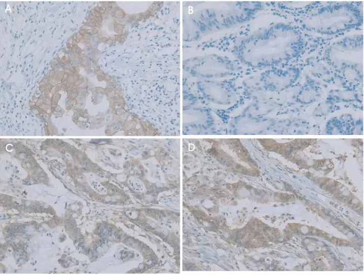
Fig. 1. Immunohistochemical staining of HER-2/neu overexpression (×200). (A) Strong complete membrane staining is observed in breast cancer cells (3+, positive control). (B) Negative control. Normal serum is used instead of primary antibody. (C) Faint membrane or cytoplasmic staining is detected in more than 10% of the tumor cells (1+). (D) Moderate complete membrane staining is observed in more than 10% of the tumor cells (2+).
148
박동일 외 14인. 대장암에서 HER-2/neu 발현에 관한 연구
시험심사위원회의 승인을 받고 시행하였다.
2. 방법
환자의 절제 조직에서 반정량적 면역화학염색법으로
HER-2/neu 단백의 과발현을 조사하였다. 면역화학염색법 은 다음의 순서로 진행하였다. 파라핀 포매 조직 슬라이드 를 xylene으로 탈파라핀화시킨 후 phosphate-buffer saline (PBS)으로 3회 세척하고, 3% hydrogen peroxide가 첨가된
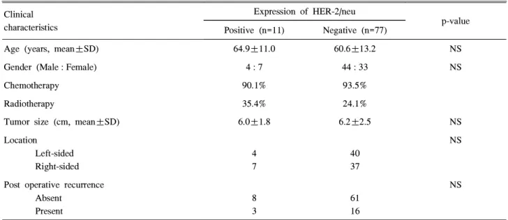
Table 1. Clinical Features according to Expression of HER-2/neu Protein in Patients with Colon Cancer Clinical
characteristics
Expression of HER-2/neu
p-value Positive (n=11) Negative (n=77)
Age (years, mean±SD) 64.9±11.0 60.6±13.2 NS
Gender (Male : Female) 4 : 7 44 : 33 NS
Chemotherapy 90.1% 93.5%
Radiotherapy 35.4% 24.1%
Tumor size (cm, mean±SD) 6.0±1.8 6.2±2.5 NS
Location
Left-sided Right-sided
4 7
40 37
NS
Post operative recurrence Absent
Present
8 3
61 16
NS
NS, not significant.
Table 2. Pathological Features according to Expression of HER-2/neu Protein in Patients with Colon Cancer Pathological
characteristics
Expression of HER-2/neu
p-value Positive (n=11) Negative (n=77)
T-stage
T1 & T2 T3 & T4
0 11
9 68
NS
N-stage N0 N1 & N2
3 8
45 32
NS
M-stage M0 M1
10 1
69 8
NS
Differentiation
Well & moderate Poor & undifferentiated
6 1
61 4
NS
Lymphovascular invasion Absent
Present
3 8
42 35
NS
Perineural invasion Absent Present
11 0
74 3
NS
NS, not significant.
149
The Korean Journal of Gastroenterology: Vol. 44, No. 3, 2004
메탄올에 15분 동안 반응시켜 고정한 후 PBS 용액으로 3회 세척하였다. 비특이적인 결합을 방지하기 위해 10배로 희석 한 정상적인 rabbit 혈청에 30분간 반응시켰다. 일차 항체인 rabbit anti-c-erbB2 (Zymed lab, South San Francisco, CA, USA)를 1:30으로 희석하여 1시간 반응시킨 후 horseradish peroxidase에 결합된 이차 항체인 goat anti-rabbit 항체 (Zymed lab, USA)를 투여하고 결합된 항체는 peroxidase chromogen substrate를 이용하여 발색하였다. 그 후 hemato- xylin으로 대조염색한 후 HER-2/neu의 발현의 강도에 따라 1+, 2+, 3+로 분류하였다.13HER-2/neu 양성 유방암 조직 을 양성 대조군으로 사용하였고 일차 항체 대신 정상 혈청 을 반응시킨 것을 음성 대조군으로 하였다. HER-2/neu 발 현을 보인 군과 보이지 않은 군 간에 여러 가지 임상적, 병 리적인 예후인자와 생존율의 차이를 분석하였다.
3. 통계 분석
통계 프로그램은 SPSS (version 11.0)를 사용하였고 두 군간에 연속형 변수들은 t-test로, 범주형 변수들은 Fisher's exact test로 분석하였으며 p값이 0.05 미만일 때 통계적으 로 유의한 차이가 있다고 판정하였다. 생존율의 차이는 Kaplan-Meier법으로 분석하였다.
결 과
88예 중 11예(12.5%)에서 HER-2/neu가 양성이었는데 1예 에서만 2+이고 나머지 10예는 1+였다(Fig. 1). HER-2/neu 양성군과 음성군 간에 연령, 성별, 항암화학요법 및 방사선 요법의 시행 빈도, 암종의 크기, 위치, TNM 병기, 분화도, 혈관 및 림프관 침범, 신경 침범, 수술 후 재발률의 차이는 관찰할 수 없었다(Table 1, 2). HER-2/neu 양성군에서 평균 생존일(2,631±141일 vs 1,646±269일)과 생존일 중앙값
이 짧고(1,589일 vs 660일), 3년 생존율(82.9% vs 67.0%) 및 5년 생존율(78.9% vs 65.5%)이 낮으며 국소 림프절 전 이가 많은 경향(41.6% vs 72.7%)을 보였으나 통계적 유의 성은 없었다(Fig. 2).
고 찰
HER-2/neu 과발현은 95% 이상에서 정상적인 HER-2/neu 유전자 복제의 수가 증가되는 유전자 증폭에 의하며 결국 유전자 전사의 증가에 의해 세포막 표면에서 HER-2/neu 수 용체의 수가 증가되어 세포의 과증식이 일어난다.4 그러므 로 HER-2/neu의 발현 상태는 DNA, RNA 및 단백질 수준 에서 다양하게 검사할 수 있으나 사용하는 병리조직의 형 태에 따라 그리고 각 실험실마다 기본적으로 실행되고, 쉽 게 판독되며, 재현성이 있게 시행될 수 있는 검사법이 다르 지만 현재 FISH (fluorescent in situ hybridization)와 면역화 학염색법이 가장 많이 사용되고 있다.
대장암에서 면역화학염색법에 의한 HER-2/neu 과발현 빈도는 0-83%로 다양한데11,14-18 이는 대상 환자군이 다른 점보다는 조직 고정 방법, 일차 항체의 선택, 항원결정인자 (epitope)의 재생, 결과의 해석 등 면역화학염색법 과정 중 의 기술적인 측면에 의한다.19HER-2/neu 유전자 검사의 기 준검사인 FISH법에 의하면 대장암의 약 0-30%에서 양성 반응을 보인다.8,14,15 대장암의 예후인자 및 생존율과의 연 관성을 종합하면 일부에선 HER-2/neu가 과발현될수록 병 기가 진행되고 생존율이 낮아진다고도 하고,14,18,20,21
다른 연구에선 생존율과 관계가 없어12,14,22 대장암 영역에서 HER-2/neu 과발현이 갖는 임상적 유용성에 대해서는 아직 정립된 것이 없다.
본 연구 결과 88예의 대장암 중 11예(12.5%)에서 HER-2/
neu의 과발현을 관찰하였고, HER-2/neu 양성군에서 평균 생존일과 생존일 중앙값이 짧고, 3년 생존율 및 5년 생존율 이 낮았으며 이는 아마도 국소 림프절 전이와 관계가 있을 것으로 생각된다. HCA-7 대장암 세포주에 Herceptin®을 투 여할 경우 암 세포주의 증식이 억제되어,23,24 이와 같은 in vitro 연구 결과와 본 연구와 같은 in vivo 연구 결과를 근거 로 대장암 분야에서도 HER-2/neu는 치료의 표적이 될 가능 성이 있다. 따라서 본 연구는 HER-2/neu 양성 대장암 환자 에서 Herceptin®을 투여하여 환자의 생존율을 향상시키고 자 하는 시도의 이론적 근거를 제시한다는 의의가 있다.
하지만 본 연구의 문제점은 HER-2/neu 양성군의 수가 적은 예비 연구로 통계적 유의성을 얻을 수 없었다는 점, 유방암에서와 같이 세포막에 강한 염색성을 보이기보다는 대부분 세포질에 염색이 되었다는 점, 그리고 HER-2/neu 양 성군 11예 중 10예에서 염색 강도가 1+로 약했다는 점 등 Fig. 2. Kaplan-Meier plot for cumulative survival in 88 patients
with colon cancer, comparing HER-2/neu-negative (n=77) and HER-2/neu-positive (n=11) tumors.
150
Park DI, et al. Clinical Significance of HER-2/neu Expression in Colon Cancer
이 있다. 하지만 염색성의 측면에서 이전의 연구에서도 세포 막보다는 세포질에 주로 염색되는 등 유방암에서와는 다른 염색성을 보인 경우15,16,18,25
도 있어 대장암에서 HER-2/neu의 발현 양상이 유방암에서와 차이가 있을 가능성을 배제할 수 없다. 따라서 본 저자 등은 현재 대상 환자의 수를 늘리 고, HER-2/neu 양성군을 대상으로 FISH를 시행해서 확증 하는 실험을 진행 중이다. 좀더 많은 환자를 대상으로 타당 성 있는 검사법 및 해석 기준을 가지고 다기관 공동 연구를 시행한다면 대장암에서 HER-2/neu 과발현과 예후인자 및 생존율과의 관계를 좀더 확실히 규명할 수 있을 것으로 생 각된다.
요 약
목적: HER-2/neu 단백은 상피성장인자 수용체와 구조가 거의 일치하기 때문에 정상 세포의 성장과 증식 과정에 관 여한다. 유방암에서 HER-2/neu의 과발현은 나쁜 생존율을 예상할 수 있는 예후인자일 뿐 아니라 항암화학요법이나 호르몬요법에 잘 반응하지 않는 등 치료에 대한 반응을 예 측할 수 있는 인자이며 HER-2/neu에 대한 단일클론 항체인 Herceptin®은 림프절에 전이된 유방암 환자에서 장기 생존 율을 향상시켜 임상에서 활발히 사용되고 있다. 본 연구에 서는 대장암 환자들을 대상으로 HER-2/neu 단백의 발현 여 부에 따른 여러 가지 임상적, 병리적 예후인자 및 생존율의 차이를 분석하여 대장암 영역에서 Herceptin®치료에 대한 이론적인 근거를 마련하고자 하였다. 대상 및 방법: 본원에 서 대장암으로 근치적 수술을 시행받은 88명의 환자를 대 상으로 면역화학염색법으로 HER-2/neu 단백의 과발현을 검사하였다. HER-2/neu 발현 유무에 따른 병리적인 예후 인자와 생존율의 차이를 비교 분석하였다. 결과: 88예 중 11예(12.5%)에서 HER-2/neu가 양성이었다. HER-2/neu 양 성 군에서 평균 생존일이 짧고(2,631±141일 vs 1,646±269 일) 3년 생존율(82.9% vs 67%)과 5년 생존율(78.9% vs 65.5%)이 낮으며 국소 림프절 전이가 많은 경향을 보였으 나 통계적 유의성은 없었다. 결론: 대상 환자 수가 적어 통 계적인 유의성은 없었지만 HER-2/neu 양성 대장암에서 생 존율이 낮은 경향을 관찰할 수 있었으며 이는 아마도 국소 림프절 전이와 관계 있을 것으로 생각된다.
색인단어: 대장암, HER-2/neu
감사의 글
이 연구는 삼성생명과학연구소 연구비(C-A4-212-1)의 보 조로 이루어졌음.
참고문헌
1. Akiyama T, Sudo C, Ogawara H, Toyoshima K, Yamamoto T. The product of the human c-erbB-2 gene: a 185- kilodalton glycoprotein with tyrosine kinase activity.
Science 1986;232:1644-1646.
2. Coussens L, Yang-Feng TL, Liao YC, et al. Tyrosine kinase receptor with extensive homology to EGF receptor shares chromosomal location with neu oncogene. Science 1985;230:1132-1139.
3. Di Fiore PP, Pierce JH, Kraus MH, Segatto O, King CR, Aaronson SA. erbB-2 is a potent oncogene when overexpressed in NIH/3T3 cells. Science 1987;237:178-182.
4. Kaptain S, Tan LK, Chen B. Her-2/neu and breast cancer.
Diagn Mol Pathol 2001;10:139-152.
5. Zarbo RJ, Hammond ME. Conference summary, Strategic Science symposium. Her-2/neu testing of breast cancer patients in clinical practice. Arch Pathol Lab Med 2003;127:549-553.
6. Hayes DF, Thor AD. c-erbB-2 in breast cancer: develop- ment of a clinically useful marker. Semin Oncol 2002;29:
231-245.
7. Uchino S, Tsuda H, Maruyama K, et al. Overexpression of c-erbB-2 protein in gastric cancer. Its correlation with long-term survival of patients. Cancer 1993;72:3179-3184.
8. Yokota J, Yamamoto T, Miyajima N, et al. Genetic alterations of the c-erbB-2 oncogene occur frequently in tubular adenocarcinoma of the stomach and are often accompanied by amplification of the v-erbA homologue.
Oncogene 1988;2:283-287.
9. Falck VG, Gullick WJ. c-erbB-2 oncogene product staining in gastric adenocarcinoma. An immunohistochemical study.
J Pathol 1989;159:107-111.
10. Ross JS, McKenna BJ. The HER-2/neu oncogene in tumors of the gastrointestinal tract. Cancer Invest 2001;19:554-568.
11. Caruso ML, Valentini AM. Immunohistochemical p53 overexpression correlated to c-erbB-2 and cathepsin D proteins in colorectal cancer. Anticancer Res 1996;16:
3813-3818.
12. Nathanson DR, Culliford AT 4th, Shia J, et al. HER 2/neu expression and gene amplification in colon cancer. Int J Cancer 2003;105:796-802.
13. Kay EW, Walsh CJ, Cassidy M, Curran B, Leader M.
C-erbB-2 immunostaining: problems with interpretation. J Clin Pathol 1994;47:816-822.
14. Osako T, Miyahara M, Uchino S, Inomata M, Kitano S, 151
대한소화기학회지: 제44권 제3호, 2004
Kobayashi M. Immunohistochemical study of c-erbB-2 protein in colorectal cancer and the correlation with patient survival. Oncology 1998;55:548-555.
15. D'Emilia J, Bulovas K, D'Ercole K, Wolf B, Steele G Jr, Summerhayes IC. Expression of the c-erbB-2 gene product (p185) at different stages of neoplastic progression in the colon. Oncogene 1989;4:1233-1239.
16. Kay EW, Mulcahy H, Walsh CB, Leader M, O'Donoghue D. Cytoplasmic c-erbB-2 protein expression correlates with survival in Dukes' B colorectal carcinoma. Histopathology 1994;25:455-461.
17. Yang JL, Ow KT, Russell PJ, Ham JM, Crowe PJ. Higher expression of oncoproteins c-myc, c-erb B-2/neu, PCNA, and p53 in metastasizing colorectal cancer than in nonmetastasizing tumors. Ann Surg Oncol 1996;3:574-579.
18. Kapitanovic S, Radosevic S, Kapitanovic M, et al. The expression of p185 (HER-2/neu) correlates with the stage of disease and survival in colorectal cancer. Gastroenterology 1997;112:1103-1113.
19. Seidal T, Balaton AJ, Battifora H. Interpretation and quan- tification of immunostains. Am J Surg Pathol 2001;25:
1204-1207.
20. Lazaris AC, Theodoropoulos GE, Anastassopoulos P,
Nakopoulou L, Panoussopoulos D, Papadimitriou K.
Prognostic significance of p53 and c-erbB-2 immuno- histochemical evaluation in colorectal adenocarcinoma.
Histol Histopathol 1995;10:661-668.
21. Saeki T, Salomon DS, Johnson GR, et al. Association of epidermal growth factor-related peptides and type I receptor tyrosine kinase receptors with prognosis of human colorectal carcinomas. Jpn J Clin Oncol 1995;25:240-249.
22. Sun XF, Carstensen JM, Stal O, Zhang H, Nordenskjold B.
c-erbB-2 oncoprotein in relation to DNA ploidy and prognosis in colorectal adenocarcinoma. APMIS 1995;103:
309-315.
23. Mann M, Sheng H, Shao J, et al. Targeting cyclooxygenase 2 and HER-2/neu pathways inhibits colorectal carcinoma growth. Gastroenterology 2001;120:1713-1719.
24. Half E, Broaddus R, Danenberg KD, Danenberg PV, Ayers GD, Sinicrope FA. HER-2 receptor expression, localization, and activation in colorectal cancer cell lines and human tumors. Int J Cancer 2004;108:540-548.
25. Natali PG, Nicotra MR, Bigotti A, et al. Expression of the p185 encoded by HER2 oncogene in normal and transformed human tissues. Int J Cancer 1990;45:457-461.
152