Mutations in the tyrosine kinase domain of the EGFR gene are rare in the Korean Oral Squamous Cell Carcinoma
1)EEEun-Ju Lee*
Abstract
The epidermal growth factor receptor(EGFR) protein kinase signaling is an important pathway in cancer development and recently reported that EGFR and its kinase domain molecules are mutated in various of cancers including head and neck cancer. Functional deregulation of EGFR due to mutations in coding exons and copy number amplification is the most common event in cancers, especially among receptor tyrosine kinases(TK). We have analyzed Korean oral squamous cell carcinomas (OSCC) cell lines for mutations in EGFRTK. Exons encoding the hot-spot regions in the TK domain of EGFR (exons 17 to 23) were amplified by using polymerase chain reaction(PCR) and sequenced directly. EGFR expression was also analyzed in 8 OSCC cell lines using western blotting. Data analysis of the EGFR exons 17 to 23 coding sequences did not show any mutations in the 8 OSCC cell lines that were analyzed. The absence of mutations indicate that protein overexpression might be responsible for activation rather than mutation.
▸Keyword : Receptor-Epidermal Growth Factor, protein-tyrosine kinase, carcinoma-squamous cell, Head and Neck Neoplasms, mouth neoplasms, exons
I. Introduction
Oral cancer(OC) is a type of head and neck cancer(HNC) that located in the oral cavity, and squamous cell carcinoma(SCC) is the most frequent histological type[1]. Recently, new strategies based on molecular targeted therapy have been developed to combat this fatal disease. The epidermal growth factor receptor (EGFR), a transmembrane protein and one of the ErbB family of receptors, is a promising target for molecular therapy.
This receptor has an intrinsic tyrosine kinase (TK) activity, and regulates cell growth in response to binding of ligands like epidermal growth factor (EGF) or transforming growth factor α[2,3]. ZD1839(Iressa), which interrupts EGFR kinase activity in the TK domain, is an orally active EGFR TK inhibitor[4-6]. ZD1839 has been
reported to reduce cellular proliferation in various tumor cell lines and tumor xenogrsfts[5,7]. However, because not all cancer cell lines show the same response, there is substantial interest in good prognostic indicators that might predict the response to ZD1839. Recent studies showed that the TK domain mutations of EGFR have become progressively obvious in cancers including head and neck cancer, brain cancer, oral cancer, lung cancer and others. Extensive studies has confirmed exon 19 and exon 21, that code for part of TK domain of EGFR in several cancers, to be the hot spot for activating mutations. These results showed that the TK domain mutations of EGFR could predict significant clinical response to ZD1839[7-9].
∙First Author: Eun-Ju Lee, Corresponding Author: Eun-Ju Lee
*Eun-Ju Lee([email protected]), Dept. of Clinical Laboratory Science, Daejeon Health Institute of technology
∙Received: 2016. 08. 09, Revised: 2016. 09. 02, Accepted: 2016. 09. 20.
∙This work was supported by Daejeon Health Institute of technology in 2014.
Cell line
(YD) Age/Sex Tumor location Pathologic diagnosis
p53 mutation exon number
HPV infection
Prior chemotherapy 8
9 10B
15 15M
17 17M
38
46/Female 56/Male 67/Male 39/Male
66/Male
67/Male
Tongue Buccal cheek
Tongue Lymph node Lymph gingiva
Lower gingiva Lymph node Lower gingiva
SCC, MD SCC, MD SCC, MD MEC, HG Metastatic SCC, PD Metastatic SCC, MD
273(VIII)
236(VII) 258(VII) 258(VII)
None None None None None None None None
None None None None None None None None Table1. Clinical and pathological characteristic of the established oral cancer cell lines.
The study of the expression and mutation status of EGFR would allow us to better comprehend the response of tumor patients to molecular targeted therapy. EGFR overexpression has been identified in various tumor types. Overexpression might lead to effects that are similar to those of mutations. However, the expression and prognostic significance in HNSCC have yielded controversial results necessitating more studies in this field. In this article, we focused on the possibility that the EGFR mutations which might be accountable to the response of ZD1839 in the Korean OSCC cell lines. To explore this possibility, we have analyzed mutation in exon 17 to 23 of EGFR occurred in 8 OSCC cell lines.
II. Materials and Methods
1. Reagents
Stock solutions for ZD1839 and antibodies were prepared as previously described [10].
2. Cell culture
Oral squamous cell carcinomas (OSCC) cell lines YD-8, YD-9, YD-10B, YD-15, YD-15M, YD-17, YD-17M, and YD-38 were obtained from Yonsei University College of Dentistry and were maintained in the manufacturer specified growth medium[10]. Clinical and pathological characteristic of the established oral cancer cell lines were listed in Table 1.
3. Growth inhibition assay
Growth inhibition was assessed by using an 3- (4,5-dimethylthiazol-2-yl)-2,5-diphenyltetrazolium bromide(MTT, Sigma, St. Louis MO) assay for the detect of viable cells. Briefly, a total of 10,000 per well in 96-well flat-bottomed plates for 24h and then treated with different concentrations of ZD1839 for 48h[11].
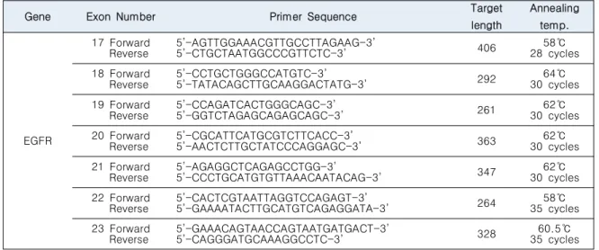
4. DNA Extraction and Mutation analysis DNA was isolated from the cell lines was isolated by a genomic DNA extraction kit(Qiagen, valencia, CA). PCR was used to amplify the 7 exons comprising the kinase domain of the EGFR gene. Intronic primers were used to amplify exon 17 to 23 of the EGFR gene, using 25ng of genomic DNA. The EGFR primer sequences, PCR fragment sizes and annealing conditions were as Table 2.
The exon 18 and 23 were amplified under the below conditions: after an initial denaturation at 94°C for 2 min, the samples were subjected to 30 cycles of denaturing for 30 sec at 95°C , annealing at for 45 sec 55°C, extension for 45 sec at 72°C, which was then a final extension for 5 min at 72°C as described earlier[12]. The PCR amplicons were run in a 1.5% agarose gel and subjected to SAP treatment before being sequenced
5. Antibodies and Western blot analysis
Lysates from cultured cells were prepared in ice-cold RIPA lysis solution(0.1% SDS, 1% NP40, 50 mM Tris-HCl (pH8.0), 150 mM NaCl and 1x protease inhibitors). Using the centrifugation in a microfuge for 15 min at 4 ℃ at 12,000 xg, debris was removed. Western blot analyses
Gene Exon Number Primer Sequence Target length
Annealing temp.
EGFR
17 Forward
Reverse 5’-AGTTGGAAACGTTGCCTTAGAAG-3’
5’-CTGCTAATGGCCCGTTCTC-3’ 406 58℃
28 cycles 18 Forward
Reverse 5’-CCTGCTGGGCCATGTC-3’
5’-TATACAGCTTGCAAGGACTATG-3’ 292 64℃
30 cycles 19 Forward
Reverse 5’-CCAGATCACTGGGCAGC-3’
5’-GGTCTAGAGCAGAGCAGC-3’ 261 62℃
30 cycles 20 Forward
Reverse 5’-CGCATTCATGCGTCTTCACC-3’
5’-AACTCTTGCTATCCCAGGAGC-3’ 363 62℃
30 cycles 21 Forward
Reverse 5’-AGAGGCTCAGAGCCTGG-3’
5’-CCCTGCATGTGTTAAACAATACAG-3’ 347 62℃
30 cycles 22 Forward
Reverse 5’-CACTCGTAATTAGGTCCAGAGT-3’
5’-GAAAATACTTGCATGTCAGAGGATA-3’ 264 58℃
35 cycles 23 Forward
Reverse 5’-GAAACAGTAACCAGTAATGATGACT-3’
5’-CAGGGATGCAAAGGCCTC-3’ 328 60.5℃
35 cycles Table 2. Primer sequences and target size
were performed as previous reported procedure[11]
using anti-EGFR and pospho-EGFR monoclonal antibody (Biogenex Co, San Ramo, CA) from the cell lysate. The membrane was incubated with an antirabbit antibody conjugated with horseradish peroxidase (Amersham, Arlington Heights, IL, USA), and the protein was measured by using the Bradford method. After detecting with each of the respective antibodies, the membraned were visualized by enhanced chemiluminescence.
6. Statistical analysis
All results describe the average of at least three separated experiments and showed as mean ± SD unless any other indicated. Student's t-tests were achieved to compare cell growth and densitometric values of Western blotting bands among the various concentrations of ZD1839. P values of <0.05 were reflected to be statistically significant.
III. Results
Eight OSCC cell lines, from surgical specimens of oral carcinoma specimens were established, of which 3 were from tongue(YD-8, -10B and –15) and 2 from lower gingiva(YD-17 and 38). The YD-15M and 17M were originated from metastatic lymph nodes and YD-9 from buccal cheek[10]. The clinicopathologic finding of patients were summarized in Table 1.
We tested the antiproli- feration effect (Fig. 1) of ZD1839 in the seven OSCC cell lines. Antiproliferative effect to ZD1839 was defined as a 50% inhibition of cell growth at a different concentration. Among the OSCC cell lines tested, only YD-38 was sensitive to the ZD1839, with an IC50 <0.01 μM; whereas the other cell lines were resistant to ZD1839. The establihed OSCC cell lines were expressed EGFR by Immuno blotting and detected that weak to moderate levels of EGFR protein expression.
Fig. 1. ZD1839 sensitivity of 8 OSCC cell lines. All cells were treated with various concentration of ZD1839 as indicated, and the antiproliferative effects of ZD1839 treatment were determined by MTT assay. Each data point represents the mean of three independent experiments. Data are presented as standard deviation. Error bar correspond to the standard error.
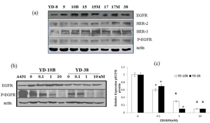
Fig. 2. EGFR expression in 8 human oral squamous cell carcinoma(OSCC) cell lines, the OSCC cell lines were evaluated for various in (a) EGFR protein, HER-2, HER-3 and p-EGFR proteins expression levels. (b) Effect of EGFR inhibitor ZD1839 on EGFR and phosphorylation of EGFR in YD-10B and YD-38 cell lines. A431 is a positive control. (c) Protein levels of p-EGFR were analyzed by densitometry, and the ratios of p-EGFR to β-actin were calculated in arbitrary unit. The control was set to a ratio of 1.0 The means ±SD of 3 experiment are shown. *p<0.05, compared with intreated cells in the absence of ZD1839
Similar amount of EGFR was detected in YD-9, YD-15, and YD-38 by western blot but showed differences in ZD1839 sensitivity. We conclude that expression of EGFR does not predictor to the antiproliferative effects of ZD1839. Thus we detected the levels of EGFR phosphorylation upon ligand addition and then compared the EGFR phosphorylation observed in serum-starved cells (Fig. 2). Among the OSCC cell lines, two cell lines showed high level of phosphorylated EGFR, and this phosphorylation was decreased in a concentration dependent fashion by ZD1839. The exons 17 to 23 of EGFR are involved in encoding kinase domain of the EGFR.
Mutations in any of these exons may be expected to affect the protein kinase function of EGFR.
The genomic DNA extracted from 8 oral squamous cell carcinoma cell lines were amplified using intronic primers flanking exon 17 and 23 of EGFR(Fig. 3). The amplified products were then subjected to direct sequencing analysis, which showed no mutations in exon 17 and 23.
Fig. 3. Results of OSCC cell lines specific PCR for these samples. A band with expected size is clearly detected. The PCR products of the exon 17 to 23 at 58℃, 30 cycles from YD 10B the representive 8 OSCC cell lines. The PCR products of the Exon 17 from the representative 8 OSCC cell lines at 58℃, 28 cycles. M is a 100 base pair size marker.
IV. Discussion
Recently, molecular targeted therapy has gained a lot of interest in the struggling against cancer. Clinical trials suggest that EGFR targeting agents can help many cancer patients, including those with Head and Neck Squamous Cell Carcinoma [2,3,13]. There are two different ways of EGFR molecular targeted drug interaction offering a more effective inhibition. The first one involves the connection of the drug to the extracellular domain of the receptor that inhibits the connection of the ligand. The second targets the intracellular portion that has tyrosine kinase activity and exerts its action by restricting ATP binding or binding to the active site of the enzyme[6, 14].
Mutations in this domain are frequent in lung cancer, especially of Asian ethnicity[9]. Interestingly, approximately 80% of patients with EGFR mutations respond to EGFR TKI, whereas only 10% of those without mutations do so[15]. These EGFR mutations affect amino acids near the ATP binding pocket, which is targeted by ZD1839, resulting in increased gene activation and sensitivity to TKI[16].
ZD1839 is an potentially active, selective inhibitor of the EGFR TK[17,18] and the EGFR TK domain mutation is known to arbitrate the ZD1839 sensitivity. Based on recent reports suggesting that EGFR mutations within the kinase domain are associated with selected OSCC patient groups and confer a greater response rate to EGFR inhibitor such as ZD1839, we determined the prevalence of mutations in exon 17 to 23 of EGFR in OSCC cell lines.
Our analysis failed to detect any mutations in exon 17 to 23 of EGFR in 8 cell lines. The present study is in agreement with some authors who have also reported the absence or low incidence of EGFR mutations in HNSCC[19-21]. It has been noticed that EGFR mutations are also rare or absent in cancers other than lung cancer.
Interestingly, one of the cell line tested was found to be sensitive to ZD1839 in vitro, whereas the other seven cell lines were resistant to the antiproliferate effects of the drug[11]. Resistance to ZD1839 was associated with uncoupling between the EGFR and mitogen-activated protein kinase, and could be predicted by analyzing the activation of GSK-3β and cyclin D1. Our data indicate that the response to ZD1839 by OSCC cells cannot be attributed solely to the status of EGFR mutations within the kinase domain and that other variables and cell properties must affect the response to ZD1839.
However, the challenge of predicting which patients will respond to ZD1839 and other tyrosin kinase inhibitors remains to be elucidated.
REFERENCES
[1] Milas, K. A. Mason, Z. Liao, K. K. Ang,
“Chemoradiotherapy: emerging treatment improve ment strategies”, Head Neck, vol. 25, pp. 152–67, 2003.
[2] C. J. Langer, “Exploring Biomarkers in Head and Neck Cancer” Cancer, vol 118, No. 16, pp. 3882- 3892, Jan, 2012
[3] E.S. Lampri, G. Chondrogiannis, E. loachim et al.,
“Biomarkers of head and neck cancer, tools or a gordian knot?”, Int J Clin Exp Med, Vol. 8, No. 7, pp. 10340-10357, 2015
[4] S. P. D’Angelo, M. C. Pietanza, M. L. Johnson et al
“Incidence of EGFR exon 19 deletions and L858R in tumor specimens from men and cigarette smokers with lung adenocarcinomas” J Clin Oncol, vol. 29, pp. 2066-2070, 2011.
[5] A. F. Gazdar, “Activating and resistance mutations of EGFR in non-small-cell lung cancer: role in clinical response to EGFR tyrosine kinase inhibitors”, Oncogene, vol. 28, pp. 24-31, 2009.
[6] F. Ciardiello and G. Tortora “A novel approach in the treatment of cancer: targeting the epidermal growth factor receptor”, Clin Cancer Res, vol. 7. pp. 2958-2970, 2001.
[7] Q. Liu, G. Ma, H. Yang et al, “Lack of epidermal growth factor receptor gene mutations in exons 19 and 21 in primary lymphoepithelioma-like carcinoma of the lung”, Thoracic Cancer, vol. 5, pp. 63-67, 2013.
[8] P. Stephens, C. Hunter, G. Bignell et al., “Lung cancer:
intragenic ERBB2 kinase mutations in tumors”, Nature, vol. 431, pp. 525–526, 2004.
[9] H. Shigematsu, L. Lin, T. Takahashi et al., “Clinical and biological features associated with epidermal growth factor receptor gene mutations in lung cancers”, J Natl Cancer Inst, vol. 97, pp. 339–462, 2005.
[10] E. J. Lee, J. Kim, S. A. Lee, E. J. Kim, Y. C. Chun, M.
H. Ryu, J. I. Yook, “Characterization of newly established oral cancer cell lines derived from six squamous cell carcinoma and two muco epidermoid carcinoma cells”, Exp Mol Med., vol. 37, pp. 379-390, 2005.
[11] N. K. Jeon, J. Kim, E. J. Lee, “The GSK-3β/Cyclin D1 Pathway is Involved in the Resistance of Oral Cancer Cells to the EGFR Tyrosine Kinase Inhibitor ZD1839”, Bio Sci Letters, vol. 20, No. 2, pp. 85-95, 2014.
[12] S. Rajendran, R. S. Muthupalani, A. Ramanathan, “Lack of RING finger domain (RFD) mutations of the c-cbl gene in oral squamous cell carcinomas in Chennai, India”, Asian Pac J Cancer Prev, vol. 14, pp. 1073-1075, 2013.
[13] E. E. Cohen, F. Rosen, and W. M. Stadler et al. “Phase II trial of ZD1839 in recurrent or metastatic squamous cell carcinoma of the head and neck”, J Clin Oncol vol.
21, pp. 1980–1987, 2003.
[14] P. M. Harari, S. M. Huang, R. S. Herbst and H. Quon,
“Molecular Targeting of the Epidermal Growth Factor Receptor. In: Harrison LB, Session RB, Hong WK, editors.
Head and Neck Cancer” A Multidisciplinary Approach.
2nded. Philadelphia: Lippincott, Williams and Wilkings, 2003.
[15] T. Fukui, T. Mitsudomi, “Gefitinib and epidermal growth factor receptor gene mutation”, Gan Kagaku Ryoho vol.34, pp.1168–1172, 2007.
[16] A. F. Gazdar, H. Shigematsu, J. Herz, J. D. Minna,
“Mutations and addiction to EGFR: the Achilles heal of lung cancers?”, Trends Mol Med, vol.10, pp.481–486, 2004.
[17] E. E. Cohen, M. W. Lingen, L. E. Martin et al. “Response of some head and neck cancers to epidermal growth factor receptor tyrosine kinase inhibitors may be linked to mutation of ERBB2 rather than EGFR”, Clin Cancer Res, vol.11, pp. 8105–8108, 2005.
[18] S. E. Wang, A. Narasanna, M. Perez-Torres et al., “HER2 kinase domain mutation results in constitutive phosphorylation and activation of HER2 and EGFR and resistance to EGFR tyrosine kinase inhibitors”, Cancer Cell, vol. 10, pp. 25–38, 2006.
[19] Y. Lemos-González, M. Páez de la Cadena, F. J.
Rodríguez-Berrocal, A. M. Rodríguez- Piñeiro, E.
Pallas, D. Valverde, “Absence of activating mutations in the EGFR kinase domain in Spanish head and neck cancer patients” Tumour Biol, vol. 28, pp. 273–279, 2007.
[20] J. Loeffler-Ragg, M. Witsch-Baumgartner, A. Tzankov et al., “Low incidence of mutations in EGFR kinase domain in Caucasian patients with head and neck squamous cell carcinoma”, Eur J Cancer, vol. 42, pp.
109–111, 2006.
[21] J. W. Lee, Y. H Soung, S. Y. Kim et al., “Absence of EGFR mutation in the kinase domain in common human cancers besides non-small cell lung cancer”, Int J Cancer, vol. 113, pp. 510–511, 2006.
Authors
Eun Ju Lee received the Ph.D. degrees in Bio-Inorganic Chemistry from Yonsei University, Seoul, Korea, in 1999.
Dr. Lee joined the faculty of the Department of Clinical Laboratory Science at Daejeon Health Institute of Technology, Daejeon, Korea, in 2007. She is currently a Professor in the Department of Clinical Laboratory Science. She is interested in head and Neck cancer prevention, cancer metabolism and signaling and drug metabolism.