Ⅰ. 서 론
구강편평세포암종은 인접조직으로의 침투 및 임파선으로의 전이 빈도가 높아 예후가 불량한 암종으로 구강 악안면 영역의 암종 가운데 가장 발생빈도가 높다1). 종양의 전이 및 인접조직으 로의 침습은 많은 단백분해효소에 의해 중재되는 기저막과 세포 외 기질의 파괴와 신생혈관의 형성을 동반한다2,3). 신생혈관형성 은 기존의 혈관으로부터 새로운 미세혈관이 형성되는 것을 말하
며 이는 발생, 치유 등의 생리학적 기전과 종양의 전이 및 침습과 같은 병리학적 기전 전반에 걸쳐 중요한 역할을 하는 것으로 알 려져 있다4). 특히 종양의 혈관형성은 종양의 성장과 전이에 있어 필수적인 과정으로 알려져 있는데, 이는 종양세포의 성장을 위 한 영양분과 산소의 공급은 물론 형성된 혈관을 통한 암세포의 이동이 필수적이기 때문이다5). 이러한 이유로 종양의 신생혈관 화에 대한 많은 연구가 이루어져 왔으며 혈관화가 잘 이루어진 종양의 경우 국소적 침습이나 원격전이가 잘 일어나고 성장하는 반면, 혈관화가 잘 되지 않는 종양은 지름 1�2 cm 이상 성장할 수 없다고 알려져 있다6,7).
본 논문의 목적은 구강편평세포암종의 전이여부 및 국소적 침 습도와 신생혈관형성도간의 상관 관계를 분석하여 알아보고, 현 재 항암치료의 한 분야로서 활발히 연구되고 있는 항신생혈관화 물질을 이용한 구강편평세포암종의 침습 및 전이에 대한 치료 가능성을 고찰하는 것이다.
김 명 진
110-744, 서울특별시 종로구 연건동 28-2
서울대학교 치과대학 구강악안면외과학 교실 Myung-Jin KimDept. of OMFS, School of Dentistry, Seoul National Univ.
28-2 Yeongun-Dong, Chongro-Gu, Seoul, 110-744, Korea Tel: 82-2-760-2632 Fax: 82-2-766-4948
E-mail: [email protected]
Abstract
(J. Kor. Oral Maxillofac. Surg. 2002;28:155-160)구강편평세포암종의 미세혈관 밀도와 악성도간 상관관계에 관한 임상병리학적 연구
이기철∙명 훈∙임성삼*∙김명진
서울대학교 치과대학 구강악안면외과학교실, 서울대학교 치과대학 치과보존학교실*
THE CLINICOPATHOLOGICAL STUDY ON THE RELATION OF MICROVESSEL DENSITY AND AGGRESSIVENESS IN ORAL SQUAMOUS CELL CARCINOMA
Gi-Cheol Lee, Hoon Myoung, Sung-Sam Lim
*, Myung-Jin Kim Department of Oral and Maxillofacial Surgery, Department of Conservative Dentistry*
College of Dentistry, Seoul National University
Oral squamous cell carcinoma (OSCC), the most common head and neck cancer, shows poor prognosis as a result of frequent local invasion and lymph node metastasis that is mediated by multiple proteolytic enzymes and angiogenesis. In recent reports, angiogenesis is known to play an important role in tumor invasion and metastasis. The purpose of this study was to determine the role of angiogen- esis in OSCCs, particularly with respect to the invasive and the metastatic potential.
The microvessel density (CD31) in 34 human OSCC cases were investigated by immunohistochemistry, and reviewed with respect to the invasiveness and the presence of lymph node metastasis and following results were obtained.
The blood vessel density (28.8±7.9) in the strong invasive cases were significantly higher than those (23.3±6.9) in the weak invasive cases. (p<0.05) In the 14 cases with lymph node metastasis, the average blood vessel density was 28.5±9.6. On the other hand, in the 20 cases without lymph node metastasis, the blood vessel density was 25.2±6.4. The blood vessel density was not statistically related to lymph node metastasis. (p>0.05)
These results suggest that angiogenesis may be related to the local invasion of OSCC and further research will be needed to investi- gate the possibility that antiangiogenic agent can be used as an anticancer agent for OSCC.
Key words : Angiogenesis, Oral squamous cell carcinoma, Lymph node metastasis, Invasion
Ⅱ. 재료 및 방법 1. 구강편평세포암종 증례 수집 및 절편제작
1998년부터 1999년까지 서울대학교 치과대학병원 구강악안면 외과에서 구강편평세포암종의 진단하에 절제 수술을 받은 34증 례의 파라핀 포매조직을 이용하였다. 환자의 임파선 전이여부와 임상병리학적 정보는 챠트와 병리보고서를 참조하여 조사하였 다. 파라핀 포매조직은 5μm두께로 잘라 triethoxysilane-coated glass slide에 부착시킨 후 Hematoxylin & Eosin (H&E)염색을 시행 하였으며 미세혈관 밀도를 측정하기 위한 면역화학적 염색을 위 해 -70℃에서 냉동 보관하였다.
2. 침습도의 평가
H&E 염색 슬라이드상에서 2인의 관찰자가 각 증례를 맹검 후 침습도를 평가하였는데 결과가 서로 다른 경우 제3의 관찰자가 재검하여 최종 결정하였다. 침습도의 평가는 다음에 제시된 바 와 같이 Yamamoto 등8)의 연구에서 제시된 방법을 기준으로 침 습이 약한 Grade a부터 침습이 강한 Grade d까지 4개의 단계로 평가하고 Grade a 와 b는 약한정도의 침습군으로, Grade c 와 d는 강한 정도의 침습군으로 구분하였다.
Grade a : nodule type growth of tumor mass into surrounding stro- mal tissue with distinct border
Grade b : cord type growth of tumor mass into surrounding stro- mal tissue with distinct border
Grade c : cord type growth of tumor mass into surrounding stro- mal tissue with indistinct border
Grade d : diffuse infiltrative growth of tumor mass into surrounding stromal tissue with indistinct border
3. CD31면역조직화학적 염색과 미세혈관밀도의 측정 및 통계분석
각 증례의 미세혈관도를 측정, 평가하기 위한 CD31의 면역화 학적 염색을 위해 각 증례의 조직편을 자일렌에서 탈파라핀하고 알코올에 함수시킨후 0.3% H2O2에서 30분, 3% normal swine serum 에서 1시간 blocking하였다. 미세혈관의 발색을 위해 CD31 (platelet/endothelial cell adhesion molecule ; PECAM) JC70 mono- clonal antibody를 1:20 으로 희석한 뒤 30 분간 상온에서 부란하고 streptavidin-biotin-peroxidase complex를 결합하였다. 3,3- diaminobenzidine hydrochloride(DAB)을 적용하여 발색 후, Mayer’s hematoxylin으로 대조염색하였다. 혈류 공급이 풍부하다 고 이미 알려져 있는 유방암종의 조직을 동일조건하에서 반응시 켜 양성대조로 삼았으며9), 음성대조는 일차항체를 정상 swine serum으로 대체하여 반응시킨 조직으로 하였다.
미세혈관 밀도는 Bosari 등10,11)이 기술한 방법으로 평가하였는 데, 종양에서 멀리 떨어진 부위와 괴사부위의 미세혈관은 제외 하고 종양주위에서 단위면적당 가장 많은 수의 미세혈관을 가진 부위를 저배율 (×100)에서 관찰하여 세 부위를 찾은 뒤 고배율 (×250)에서 모세혈관의 수를 세어 그 평균값으로 하였다.
임파선 전이여부 및 침습도에 따른 미세혈관 밀도의 차이는 t-test를 이용하여 통계 분석하였고 유의수준은 p<0.05로 정하였 다.
Ⅲ. 결 과 1. 구강편평세포암종 증례 분석
1998년부터 1999년까지 서울대학교 치과대학 구강악안면외과 에서 구강편평세포암종 진단하에 절제 수술을 받은 총 34증례를 분석한 결과, 임파선 전이를 보인 증례가 14례(41.2%)였으며 20 례에서 임파선 전이를 보이지 않았다. H&E염색상에서 침습도를 평가한 결과 침습도a, b, c, d는 각각 9, 5, 10, 10례였고 Grade a 와 b는 약한정도의 침습군으로, Grade c 와 d는 강한 정도의 침습군 으로 구분한 결과, 약한 정도의 침습을 보인 증례는 14례, 강한 정도의 침습을 보인 증례는 20례로 나타났다 (Table 1).
2. CD31 발현과 미세혈관 분포양상
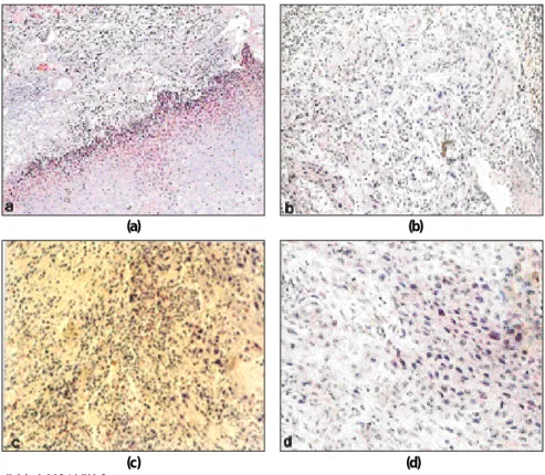
증례에서 CD31의 면역조직 화학반응은 주로 종양괴 주변의 혈 관에 국한되어 갈색으로 나타났다. 반면 같은 조건하에서 반응 시킨 음성대조군에서는 발현이 관찰되지 않았다. CD31 은 종양 세포가 밀집한 부위에 주로 발현되었으며 종양이 성장하는 주변 부위에도 발현을 관찰 할 수 있었다 (Fig. 2).
3. 임파선전이여부와 미세혈관밀도간 상관관계
34증례 중 임파선 전이가 있었던 14례에서의 평균 혈관밀도는 28.5±9.6이었던 반면, 임파선 전이가 없었던 20례의 평균 혈관밀 도는 25.2±6.4로 나타나 임파선 전이가 있는 증례에서의 미세혈 관밀도가 높았던 것으로 나타났으나 통계적 유의성은 없었다 (p>0.05, Fig. 3).
4. 침습도와 미세혈관밀도간 상관관계
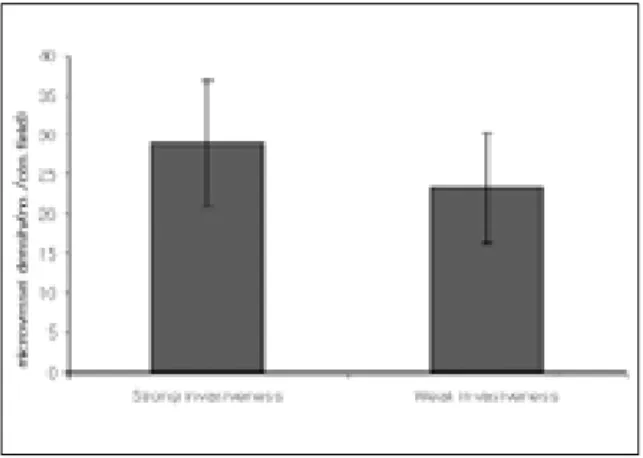
강한 침습도를 보인 20증례 (invasive grade c or d)에서의 평균 미세혈관밀도는 28.8±7.9로서 침습도가 약했던 14증례 (invasive grade a or b)의 평균 미세혈관밀도인 23.3±6.9보다 높았으며 통 계적 유의성이 있었다(p<0.05, Fig. 4).
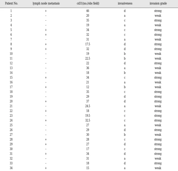
Table 1. Clinicopathological profiles and immunohistochemical expression of CD31 in 34 oral squamous cell carcinomas
1 + 46 d strong
2 - 20 a weak
3 - 35 c strong
4 - 19 a weak
5 + 34 c strong
6 + 32 c strong
7 - 31 a weak
8 + 17.5 d strong
9 + 32 d strong
10 - 19 b weak
11 - 22.5 b weak
12 - 22 d strong
13 - 36 a weak
14 - 18 b weak
15 + 34 c strong
16 - 21 a weak
17 + 12 b weak
18 - 35 c strong
19 - 29 d strong
20 + 37 d strong
21 - 24.5 a weak
22 + 18 c strong
23 - 19.5 c strong
24 + 32.5 c strong
25 - 27 a weak
26 - 29 d strong
27 - 30 b weak
28 + 28 c strong
29 + 27 d strong
30 - 17 c strong
31 + 34 d strong
32 - 31 a weak
33 - 18 d strong
34 + 15 a weak
+: lymph node metastasis positive -: lymph node metastasis negative Invasion Grade
a: nodule type growth of tumor mass into surrounding stromal tissue with distinct border b: cord type growth of tumor mass into surrounding stromal tissue with distinct border c: cord type growth of tumor mass into surrounding stromal tissue with indistinct border d: diffuse infiltrative growth of tumor mass into surrounding stromal tissue with indistinct border
*Grade a and b were interpreted as weak invasive cases and Grade c and d were as strong cases.
Patient No. lymph node metastasis cd31(no./obs field) invasiveness invasion grade
(b) (a)
(d) (c)
Fig. 1. Various type of invasion
a. Grade a: nodule type growth of tumor mass into surrounding stromal tissue with distinct border b. Grade b: cord type growth of tumor mass into surrounding stromal tissue with distinct border c. Grade c: cord type growth of tumor mass into surrounding stromal tissue with indistinct border d. Grade d: diffuse infiltrative growth of tumor mass into surrounding stromal tissue with indistinct border (All photographs are at a magnification x 100).
(b) (a)
(d) (c)
Fig. 2. Photomicrography of CD31 expression
a. the case of CD31expression with low level (x100 fields). The areas of highest vascularization were chosen.
b. the case of CD31expression with low level (x200 fields).
c. the case of CD31expression with high level (x100 fields). The areas of highest vascularization were chosen.
Ⅳ. 총괄 및 고안
종양과 같은 질병에서 신생 혈관화가 많다고 알려진 이래로, 고형암의 성장 및 전이와 신생혈관화간의 관계에 대한 많은 연 구가 이루어져 현재 고형성 종양의 성장, 침투 및 전이에 있어서 신생혈관 형성이 필수적이라는 것은 잘 알려져 있다12,13). 또한 각 종 종양의 혈관화정도에 따른 전이 및 생존율의 차이가 있다는 보고도 있다14-17). Gasparini 등18)은 유방암에서 종양의 혈관화는 임 상적 예후와 밀접한 관계가 있으며 항신생혈관화개념은 암종치 료에 있어 전향적인 치료개념으로서 중요하다고 보고한 바 있 다.
두경부종양에서도 미세혈관화정도와 임상적 예후간의 관련성 에 대한 몇 가지 연구가 이루어졌다. Leedy 등19)의 연구와 같이 구 강암에서 혈관화와 종양의 임파선전이사이에는 유의한 관계를 보이지 못했다고 보고된 바도 있었으나, Williams 등20)은 신생혈 관화가 두경부종양의 재발과 유의한 연관이 있으며 독립적인 예 후인자로서의 가치를 가진다고 보고하였고, Pazouki 등21)은 혈관 화와 두경부종양의 진행 (progression)간에 유의한 관련이 있다고 보고하였다. Sion-Vardy 등22)도 59례의 원발부 후두암종에서 fac- tor VIII-related antigen 과 CD-31의 발현도가 종양의 침습도와 유 의한 상관관계를 보였다고 하였다. Shipitzer 등23)도 초기 설암의 혈관화는 임파선전이와 유의한 관계를 보였다고 보고하였다. 이 처럼 두경부종양에서도 신생혈관화는 종양의 진행, 침습, 전이 와 밀접한 관련이 있는 것으로 보는 것이 일반적이며 Gasparini 등24)이 주장한 바와 마찬가지로 두경부 악성종양의 전이와 미세 혈관밀도는 양의 관계를 가지는 것으로 보인다.
본 연구에서는 혈관수의 측정 방법에 따른 측정결과 변이를 최 소화 하기위해 종양에서 멀리 떨어진 부위와 괴사부위의 미세혈 관은 제외하였고 미세혈관 계산 시 근육층을 가지거나, 적혈구 보다 8배 이상의 직경을 가진 큰 혈관은 제외하였으며, 혈관내강
의 유무와 상관없이 CD31 항체에 양성이고 인접세포군과 명백 하게 분리되어 있는 내피세포 집락은 한 개의 혈관으로 인정하 였다. 그리고 염증에 의한 혈관증식을 배제하기 위해 종양근처 에서 혈관수가 많더라도 염증이 심한 부분은 혈관수 측정부위에 서 제외시켰다. 본 연구에서의 분석결과, 미세혈관밀도는 임파 선 전이가 있는 증례에서 높은 수치를 보였으나 통계적 유의성 을 보이지 못해, 보다 많은 증례의 분석을 통한 재검증이 필요할 것으로 사료되었다. 한편, 침습도가 높은 증례에서의 미세혈관 밀도는 약한 침습도를 보인 증례에 비해 유의하게 높게 나타났 는데, 이는 구강편평세포암종에서의 미세혈관화가 종양의 침습 에 매우 중요한 인자라는 것을 시사한다고 할 수 있으며, CD31 면역화학적 염색방법을 이용한 미세혈관밀도의 측정이 종양의 침습도를 어느 정도 예측할 수 있는 유용한 종양표지자로서 이 용될 수 있음을 보여주었다고 생각된다. 미세혈관을 형성하는 형질을 보이는 종양세포는 혈관형성인자 (angiogenic factors)들을 분비하여 혈관형성을 유도하는 것으로 알려져 있으며25)미세혈 관을 형성하기 위해 종양세포와 간질섬유아세포에서 분비되는 이러한 혈관형성인자와의 연계된 연구가 필요할 것으로 보인다.
이러한 혈관형성인자에는 aFGF(acidic fibroblast growth factor), bFGF(basic fibroblast growth factor), TGF-α(transforming growth fac- tor-α), TGF-β(transforming growth factor-β), VEGF(vascular endothe- lial growth factor), interleukin-8, TNF(tumor necrosis factor)와 PD- ECGF(platelet-derived endothelial cell growth factor) 등이 있다26). 다 만 종양마다 주된 혈관형성인자가 있기 때문에, 보다 명확한 관 계를 얻기 위해서는 종양의 혈관형성을 우세하게 조절하는 주된 혈관형성인자를 평가할 필요가 있다. 예를 들면 유방암, 전립선 암등 타암종에서는 미세혈관형성에 있어 주된 역할을 담당하는 것으로 알려진 VEGF는 구강편평상피세포암종의 주된 혈관형성 인자가 아닐 가능성이 시사된 바 있다27). 따라서 구강편평세포암 종의 주된 혈관형성인자를 찾아내는 연구가 필요할 것으로 보인
Fig. 4. Relationship between microvessel density and
histologic lymph node metastasisMicrovessel density (number/observation field) of the cases with lymph node metastasis were mea- sured and compared with those of the cases with- out lymph node metastasis.
Fig. 3. Relationship between microvessel density and
OSCC invasivenessMicrovessel density (number/observation field) of strong invasive cases were measured and com- pared with those of weak invasive cases.
다. 한편, 두경부 영역은 해부학적으로 혈류 공급이 풍부하기 때 문에 구강암의 성장은 다른 부위의 종양보다 혈관형성에 덜 의 존적이고, 종양세포의 유전적 변이로 인해 종양세포는 저산소 환경에서도 일정 수준까지는 성장가능할 것이라는 연구결과가 있으며28), 혈관형성이외에도 종양의 성장을 조절하는 많은 인자 가 있으므로 현 시점에서는 혈관형성의 억제를 주된 암치료 전 략으로 하기 보다는,수술등의 주된 치료방법의 적용 후 부차적 인 방법으로서 고려해야 한다는 의견이 있으므로 보다 두경부 종양에서의 이에 대한 많은 연구가 뒤따라야 할 것으로 사료된 다27-29).
Ⅴ. 결 론
34증례의 구강편평세포암종에서 임파선전이가 있는 14예 (41.2%)에서는 28.5(±9.6)의 미세혈관밀도를 보인 반면, 임파선 전이가 없었던 20예(58.8%)에서는 25.2(±6.4)의 낮은 미세혈관밀 도를 보였으나 통계적 유의성은 없었다(p>0.05). 인접조직으로의 침습이 강한 증례에서 미세혈관밀도는28.8(±7.9)로서 침습이 약 한 증례의23.3(±6.9)보다 유의하게 높은 경향을 나타냈다.
(p<0.05)
이러한 결과로 보아 신생혈관화는 구강편평세포암종의 침습 과 유의한 연관이 있으며 향후 효과적인 항신생혈관화 제제의 개발에 대한 연구가 필요할 것으로 사료된다.
References
1. Spiro RH, Alfonso AE, Farr HW, Strong EW: Cervical node metasta- sis from epidermoid carcinoma of the oral cavity and oropharynx. A critical assessment of current staging. Am J Surg 128(4):562-567, 1974.
2. Weidner N, Folkman J, Pozza F, Bevilacqua P, Allred EN, Moore DH, Meli S, Gasparini G: Tumor angiogenesis: A new significant and independent prognostic indicator in early-stage breast carcino- ma. J Natl Cancer Inst 84:1875-1887, 1992.
3. Fox SB: Tumor angiogenesis and prognosis. Histopathol 30:294-301, 1997.
4. Battegay: Angiogenesis: mechanic insights, neovascular diseases, and therapeutic prospects. J Mol Med 73(7):333-346, 1995.
5. Folkman J, Cotran RS : Relationship of vascular proliferation to tumor growth. Int Rev Exp Pathol 16 : 207-248, 1976.
6. Schiller JH, Bittner G : Potentiation of platinum antitumor effects in human lung tumor xenografts by the angiogenesis inhibitor squalamine : effects on tumor neovascularization. Clin Cancer Res 5 : 4287-4294, 1999.
7. Kenyon BM, Browne F, D’Amato RJ : Effects of thalidomide and related metabolites in a mouse corneal model of neovascularization.
Exp Eye Res 64 : 971-978, 1997.
8. Yamamoto E, Miyakawa A, Kohama G: Mode of invasion and lymph node metastasis in squamous cell carcinoma of the oral cavity. Head Neck Surg 6:938-947, 1984.
9. Sakamoto A, Oda Y, Iwamoto Y, Tsuneyoshi M. Expression of MT1- MMP, MMP-2, and TIMP-2 in human cartilagenous tumors with spe- cial emphasis on mesenchymal and dedifferentiated chondrosarco-
ma. J Cancer Res Clin Oncol 125: 541-548, 1999.
10. Ferriola PC, Cody V, Middleton EJ: Protein kinase C inhibition by plant flavonoids. Biochemical Pharmacology 38:1617-1624, 1989.
11. Akiyama T, Ishida J, Nakagawa S, Ogawara H, Watanabe S, Itoh N, Shibuya M, Fukami Y: Genistein, a specific inhibitor of tyrosine-spe- cific protein kinases. J Biol Chem 25;262(12):5592-5595, 1987.
12. Hill TD, Dean NM, Mordan LJ, Lau AF, Kanemitsu MY, Boynton AL:
PDGF-induced activation of phospholipase C is not required for induction of DNA synthesis. Science 248:1660-1663, 1990.
13. Fox SB : Tumor angiogenesis and prognosis. Histopathol 30:294- 301, 1997.
14. Dickinson AJ, Fox SB, Persad J : Quantification of angiogenesis as an independent predictor of prognosis in invasive bladder carcino- mas. Br J Urol 74 : 762-766, 1994.
15. Jaeger TM, Weidner N, Chew K : Tumor angiogenesis correlates with lymph node metastasis in invasive bladder cancer. J Urol 154 : 69-71, 1995.
16. Takahashi Y, Kitadai Y, Bucana CD : Expression of vascular endothelial cell growth factor and its receptor, KDR, correlates with vascularity, metastasis, and proliferation of human colon cancer.
Cancer Res 55 : 3964-3968, 1995.
17. Tanigawa N, Amaya H, Matsumura M : Extent of tumor vasculariza- tion correlates with the prognosis and hematogenous metastasis in gastric carcinomas. Cancer Res 56 : 2671-2676, 1996.
18. Gasparini G, Harris AL: Clinical importance of the determination of tumor angiogenesis in breast carcinoma: Much more than a new prognostic tool. J Clin Oncol 13:765-782, 1995.
19. Leedy DA, Trune DR, Kronz JD, Weidner N, Cohen JI: Tumor angio- genesis, the p53 antigen, and cervical metastasis in squamous cell carcinoma of the tongue. Otolaryngol Head Neck Surg 111:417-422, 1994.
20. Williams JK, Carlson GW, Cohen C, Derose PB, Hunter S, Jurkiewicz MJ: Tumor angiogenesis as a prognostic factor in oral cavity tumor.
Am J Surg 168:373-380, 1994.
21. Pazouki S, Chisholm DM, Adi MM, Carmichael G, Farquharson M, Ogden GR, Schor SL, Schor AM: The association between tumour progression and vascularity in the oral mucosa. J Pathol 183:39-43, 1997.
22. Sion-Vardy N, Fliss DM, Prinsloo I, Shoham-Vardi I, Benharroch D:
Neoangiogenesis in squamous cell carcinoma of the larynx - biolog- ical and prognostic associations. Pathol Res Pract 197(1):1-5, 2001.
23. Shpitzer T, Chaimoff M, Gal R, Stern Y, Feinmesser R, Segal K:
Tumor angiogenesis as a prognostic factor in early oral tongue can- cer. Arch Otolaryngol Head Neck Surg 122:865-868, 1996.
24. Gasparini G, Weidner N, Maluta S, Pozza F, Boracchi P, Mezzetti M, Testolin A, Bevilacqua P: Intratumoral microvessel density and p53 protein: correlation with metastasis in head and neck carcinoma. Int J Cancer 55:739-744,1993.
25. Folkman J, Shing Y : Angiogenesis. J Biol Chem 267 : 10931-10934, 1992.
26. Tokumo K, Kodama J, Seki N, Nakanishi Y, Miyagi Y, Kamimura S, Yoshinouchi M, Okuda H, Kudo T : Different angiogenic pathways in human cervical cancers. Gynecol Oncol 68 : 38-44, 1998.
27. Myoung H, Hong SD, Kim YY, Hong SP, Kim MJ: Evaluation of the anti-tumor and anti-angiogenic effect of paclitaxel and thalidomide on the xenotransplanted oral squamous cell carcinoma. Cancer Letters 163:191-200, 2001.
28. Gleich LL, Biddinger PW, Pavelic ZP, Gluckman JL : Tumor angio- genesis in T1 oral cavity squamous cell carcinoma: role in predict- ing tumor aggressiveness. Head Neck 18 : 343-346, 1996.
29. Gleich LL, Zimmerman N, Wang YO, Gluckman JL : Angiogenic inhibition for the treatment of head and neck cancer. Anticancer Res 18 : 2607-2609, 1998.