Oral squamous cell carcinoma (OSCC) is treated with surgery, radiotherapy, and chemotherapy, often in combina- tion. For patients in early TNM stages I and II, the effect of a single surgical treatment or radiotherapy was not statistically significant9. However, for patients in a more advanced TNM stage (III or IV), the combination of surgical and radiotherapy showed the best therapeutic effect10.
The TNM staging, especially for lymph node metastasis, is the scoring system most widely used among prognostic factors for patient survival11,12. Several biomarkers have been studied as serologic markers, but their specificity is low and clinical applications are difficult13. It is essential that clini- cians discover effective and clinically efficient biomarkers as prognostic factors for OSCC.
In studies conducted over the last few decades, platelet ac- tivation has been considered an important biological process for cancer occurrence and metastasis14-16. Platelet (PLT) and mean platelet volume (MPV) are the most common measures
I. Introduction
The number of cases of carcinoma of the lips and oral cav- ity worldwide was about 300,000 in 2012, or 2.1% of all car- cinomas that year1,2. Among them, squamous cell carcinoma was most common among males aged over 60 years who drank and smoked3-5. In recent years, however, the incidence has been increasing in women 40 years and below who do not have these same cancer risk factors6-8.
Chul-Hwan Kim
Department of Oral and Maxillofacial Surgery, College of Dentistry, Dankook University, 119 Dandae-ro, Dongnam-gu, Cheonan 31116, Korea TEL: +82-41-550-0271 FAX: +82-41-551-8988
E-mail: [email protected]
ORCID: http://orcid.org/0000-0002-5199-2420
This is an open-access article distributed under the terms of the Creative Commons Attribution Non-Commercial License (http://creativecommons.org/
licenses/by-nc/4.0/), which permits unrestricted non-commercial use, distribution, and reproduction in any medium, provided the original work is properly cited.
CC
Count of platelet and mean platelet volume score: serologic prognostic factor in patients with oral squamous cell carcinoma
Jae Woo Park, Chul-Hwan Kim, Yong Chan Ha, Moon Young Kim, Sung Min Park Department of Oral and Maxillofacial Surgery, College of Dentistry, Dankook University, Cheonan, Korea
Abstract(J Korean Assoc Oral Maxillofac Surg 2017;43:305-311)
Objectives: TNM staging, especially for lymph node metastasis, is the scoring system most widely used among prognostic factors for cancer survival.
Several biomarkers have been studied as serologic markers, but their specificity is low and clinical applications are difficult. This study aimed to estab- lish a scoring system for patients with oral squamous cell carcinoma (OSCC) using platelet (PLT) and mean platelet volume (MPV) levels measured postoperatively and to evaluate their significance as prognostic factors.
Materials and Methods: We studied 40 patients admitted to the Department of Oral and Maxillofacial Surgery of Dankook University Hospital who were diagnosed with primary OSCC histopathologically between May 2006 and May 2012. Clinical pathological information obtained from the medi- cal records of each patient included age, sex, height, weight, tumor location, degree of differentiation, tumor diameter, lymph node metastasis, TNM stage, and other test values including white blood cell, MPV, PLT, C-reactive protein (CRP), and albumin obtained through a test conducted within 7 days before surgery. Count of platelet (COP)-MPV Score: Patients with both PLT and MPV values below the cut-off values were defined as score 0 (group A). Patients with at least one of the two higher than the cut-off value were defined as score 1 (group B).
Results: Univariate analyses showed N-metastasis, COP-MPV (A vs B), PLT, platelet-lymphocyte ratio, and CRP were statistically significant prog- nostic factors. A multivariate Cox proportional hazards model showed N-metastasis (hazard ratio [HR] 6.227, P=0.016) and COP-MPV (A vs B) (HR 18.992, P=0.013) were independent prognostic factors with a significant effect on survival.
Conclusion: COP-MPV score is a simple and cost-effective test method and is considered a more effective prognostic factor than other considered factors in predicting the prognosis of OSCC patients.
Key words: Oral squamous cell carcinoma, Prognostic factor, Count of platelet, Mean platelet volume
[paper submitted 2017. 3. 2 / revised 1st 2017. 4. 6, 2nd 2017. 5. 8 / accepted 2017. 5. 26]
Copyright © 2017 The Korean Association of Oral and Maxillofacial Surgeons. All rights reserved.
263.5 ×10⁹ L–1 and for MPV was 10.15 fL. Patients with both PLT and MPV values below cut-off were defined as score 0 (group A). Patients with at least one of the two higher than cut-off were defined as score 1 (group B).
Neutrophil-lymphocyte ratio (NLR) and platelet-lympho- cyte ratio (PLR) were calculated by dividing absolute neutro- phil and PLT by absolute lymphocyte count.
3. Statistical analysis
Overall survival was defined as the period of time to the patientʼs death or the period of time to the date when last follow-up was performed. In order to compare significance between groups, categorical variables were expressed as a percentage using the χ2 test and continuous variables that were normally distributed were expressed through regu- larity tests. For continuous variables that did not follow a standard distribution, the mean of the two groups was com- pared using the Mann-Whitney U test and then expressed as mean±standard deviation. Cut-off values for all continuous variables were determined using ROC curves. Variables with P-values less than 0.05 were considered significant prognos- tic factors through univariate analysis. The effect of these variables on survival rate was analyzed using a multivariate Cox proportional hazards model. Kaplan-Meier curves and log-rank tests were used to determine the difference in sur- vival rate between groups. IBM SPSS Statistics 21.0 software (IBM Co., Armonk, NY, USA) was used to conduct statistical analyses. P-values less than 0.05 were considered statistically significant.
III. Results
1. Patient characteristics
A total of 40 patients (28 males and 12 females) were in- cluded in this study. The mean age at the time of diagnosis was 64.08±11.59 years (median age, 66 years; range, 35-88 years). The mean follow-up period was 32.58±22.49 months (range, 3.7-82.0 months) and 11 patients died during the ob- servation period. For TNM stage, 10 patients (25.0%) were stage I, 7 patients (17.5%) were stage II, 6 patients (15.0%) were stage III, and 17 patients (42.5%) were stage IV. Pri- mary tumors were located on the lips in 2 patients (5.0%), on the buccal mucosa in 3 patients (7.5%), on the floor of the mouth in 3 patients (7.5%), on the gum in 12 patients (30.0%), in the maxillary sinus in 2 patients (5.0%), and on the retro- of platelet activation17, and high MPV levels indicate abnor-
mal platelet production and activation18. Recent studies have shown that MPV levels are relatively high in tumor patients, and that prognosis was poor in patients with gastric tumors showing relatively high MPV levels19-21. There have been no studies conducted on the application of both PLT and MPV level in predicting postoperative survival in patients with OSCC.
This study aimed to establish a scoring system for OSCC patients using PLT and MPV levels measured postoperatively and to evaluate their significance as prognostic factors.
II. Materials and Methods
1. Patients
We studied 40 patients admitted to the Department of Oral and Maxillofacial Surgery of Dankook University Hospital (Cheonan, Korea) who were diagnosed with primary OSCC histopathologically between May 2006 and May 2012. Pa- tient cases were excluded for any of the following reasons:
(1) experienced severe complications or died within 30 days after surgery, (2) experienced preoperative systemic inflam- matory response syndrome, (3) had infections or autoimmune diseases, or (4) had recently undergone radiotherapy or che- motherapy. A total of 40 patient cases were included in the study.
Clinical pathological information obtained from the medi- cal records of each patient included age, sex, height, weight, tumor location, degree of differentiation, tumor diameter, lymph node metastasis, and TNM stage. TNM stage was diagnosed according to the American Joint Committee on Cancer (AJCC) 7th edition and other test values including white blood cell (WBC), MPV, PLT, C-reactive protein (CRP), and albumin obtained through a test conducted within 7 days before surgery. We conducted this study in compliance with the principles of the Declaration of Helsinki. Due to the retro- spective nature of this study, it was granted written exemption by the Dankook University Dental Hospital’s Institutional Review Board. The informed consent was waived.
2. Count of platelet and mean platelet volume (COP- MPV) score system
The cut-off values of PLT and MPV were determined by receiver operating characteristic (ROC) curves. Groups were divided as described below. The cut-off value for PLT was
and that of the COP-MPV group B was higher 260.48±70.50 (P=0.008). The MPV level of the COP-MPV group A (9.68±0.34) was lower than that of COP-MPV group B (10.27±0.72) (P=0.005). There was no statistically significant relationship with NLR (P=0.679), PLR (P=0.267), WBC (P=0.525), CRP (P=0.600), or albumin (P=0.738).(Table 2)
3. Univariate analyses
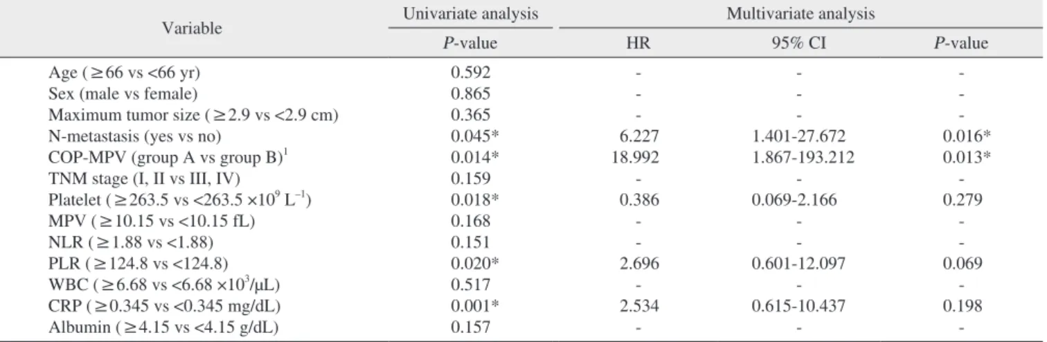
Clinical and laboratory variables were divided into groups based on their respective cut-off values and the relationship between overall survival and each respective variable was an- alyzed. Univariate analyses showed N-metastasis (P=0.045), COP-MPV (A vs B) (P=0.014), PLT (≥263.5 vs <263.5 ×10⁹ L–1) (P=0.018), PLR (≥124.8 vs <124.8) (P=0.020), and CRP (≥0.345 vs <0.345 mg/dL) (P=0.001) were statistically sig- nificant prognostic factors.(Table 3)
4. Multivariate analyses
A multivariate Cox proportional hazards model was used with univariate analyses to determine the significance of variables with a P-value less than 0.05 as independent prog- nostic factors for OSCC patients. Results were as follows: N- metastasis (HR 6.227, 95% confidence interval [CI] 1.401- 27.672, P=0.016); COP-MPV (A vs B) (HR 18.992, 95% CI 1.867-193.212, P=0.013), indicating both N-metastasis and COP-MPV were significant independent prognostic factors for OSCC patient survival.(Table 3)
molar pad in 4 patients (10.0%). Mean PLT and MPV were 243.93±61.382 ×10⁹ L–1 (103-389 ×109 L–1) and 10.05±0.67 fL (8.8-11.4 fL), respectively. Other parameter values and ranges were as follows: WBC, 6.44±1.79 ×10³/μL (3.52- 10.83 ×10³/μL); albumin, 4.14±0.45 g/dL (2.6-4.8 g/dL);
CRP, 0.52±0.79 mg/dL (0.02-4.61 mg/dL); PLR, 127±39.51 (37.73-252.46); NLR, 1.92±0.76 (0.53-3.89); and maximum tumor size, 2.75±1.36 cm (0.8-5.9 cm).
2. Clinicopathological characteristics between COP- MPV groups
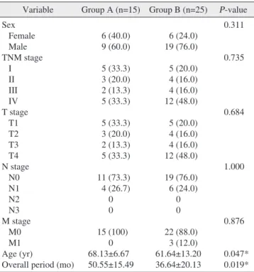
The mean age at the time of clinicopathological char- acteristic measurement for COP-MPV group A was older (68.13±6.67 years) than group B (61.64±13.20 years) (P=0.047). The COP-MPV group A was in the study longer on average (50.55±15.49 months) than the COP-MPV group B (36.64±20.13 months) (P=0.019). There was no statistical- ly significant relationship with age (P=0.311), N-metastasis (P=1.000), or TNM stage (P=0.735).(Table 1)
The PLT of the COP-MPV group A was 216.33±25.87
Table 1. Clinical and pathological characteristics between COP- MPV groups A and B
Variable Group A (n=15) Group B (n=25) P-value Sex
Female Male TNM stage I II III IV T stage T1 T2 T3 T4 N stage N0 N1 N2 N3 M stage M0 M1 Age (yr)
Overall period (mo)
6 (40.0) 9 (60.0) 5 (33.3) 3 (20.0) 2 (13.3) 5 (33.3) 5 (33.3) 3 (20.0) 2 (13.3) 5 (33.3) 11 (73.3) 4 (26.7)
0 0 15 (100)
0 68.13±6.67 50.55±15.49
6 (24.0) 19 (76.0) 5 (20.0) 4 (16.0) 4 (16.0) 12 (48.0) 5 (20.0) 4 (16.0) 4 (16.0) 12 (48.0) 19 (76.0) 6 (24.0)
0 0 22 (88.0)
3 (12.0) 61.64±13.20 36.64±20.13
0.311 0.735
0.684
1.000
0.876
0.047*
0.019*
(COP-MPV: count of platelet and mean platelet volume)
*P<0.05.
Values are presented as number (%) or mean±standard deviation.
Group A: COP-MPV=0, Group B: COP-MPV=1.
Jae Woo Park et al: Count of platelet and mean platelet volume score: serologic prognostic factor in patients with oral squamous cell carcinoma. J Korean Assoc Oral Maxillofac Surg 2017
Table 2. Serological characteristic between COP-MPV groups A and B
Variable Group A Group B P-value
Platelet (×109 L–1) MPV (fL) NLR PLR
WBC (×103/μL) CRP (mg/dL) Albumin (g/dL)
216.33±25.87 9.68±0.34 1.97±0.75 117.94±28.42
6.27±1.91 0.73±1.24 4.11±0.56
260.48±70.50 10.27±0.72
1.90±0.78 132.43±44.54
6.54±1.75 0.40±0.29 4.16±0.38
0.008*
0.005*
0.679 0.267 0.525 0.600 0.738 (COP-MPV: count of platelet and mean platelet volume, NLR:
neutrophil-lymphocyte ratio, PLR: platelet-lymphocyte ratio, WBC:
white blood cell, CRP: C-reactive protein)
*P<0.05.
Values are presented as mean±standard deviation.
Group A: COP-MPV=0, Group B: COP-MPV=1.
Jae Woo Park et al: Count of platelet and mean platelet volume score: serologic prognostic factor in patients with oral squamous cell carcinoma. J Korean Assoc Oral Maxillofac Surg 2017
IV. Discussion
Recent studies have provided evidence that platelet activa- tion is clinically significant in some malignant tumors. In par- ticular, in a series of processes in which cancer cells spread to other organs through blood circulation, platelets are important for cancer cells to aggregate and be discharged from blood vessels22,23. Some studies have reported that, in cancer pa- tients, platelet activation indexes (soluble P-selectin, soluble CD40 ligand, and platelet factor 4) are relatively higher than in normal patients24-26. Some studies have reported that MPV 5. Kaplan-Meier analyses
Kaplan-Meier analyses were used to determine the dif- ference in overall survival between COP-MPV groups. The prognosis of COP-MPV group B was worse than that of COP-MPV group A (P=0.014). The prognosis of the group with lymph node metastasis was worse than that of the non- metastasis group (P=0.018).(Fig. 1, 2)
Table 3. Univariate and multivariate analyses of overall survival for oral squamous cell carcinoma patients
Variable Univariate analysis Multivariate analysis
P-value HR 95% CI P-value
Age (≥66 vs <66 yr) Sex (male vs female)
Maximum tumor size (≥2.9 vs <2.9 cm) N-metastasis (yes vs no)
COP-MPV (group A vs group B)1 TNM stage (I, II vs III, IV)
Platelet (≥263.5 vs <263.5 ×10⁹ L–1) MPV (≥10.15 vs <10.15 fL) NLR (≥1.88 vs <1.88) PLR (≥124.8 vs <124.8) WBC (≥6.68 vs <6.68 ×10³/μL) CRP (≥0.345 vs <0.345 mg/dL) Albumin (≥4.15 vs <4.15 g/dL)
0.592 0.865 0.365 0.045*
0.014*
0.159 0.018*
0.168 0.151 0.020*
0.517 0.001*
0.157
- - - 6.227 18.992
- 0.386
- - 2.696
- 2.534
-
- - - 1.401-27.672 1.867-193.212
- 0.069-2.166
- - 0.601-12.097
- 0.615-10.437
-
- - - 0.016*
0.013*
- 0.279
- - 0.069
- 0.198
-
(HR: hazard ratio, CI: confidence interval, COP-MPV: count of platelet and mean platelet volume, NLR: neutrophil-lymphocyte ratio, PLR:
platelet-lymphocyte ratio, WBC: white blood cell, CRP: C-reactive protein)
1Group A: COP-MPV=0, Group B: COP-MPV=1.
*P<0.05.
Jae Woo Park et al: Count of platelet and mean platelet volume score: serologic prognostic factor in patients with oral squamous cell carcinoma. J Korean Assoc Oral Maxillofac Surg 2017
0 20 40 60 80 100
0 0.8 0.6 0.4 survivalCumulative 0.2
Overall period (mo) Survival functions
0 1
0-censored 1-censored COP-MPV 1.0
Fig. 1. Kaplan-Meier curves of overall survival between count of platelet and mean platelet volume (COP-MPV) scores 0 and 1.
Jae Woo Park et al: Count of platelet and mean platelet volume score: serologic prognostic factor in patients with oral squamous cell carcinoma. J Korean Assoc Oral Maxillofac Surg 2017
0 20 40 60 80 100
0 0.8 0.6 0.4 0.2
Survival functions
No Yes
-censored No
Yes-censored N-metastasis 1.0
Cumulativesurvival
Overall period (mo)
Fig. 2. Kaplan-Meier curves of overall survival between N-metas- tasis yes and no.
Jae Woo Park et al: Count of platelet and mean platelet volume score: serologic prognostic factor in patients with oral squamous cell carcinoma. J Korean Assoc Oral Maxillofac Surg 2017
that of the non-metastasis group (Fig. 2), and there was no statistically significant difference in survival rates between the group with maximum tumor size larger than the cut-off value and the group with maximum tumor size not larger than the cut-off value (P=0.365). Zhang et al.39 reported a 5-year survival rate of 20.5% in the N-metastasis group for OSCC patients and 72.3% in the non-metastasis group, claiming that N-metastasis was a more important prognostic factor for predicting survival rate than T stage. Through Cox regres- sion, this study has shown that COP-MPV score (P=0.013) is a more meaningful prognostic factor than N-metastasis (P=0.016) for predicting the survival rate of OSCC patients.
These results are similar to those of Zhang et al.40 regarding esophageal squamous cell carcinoma patients.
Khandavilli et al.41 reported that patients with OSCC with high preoperative CRP levels had poor prognosis, and that patients in TNF stage III and IV had relatively higher preop- erative CRP levels than patients in stage I and II. Gockel et al.42 reported that lymph node metastasis and tumor extension increased in patients with higher CRP levels, and Park et al.43 reported that survival rate was low in patients with a high CRP/albumin ratio. Similar to these results, CRP (≥0.345 vs
<0.345) (P=0.001) was a statistically significant prognostic factor on univariate analysis in this study. However, on mul- tivariate analysis, CRP (P=0.198) was not an independent prognostic factor. These results indicate that the stratifying ability of CRP was likely incorporated in the COP-MPV score.
Kaplan-Meier analysis showed the survival rate of COP- MPV group A was higher than group B indicating that, if both COP and MPV were lower than the cut-off value, post- operative prognosis would be better.
Univariate analysis showed the P-value for PLT was 0.018 and the P-value for MPV was 0.168. PLT was a statistically significant prognostic factor alone but MPV alone was not.
However, COP-MPV score (P=0.014) combining PLT and MPV was a more significant prognostic factor than PLT (P=0.018). These results suggest that COP-MPV score, which considers PLT and MPV in a comprehensive manner, is help- ful for predicting the prognosis for OSCC patients.
This study has several limitations. First, the sample size is relatively small and it was a retrospective study conducted at a single institution. The initial hypothesis predicted that prognosis would be worse if both COP and MPV were higher than the cut-off value, but it was difficult to confirm this statistically because of the small sample size. Furthermore, the cut-off values for COP and MPV set in this study are not is also closely related to various thromboembolic disorders
including ischemic cardiovascular disease27,28, and some stud- ies have reported that MPV is related to malignant tumors19,29. However, whether the prognosis of patients with relatively high MPV is worse than those with low MPV remains a subject of debate. Osada et al.30 reported that MPV level rose in malicious tumors such as gastric cancer. In contrast, Mutlu et al.31 did not find any elevation in MPV level in vari- ous cancer patients and, compared with time of first cancer diagnosis, MPV level was significantly reduced at the time of thrombotic events. However, larger platelets secrete more chemical mediators upon stimulation than smaller ones, and the increase in number of larger platelets suggests that stimu- lation exists from diseases such as cardiovascular disease or malignant tumors27,28,30. PLT and MPV are important indexes for assessing platelet activation state17. It is essential to pre- dict the prognosis of OSCC patients using a scoring system that includes comprehensive consideration of PLT and MPV.
The mean age of group A was higher (68.13 years) than group B (61.64 years), indicating that age was lower in group B with poor prognosis (P=0.047).(Table 1) Goldstein and Irish32 and Sarkaria and Harari33 found in their study that squamous cell carcinoma in the head and neck showed a more aggressive tendency in younger age groups. In contrast, Veness et al.34 reported that it was difficult to predict progno- sis by age35. However, in recent years, the incidence of OSCC is increasing in younger age groups36-38.
In overall period, the COP-MPV group A was longer (50.55 months) than the COP-MPV group B (36.64 months) (P=0.019). There was no statistically significant relationship between COP-MPV score and sex, tumor location, N-metas- tasis, and TNM stage.(Table 1)
Multivariate analysis showed N-metastasis and COP- MPV were statistically significant independent prognostic factors. The N-metastasis group had a 6.227 times higher risk of death than the non-metastasis group (HR 6.227, 95%
CI 1.401-27.672) and the risk of death in COP-MPV group B was 18.992 times higher than COP-MPV group A (HR 18.992, 95% CI 1.867-193.212, P=0.013). These results in- dicate that COP-MPV score had a greater effect on survival prognosis for OSCC patients than N-metastasis.
During the observation period, the survival rate of COP- MPV group B (56%) was significantly lower than that of COP-MPV group A (93.4%). The survival rate of the N- metastasis group was 50% and the survival rate of the non- metastasis group was 76.7%. Kaplan-Meier analysis showed the survival rate in the N-metastasis group was not better than
6. Llewellyn CD, Johnson NW, Warnakulasuriya KA. Risk factors for squamous cell carcinoma of the oral cavity in young people--a comprehensive literature review. Oral Oncol 2001;37:401-18.
7. Singh B, Bhaya M, Zimbler M, Stern J, Roland JT, Rosenfeld RM, et al. Impact of comorbidity on outcome of young patients with head and neck squamous cell carcinoma. Head Neck 1998;20:1-7.
8. Schantz SP, Yu GP. Head and neck cancer incidence trends in young Americans, 1973-1997, with a special analysis for tongue cancer. Arch Otolaryngol Head Neck Surg 2002;128:268-74.
9. Lydiatt DD, Robbins KT, Byers RM, Wolf PF. Treatment of stage I and II oral tongue cancer. Head Neck 1993;15:308-12.
10. Layland MK, Sessions DG, Lenox J. The influence of lymph node metastasis in the treatment of squamous cell carcinoma of the oral cavity, oropharynx, larynx, and hypopharynx: N0 versus N+. La- ryngoscope 2005;115:629-39.
11. Forastiere A, Koch W, Trotti A, Sidransky D. Head and neck can- cer. N Engl J Med 2001;345:1890-900.
12. Woolgar JA, Rogers S, West CR, Errington RD, Brown JS, Vaughan ED. Survival and patterns of recurrence in 200 oral cancer patients treated by radical surgery and neck dissection. Oral Oncol 1999;35:257-65.
13. Yotsukura S, Mamitsuka H. Evaluation of serum-based cancer biomarkers: a brief review from a clinical and computational view- point. Crit Rev Oncol Hematol 2015;93:103-15.
14. Lip GY, Chin BS, Blann AD. Cancer and the prothrombotic state.
Lancet Oncol 2002;3:27-34.
15. Nash GF, Turner LF, Scully MF, Kakkar AK. Platelets and cancer.
Lancet Oncol 2002;3:425-30.
16. Gay LJ, Felding-Habermann B. Contribution of platelets to tumour metastasis. Nat Rev Cancer 2011;11:123-34.
17. Kamath S, Blann AD, Lip GY. Platelet activation: assessment and quantification. Eur Heart J 2001;22:1561-71.
18. Kai H, Kitadai Y, Kodama M, Cho S, Kuroda T, Ito M, et al. In- volvement of proinflammatory cytokines IL-1beta and IL-6 in pro- gression of human gastric carcinoma. Anticancer Res 2005;25:709- 19. Kurt M, Onal IK, Sayilir AY, Beyazit Y, Oztas E, Kekilli M, et al. 13.
The role of mean platelet volume in the diagnosis of hepatocellular carcinoma in patients with chronic liver disease. Hepatogastroen- terology 2012;59:1580-2.
20. Karaman K, Bostanci EB, Aksoy E, Kurt M, Celep B, Ulas M, et al. The predictive value of mean platelet volume in differential di- agnosis of non-functional pancreatic neuroendocrine tumors from pancreatic adenocarcinomas. Eur J Intern Med 2011;22:e95-8.
21. Kılınçalp S, Ekiz F, Başar O, Ayte MR, Coban S, Yılmaz B, et al.
Mean platelet volume could be possible biomarker in early diagno- sis and monitoring of gastric cancer. Platelets 2014;25:592-4.
22. Connolly GC, Phipps RP, Francis CW. Platelets and cancer-associ- ated thrombosis. Semin Oncol 2014;41:302-10.
23. Stegner D, Dütting S, Nieswandt B. Mechanistic explanation for platelet contribution to cancer metastasis. Thromb Res 2014;133 Suppl 2:S149-57.
24. Caine GJ, Lip GY, Stonelake PS, Ryan P, Blann AD. Platelet acti- vation, coagulation and angiogenesis in breast and prostate carci- noma. Thromb Haemost 2004;92:185-90.
25. Li L, Li P, Yang YQ, Zhang H, Ai P, Wang F, et al. sCD40L, sP- selectin and sICAM-1 plasma levels in nasopharyngeal carcinoma.
Sichuan Da Xue Xue Bao Yi Xue Ban 2009;40:513-6.
26. Peterson JE, Zurakowski D, Italiano JE Jr, Michel LV, Connors S, Oenick M, et al. VEGF, PF4 and PDGF are elevated in platelets of colorectal cancer patients. Angiogenesis 2012;15:265-73.
27. Azab B, Torbey E, Singh J, Akerman M, Khoueiry G, McGinn JT, et al. Mean platelet volume/platelet count ratio as a predictor of long-term mortality after non-ST-elevation myocardial infarction.
Platelets 2011;22:557-66.
28. Slavka G, Perkmann T, Haslacher H, Greisenegger S, Marsik C, Wagner OF, et al. Mean platelet volume may represent a predictive
generally applicable to all patients with OSCC. In this study, COP-MPV score had more statistically significant results than other prognostic factors in determining OSCC patient prognosis. Future prospective studies with more subjects using COP-MPV score as a prognostic factor are needed to identify the applicable cut-off value for all patients with OSCC.
V. Conclusion
This study established that COP-MPV score was statisti- cally significant for predicting the prognosis of 40 patients with OSCC. Multivariate analysis showed the survival rate of COP-MPV group B was lower than group A and COP- MPV score (HR 18.992, P=0.013) was found to be a more significant independent prognostic factor than N-metastasis (HR 6.227, P=0.016). COP-MPV score is a simple and cost- effective test method, and is considered a more effective fac- tor relative to other considered factors in predicting OSCC patient prognosis.
Conflict of Interest
No potential conflict of interest relevant to this article was reported.
ORCID
Jae Woo Park, http://orcid.org/0000-0002-5549-5202 Chul-Hwan Kim, http://orcid.org/0000-0002-5199-2420 Yong Chan Ha, http://orcid.org/0000-0002-0069-3797 Moon Young Kim, http://orcid.org/0000-0001-9596-7481 Sung Min Park, http://orcid.org/0000-0001-8573-9071
References
1. Siegel R, Naishadham D, Jemal A. Cancer statistics, 2013. CA Cancer J Clin 2013;63:11-30.
2. Ferlay J, Soerjomataram I, Dikshit R, Eser S, Mathers C, Rebelo M, et al. Cancer incidence and mortality worldwide: sources, methods and major patterns in GLOBOCAN 2012. Int J Cancer 2015;136:E359-86.
3. Johnson N. Tobacco use and oral cancer: a global perspective. J Dent Educ 2001;65:328-39.
4. Reibel J. Tobacco and oral diseases. Update on the evidence, with recommendations. Med Princ Pract 2003;12 Suppl 1:22-32.
5. Lewin F, Norell SE, Johansson H, Gustavsson P, Wennerberg J, Biörklund A, et al. Smoking tobacco, oral snuff, and alcohol in the etiology of squamous cell carcinoma of the head and neck: a popu- lation-based case-referent study in Sweden. Cancer 1998;82:1367- 75.
parameter for overall vascular mortality and ischemic heart disease.
Arterioscler Thromb Vasc Biol 2011;31:1215-8.
29. Cho SY, Yang JJ, You E, Kim BH, Shim J, Lee HJ, et al. Mean platelet volume/platelet count ratio in hepatocellular carcinoma.
Platelets 2013;24:375-7.
30. Osada J, Rusak M, Kamocki Z, Dabrowska MI, Kedra B. Platelet activation in patients with advanced gastric cancer. Neoplasma 2010;57:145-50.
31. Mutlu H, Artis TA, Erden A, Akca Z. Alteration in mean platelet volume and platicrit values in patients with cancer that developed thrombosis. Clin Appl Thromb Hemost 2013;19:331-3.
32. Goldstein DP, Irish JC. Head and neck squamous cell carcinoma in the young patient. Curr Opin Otolaryngol Head Neck Surg 2005;13:207-11.
33. Sarkaria JN, Harari PM. Oral tongue cancer in young adults less than 40 years of age: rationale for aggressive therapy. Head Neck 1994;16:107-11.
34. Veness MJ, Morgan GJ, Sathiyaseelan Y, Gebski V. Anterior tongue cancer: age is not a predictor of outcome and should not alter treat- ment. ANZ J Surg 2003;73:899-904.
35. Popovtzer A, Shpitzer T, Bahar G, Marshak G, Ulanovski D, Fein- messer R. Squamous cell carcinoma of the oral tongue in young patients. Laryngoscope 2004;114:915-7.
36. Patel SC, Carpenter WR, Tyree S, Couch ME, Weissler M, Hack- man T, et al. Increasing incidence of oral tongue squamous cell carcinoma in young white women, age 18 to 44 years. J Clin Oncol
2011;29:1488-94.
37. Marocchio LS, Lima J, Sperandio FF, Corrêa L, de Sousa SO. Oral squamous cell carcinoma: an analysis of 1,564 cases showing ad- vances in early detection. J Oral Sci 2010;52:267-73.
38. Chow CW, Tabrizi SN, Tiedemann K, Waters KD. Squamous cell carcinomas in children and young adults: a new wave of a very rare tumor? J Pediatr Surg 2007;42:2035-9.
39. Zhang WB, Wang Y, Mao C, Guo CB, Yu GY, Peng X. Cervical metastasis of maxillary squamous cell carcinoma. Int J Oral Maxil- lofac Surg 2015;44:285-91.
40. Zhang F, Chen Z, Wang P, Hu X, Gao Y, He J. Combination of platelet count and mean platelet volume (COP-MPV) predicts post- operative prognosis in both resectable early and advanced stage esophageal squamous cell cancer patients. Tumour Biol 2016;37:
9323-31.
41. Khandavilli SD, Ceallaigh PO, Lloyd CJ, Whitaker R. Serum C- reactive protein as a prognostic indicator in patients with oral squa- mous cell carcinoma. Oral Oncol 2009;45:912-4.
42. Gockel I, Dirksen K, Messow CM, Junginger T. Significance of preoperative C-reactive protein as a parameter of the periopera- tive course and long-term prognosis in squamous cell carcinoma and adenocarcinoma of the oesophagus. World J Gastroenterol 2006;12:3746-50.
43. Park HC, Kim MY, Kim CH. C-reactive protein/albumin ratio as prognostic score in oral squamous cell carcinoma. J Korean Assoc Oral Maxillofac Surg 2016;42:243-50.