Ⅰ. 서 론
구강내 암종은 인체에 발생되는 전체 암종의 약 3%를 차지하 고 있으며 발생되는 암종의 95% 이상이 편평세포암종으로 이들
의 발생은 흡연, 음주, 자외선과 방사선 조사 및 human papilloma virus 감염등과 밀접한 관계가 있다
1-4).
일반적인 암의 발생기전은 종양유전자(oncogene)와 종양억제 유전자(antioncogene)의 비정상적인 작용에 의한 것으로 알려졌 다. 즉 종양유전자의 활성증가와 돌연변이에 의한 종양억제유전 자의 활성감소는 정상세포를 비정상적으로 활성화시켜 세포분 열이 제어되지 않고 지속적으로 촉진되어 결국 세포가 급성장하 여 종양으로 발전하게 된다
5-7).
현재까지 알려진 가장 중요한 종양억제유전자는 망막모세포 종(retinoblastoma : Rb) gene과 p53 gene들로 이들에 대한 연구는 많이 이루어지고 있다.
구강 편평세포암종에서 Differential Polymerase Chain Reaction에 의한 Cyclin D1 유전자의 증폭에 대한 연구
김기순∙김경욱∙이재훈∙김창진*
단국대학교 치과대학 구강악안면외과학교실, 순천향대학교 의과대학 병리학교실*
Abstract
CYCLIN D1 GENE AMPLIFICATION IN ORAL SQUAMOUS CELL CARCINOMA USING DIFFERENTIAL POLYMERASE CHAIN REACTION
Kee-Soon Kim, Kyung-Wook Kim, Jae-Hoon Lee, Chang-Jin Kim * Department of Oral and Maxillofacial Surgery, College of Dentistry, Dankook University
Department of Pathology College of Medicine, Soon Chun Hyang University*
Neoplastic growth is characterized by alterations of oncogenes and antioncogenes. The interaction between activated oncogenes and functional deletion of antioncogene appears to be the driving force directing normal cells to uncontrolled growth resulting in tumor.
In addition to those genes mentioned, other genes controlling the entry of cells into the cell cycle have recently been implicated in cancer development. The overexpression of the cyclin D1 gene, which has been mapped to 11q13, either by gene rearrangement or amplification has been noted in various malignant tumors. The product of the cyclin D1 gene forms a complex with cyclin-dependent protein kinases(CDK4) that governs a key transition in the cell cycle.
The relationships between the overexpression of cyclin D1 assessed by immunihistochemistry and the amplification of the cyclin D1 gene by differential polymerase chain reaction(DPCR) using primers for dopamin D2 receptor gene in 13 cases of squamous cell carci- nomas of the oral cavity have been studied.
The semiquantitative assay of cyclin D1 amplification has been made by cyclin D1/dopamin D2 receptor(CD/DR) ratio.
The results were as follows;
1. In the normal tissue and the tumor, the CD/DR ratios were 0.82 and 1.36 respectively. This implicates 1.65-fold amplification of cyclin D1 gene in tumor compared to that in normal tissue.
2. The tumor tissue which showed overexpression of cyclin D1 by immunohistochemistry revealed 2-fold amplification of cyclin D1 compared to the normal tissue.
3. The tumor tissue which showed mild expression of cyclin D1 by immunihistochemistry revealed 1.7-fold amplification of cyclin D compared to the normal tissue.
4. The cyclin D1 was overexpressed in the tumor tissue at the rate of 38%.
Above results suggest that cyclin D1 has close correlation with the development of carcinoma in the oral cavity. But further studies were needed to elucidate the carcinogeneic mechanisms by comparative studies among cyclin D1, pRb and p53.
김 경 욱
330-716, 충남 천안시 신부동 산7-1
단국대학교 치과대학 구강악안면외과학교실 Kyung-Wook KimDept. of OMFS, College of Dentistry, Dankook University san7-1, Shinbu-Dong, Chon-An, Chung-Nam, 330-716, Korea Tel:82-417-550-1991 Fax:82-417-551-8988
E-mail : [email protected]
그러나 근자에는 이들 종양유전자와 종양억제유전자들 외에 세포주기(cell cycle)를 조절하는 물질들이 종양발생과 밀접한 관 련이 있다는 보고가 나오고 있다
8).
세포주기 중 G
1에서 S기로 이행되는 부위는 세포주기의 검문 소(check point)로서 이 부위의 조절은 전체적인 세포주기를 제어 하는 중요한 부위인데 cyclin D1은 세포주기의 G
1에서 S기로 이 행되는 부위를 조절해주는 중요한 인자로 유방암
9), 식도암
10), 두 경부의 편평상피암
11), 간암
12,13), 폐암
14), 대장암
15), 전립선암
16)및 자 궁경부암
17)등의 다양한 종양유발에 관여한다.
Cyclin D1유전자는 염색체의 11q13 부위에 위치하고 있으며 이 유전자의 재배열 혹은 증폭(rearrangement or amplification)이 일 어나면 cyclin dependent kinase(CDK)4나 CDK6와 결합하여 암억 제 유전자의 하나인 retinoblastoma 유전자의 활동을 억제시켜 S 기로 진입을 촉진시켜 세포주기 조절능력을 잃고 과발현되면서 종양이 발생하게 된다
18-20).
식도암에서 cyclin D1의 증폭을 조사하였더니 약 20% 이상에서 발견되었다는 보고가 있고
21-26)최근에는 Gramlich 등
10)이 파라핀 포매조직을 시료로 구강상피세포의 생명현상과 관련이 없는 dopamin D
2receptor 유전자(DR)를 기준으로 하여 differential polymerase chain reaction(DPCR) 기법으로 식도암조직에서 cyclin D1 유전자의 증폭을 조사하여 보고하였다.
본 연구에서는 13례의 구강 편평세포암종을 대상으로 정상 및 암 조직 세포에서 DPCR기법에 의한 cyclin D1 유전자의 증폭과 면역조직화학적염색을 통한 cyclin D1의 발현을 검사하여 구강 편평세포암종 발생과 cyclin D1유전자의 증폭과의 관계를 조사 하여 다소의 지견을 얻었기에 보고하는 바이다.
Ⅱ. 연구재료 및 방법 1. 연구재료
구강내 병변 중 조직검사로 편평세포암종으로 진단된 13례를 대상으로 하였다. 생검 및 수술로 적출한 조직을 10% neutral buffered formalin으로 24시간 고정 후 통상적인 방법으로 파라핀 포매조직을 만들고 이에서 6㎛ 절편을 제작하여 hematoxylin- eosin염색을 시행한 후 판독하여 편평세포암종의 조직학적 유형 을 판독하였다. 13례의 편평세포암종 중 3례는 분화도가 높은 편평세포암종이었고 10례는 중등도 분화를 보이는 편평세포암 종이었다.
유방암조직을 양성대조군으로 하였다.
2. 연구방법
가) DNA추출
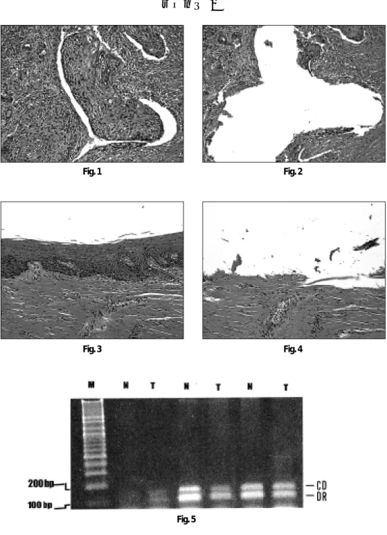
암세포와 정상세포에서 각각 DNA를 추출하기 위하여 microdissection 방법으로 암 조직과 정상조직을 구분하였다(Fig.
1, 2, 3&4). 이를 위하여 파라핀 포매조직에서 5개의 7㎛절편을 채취하여 통상적인 방법으로 탈파라핀하고 degraded alcohol로
함수시킨 후 조직을 건조시켰다. 건조된 조직을 10× 현미경하 에 28 guage 바늘로 암조직과 정상조직을 긁어 300㎕의 digestion buffer (50 mM Tris HCl, pH 8.5, 1mM EDTA, 0.5% Tween 20, 400㎍
proteinase K)가 들어있는 1.5㎖ Eppendorf tube에 넣어 55℃에서 24시간 부란하였다. 부란 후 proteinase K의 활성을 억제하기 위 하여 97℃에서 5분간 처리하였고 그 후 phenol/chloroform으로 DNA를 추출하였다. 추출과정은 65℃의 0.5% sodium sarcosin의 buffer-saturated phenol을 300㎕씩 시료에 넣어 vortex시켜 homogenous emulsion을 만든 후 3,500g로 원심분리 하였다. 그 후 상층액을 깨끗한 Eppendorf tube에 옮긴 후 300㎕의 chloro- form isoamylalcohol을 첨가하여 vortex로 잘 섞은 후 역시 3,500g 로 원심분리 하였고, 이 과정을 3회 반복하였다. 그 후 상층액을 새 tube에 옮긴 후 1/10 volume의 3M sodium acetate와 2X volume 의 100% ethanol을 첨가하여 조심스럽게 섞은 후 -70℃에 1시간 방치하여 DNA를 침전 시켰다. 그 후 3,500g로 5분간 원심분리 한 후 상층액을 버리고 침전물을 70% cold ethanol로 수세 후 재 원 심분리하여 DNA를 침전시키고 DNA pellet을 공기 중에 건조시 킨 후 멸균 증류수로 용해시켰다.
추출한 DNA의 순도는 photometer로 260nm와 280nm의 파장에 서 수치를 읽어 이 수치의 비 260/280가 1.8 이상인 DNA를 시료 로 사용하였고 순도가 이 수치 이하인 경우에는 phenol/chloro- form추출을 다시 하였다.
이렇게 추출한 DNA는 1% agarose gel에 전기영동하여 DNA를 확인하였다.
나) Differential Polymerase Chain Reaction (DPCR)
Differential polymerase chain reaction(DPCR)을 위하여 152 bp크 기의 cyclin D1유전자와 112 bp크기의 dopamine D
2receptor(DR) 유전자를 동시에 증폭하였다. Cyclin D1유전자와 DR유전자는 11q에 같이 있기 때문에 이를 동시에 증폭하여 비교함으로 cyclin D1유전자의 증폭량의 비교를 알 수 있었다.
이들의 Primer는 다음과 같았다
27,28). Cyclin D1(CD);
Up stream : 5′ACCAGCTCCTGTGCTGCGAA3′
Down stream : 5′CAGGACCTCCTTC6TGCACAC3′
Dopamine D2 receptor (DR);
Up stream : 5′CCACTGAATCTGTCCTGGTATG3′
Down stream : 5′GCGTGGCATAGTAGTTGTAGTGG3′
PCR조건은 총 25㎕로 template DNA 1㎕, 50 mmol/L KCl, 10
mmol/L Tris-HCl (pH 8.3), 1.5 mmol/L MgCl2, 0.2 mmol/L of each
dNTP, 0.5 μmol/L each primer의 조성에 0.625 U of Taq poly-
merase(Perkin-Elmer Cetus)를 넣어 반응시켰다. 반응은 95℃에서
10분, 그 후 95℃, 50℃ 및 72℃에서 각각 1분씩, 이 과정을 35회
반복하였다. PCR산물을 3% SeaKem agarose gel에 전기영동 후
ethidium bromide로 염색하였고 polaroid film으로 사진 촬영하여
화상분석기(Optimas Corp., Bothell, USA)로 Optimas Ver.6 System
(Media Cybernetics
�, Silver Spring, MD, USA)을 이용하여 밀도분석
(densitometric analysis)을 시행하여 CD/DR 비율을 구하였다.
다) Cyclin D1의 면역조직화학적 염색
조직세포에서의 cyclin D1발현을 검색하기 위하여 cyclin D1 단 일클론항체 (Novocastra, UK)를 이용하였다. 면역조직화학적 염 색을 시행하기 위하여 파라핀 포매조직에서 6㎛의 절편을 만들 어 탈파라핀한 후 graded alcohol로 함수시킨 후 내인성 peroxi- dase를 차단하기 위하여 3% 과산화 수소용액을 methanol에 만들 어 실온에서 30분간 부란하였다. 그 후 phosphate buffered saline(PBS, pH 7.4)으로 수세하고, 다시 비 특이성 결합을 방지하 기 위하여 정상 goat serum에 조직을 실온에서 30분간 부란하였 다. 다시 PBS로 3분간 3회 수세 후 항원을 노출시키기 위하여 10mM의 citrate buffer (pH 6.0)에 조직을 넣고 microwave oven에 서 15분간 끓인 후 실온에서 서서히 식힌 후 PBS로 3회 수세하였 고, cyclin D1에 대한 단일클론항체를 1:50으로 희석하여 실온에 서 1시간 부란하였다. 다시 PBS로 3회 수세하였고, biotin-labeled antimouse immune goat serum과 실온에서 30분 부란하였고 이를 다시 3회 PBS로 수세한 후 peroxidase labeled streptavidin과 실온 에서 30분 반응시킨 후 PBS로 수세하였고, diaminobenzidine으로 발색하였다. 물로 수세 후 2% methyl green으로 대조염색을 시행 하고 염색이 진하게 나타나는 부위를 기준으로 하여 광학현미경 으로 관찰하여(×200) 갈색의 염색상으로 나타나는 핵이 5% 미 만은 - , 5%�30%는 + , 30%�70%는 ++ 그리고 70% 이상은 +++로 표시하였다.
Ⅲ. 연구결과
1. Differential Polymerase Chain Reaction
Cyclin D1과 dopamine D
2receptor에 대한 각각의 primer로 증폭 한 산물은 각각 152bp와 112bp에 뚜렷한 2개의 띠가 관찰되었다 (Fig. 5).
정상조직과 종양조직의 띠의 발현강도를 비교해보면 정상에 비하여 종양조직에서 CD띠의 강도가 높은 것이 관찰되었다.
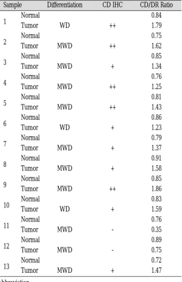
띠의 강도를 밀도분석한 결과는 Table 1에서와 같다.
13례의 시료에서 정상조직에서의 CD/DR ratio는 0.82였고 종양 조직에서의 CD/DR ratio는 1.36으로 종양조직에서의 CD증폭은 정상에 비하여 1.65배 높았다. 면역조직화학적 염색결과에 의한 cyclin D1발현과 유전자 증폭과의 관계를 보면, cyclin D1발현이 (++)인 5례에서의 정상조직의 CD/DR ratio는 0.80이며, 종양조직 에서의 CD/DR ratio는 1.59로 약 2배의 CD유전자가 증폭이 관찰 되었다. 또한 cyclin D1 발현이 (+)인 6례에서의 정상조직의 CD/DR ratio는 0.83이고 종양조직에서의 CD/DR ratio는 1.43으로 약 1.7배의 CD유전자의 증폭이 관찰되었다. 반면 cyclin D1의 발 현이 (-)인 2례에서의 정상조직의 CD/DR ratio는0.83이고 종양조 직에서의 CD/DR ratio는 0.55로 정상조직에 비하여 유전자의 증 폭이 낮은 결과를 보였다.
2. Expression of Cyclin D1 by Immunohistochemistry
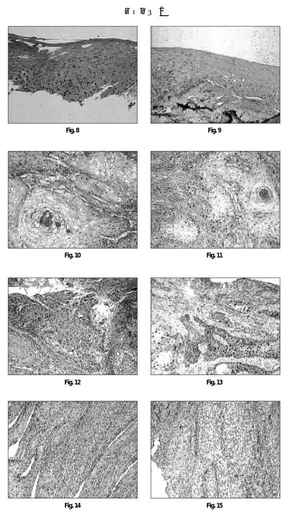
양성 대조군인 유방암조직에서의 cyclin D1발현은 암세포의 핵 에 진갈색의 염색상으로 관찰되었다(Fig. 6, 7).
정상조직에서의 cyclin D1 발현은 세포증식이 활발한 중층편평 상피층의 기저세포에 아주 드물게 관찰되었다(Fig. 8, 9). 종양조 직에서의 cyclin D1발현은 (-)부터 (++)까지 다양한 염색상을 보 였고(Fig. 10, 11, 12, 13, 14 & 15), 조직학적 분화도에 따른 cyclin D1의 발현의 차이는 관찰되지 않았다(Table 1).
Table 1. Results of cyclin D
1Immunohistochemistry and CD/DR Ratio of the Samples.
Normal 0.84
1 Tumor WD ++ 1.79
Normal 0.75
2 Tumor MWD ++ 1.62
Normal 0.85
3 Tumor MWD + 1.34
Normal 0.76
4 Tumor MWD ++ 1.25
Normal 0.81
5 Tumor MWD ++ 1.43
Normal 0.86
6 Tumor WD + 1.23
Normal 0.79
7 Tumor MWD + 1.37
Normal 0.91
8 Tumor MWD + 1.58
Normal 0.85 9 Tumor MWD ++ 1.86
Normal 0.83
10 Tumor WD + 1.59
Normal 0.76 11 Tumor MWD - 0.35 Normal 0.89 12 Tumor MWD - 0.75 Normal 0.72 13 Tumor MWD + 1.47
Abbreviation :
WD : Well Differentiated
MWD : Moderately Well Differentiated CD IHC : Cyclin D1 Immunohistochemistry CD/DR Ratio : Cyclin D1/Dopamine D2 receptor Ratio - : Below 5% positivity of total expressed nuclei + : 5%�30% positivity of total expressed nuclei ++ : 30%�70% positivity of total expressed nuclei
Sample Differentiation CD IHC CD/DR Ratio
Ⅳ. 총괄 및 고찰
분자생물학의 발달로 인해 암종의 발암과정은 유전의 변이 즉 유전자의 손상에 의해 발생된다는 것이 규명되었으며, 두경부영 역에서 임상적으로 암종형태로 발현되기 위해서는 적어도 6�10 개의 유전자 변이가 필요한 것으로 보고되고 있다
29).
암의 발생기전에 중요한 역할을 하는 유전자들은 크게 두가지 의 형태, 전종양유전자(proto-oncogenes)와 종양억제유전자 (tumor suppressed genes)로 대별된다. 전종양유전자들은 발암과 정에서 증폭(amplification)되어 작용을 나타내며 C-erbB-1, Cyclin D1, int-2, hst-1, emi-1, C-myc, ras, HER-2/neu and E-cadherin 등이 여기에 속한다. 종양억제유전자들은 세포의 성장과 게놈 (genome)의 돌연변이를 억제시킴으로 클론(clone)의 팽창을 저 지하는 기능을 갖는데, 이 중 Rb와 P53 유전자는 현재까지 알려 진 가장 대표적인 종양억제유전자들이다. 전종양유전자들과 종 양억제유전자들은 서로 다른 기능을 나타내는 단백질의 암호화 (encode)에 작용하며 그 기능에 따라, 세포주기 조절군-cyclins, P53 & Rb, 신호의 인공적 변환군(signal transduction)-ras, 유전정보 의 전사조절군(transcriptional regulation)-junfos 등으로 구분할 수 있으며 이와 같은 전종양유전자들의 활성화와 종양억제유전자 들의 비활성화의 유전적 변화로 인해 발암은 발생되고 진행되어 진다
30).
Cyclin D1유전자는 염색체의 11q13 부위에 위치하고 있으며 이 유전자의 재배열(rearrangement) 혹은 증폭이 일어나면 cyclin dependent kinase(CDK)4나 CDK6와 결합하여 암억제 유전자의 하나인 retinoblastoma 유전자의 활동을 억제시켜 S기로 진입을 촉진시켜 세포주기 조절능력을 잃고 과표출되면서 종양이 발생 하게 된다
18-20).
Cyclin D1은 처음으로 부갑상선의 선종에서 발견된 것으로 부 갑상선 호르몬 유전자의 5′-regulatrory region이 부갑상선 선종 유전자(Parathyroid adenoma 1 = PRAD1)의 promoter에 인접하여 전위(trans location)되어 PRAD1 mRNA와 단백이 극적으로 증가하 여 부갑상선에 선종이 발생하는 것으로 밝혀졌고 후에 PRAD1유 전자가 cyclin D1유전자임이 입증되었다
31). 또한 염색체의 11q13 부위는 B cell 중심세포성 림프 종(centrocytic lymphoma)과 백혈 병(leukemia)에서 상호전위(reciprocal trans location, 11:14, q13:
q32)가 흔히 동반되는데 전위된 부위를 BCL-1이라 부르고 이 부 위가 immunoglobulin heavy chain enhancer element가 있는 14q32 에 인접하여 전위되면 활성화되고 이러한 활성화가 암발생과 밀 접한 관련이 있는 것으로 알려졌다. 후에 BCL-1 protooncogene이 cyclin D1유전자로 밝혀졌다. Cyclin D1 유전자가 존재하는 염색 체 11q13 부위의 증폭은 다양한 종양에서 관찰되고 있다. 이러한 유전자의 증폭은 cyclin D1 mRNA증가뿐 아니라 cyclin D1단백의 과발현도 동반하고 있다. Cyclin D1이 과발현된 종양은 조직학적 분화도가 낮을 뿐 아니라 예후도 나쁘다고 한다. 한편 cyclin D1 유전자의 증폭이 없으면서도 cyclin D1의 과발현이 유방암에서 45% 직장암에서 40% 관찰된다고 하는데 이의 기전으로는 cyclin D1 mRNA가 안정화되어 파괴되지 않기 때문에 이 단백의 발현
이 증가하는 것으로 알려지고 있다
32-33).
Cyclin D1의 증폭이 종양을 일으키는 기전은 망막세포종 암억 제유전인자의 산물인 pRb와의 관계로 설명할 수 있다. pRb는 105-110 Kd의 핵인산단백으로 모든 세포에서 표출되고 있다.
pRb는 전사(transcription)요소의 일종인 E2F와 결합하여 E2F의 기 능을 억제한다. E2F는 세포주기의 S기로 돌아가는데 필요한 c- myc, cyclin A, dihydrofolate reductase 및 thymidylate synthetase등을 합성하는데 중요한 전사요소이다. pRb의 종양억제기전은 세포 주기와 관련된 인산화에 의하여 조절된다. 즉 pRb는 G
1기의 초 기와 중기에는 저인산화상태로 존재하며, G
1기의 말기에는 인산 화된 상태로 되고 이러한 상태는 세포분열의 말기까지 지속된 다. 저인산화된 pRb는 E2F와 강한 결합을 하여 G
1기에서 S기로 세포주기가 넘어가는것을 억제해준다. 그러나 G
1후기에 cyclin D-CDK4 복합체는 pRb를 인산화시키고, 따라서 pRb는 결합된 E2F를 이탈시켜 핵에서의 전사요소로 작용하게 함으로 세포 주 기를 돌게 한다. 이러한 기전으로 cyclin D1의 증가는 세포주기를 제어 없이 촉진시켜 주기 때문에 암이 발생할 수 있다
34).
일반적으로 유전자의 증폭은 Southern blot으로 확인할 수 있으 나 이 방법은 복잡한 과정을 거쳐야 하고 radioisotope을 사용해 야 하는 단점이 있다. Gramlich 등
10)과 Suzuki 등
35)은 DPCR기법으 로 식도암종과 식도암육종에서 cyclin D1유전자 증폭을 발표하 였다. 이 기법은 연구하고자 하는 조직세포의 핵에 존재하고 있 으나 이 조직세포의 생명현상과 무관하기 때문에 전혀 발현되지 않는 유전자를 증폭 대상의 유전자와 함께 PCR기법으로 증폭하 여 이 두 유전자의 비를 비교함으로 대상 유전자의 증폭을 비교 하여 알 수 있는 기법이다. 본 연구에서는 대상 유전자인 cyclin D1(CD) 유전자를 구강상피세포의 생명현상과 전혀 관련이 없는 dopamin D2 receptor유전자(DR)와 함께 증폭하여 CD/DR ratio를 화상분석 program으로 얻어 이 유전자의 증폭을 연구하였다. 따 라서 cyclin D1의 증폭이 없는 정상 조직에서의 CD/DR ratio는 1 에 가깝다. 그러나 본 연구에서는 cyclin D1이 증폭되지 않은 정 상조직에서의 평균 CD/DR ratio가 1 이하인 0.82이었는데 이러한 결과는 사용한 primer의 효율성과 조직의 고정에 따른 DNA의 변 화에 의한 것으로 해석될 수 있다. 그러나 이러한 수치는 Gramlich 등
10)의 연구 결과와 유사한 것이었다.
종양조직에서의 CD/DR ratio는 1.36으로 종양조직에서의 CD증
폭은 정상에 비하여 1.65배 높았다. 면역조직화학적 염색결과에
의한 cyclin D1발현과 유전자 증폭과의 관계를 보면, cyclin D1발
현이 (++)인 5례에서의 정상조직의 CD/DR ratio는 0.80이었으며,
종양조직에서의 CD/DR ratio는 1.59로 약 2배의 CD유전자의 증
폭이 관찰되어 면역조직학적 염색에 의한 cyclin D1발현과 유전
자 증폭과 상관 관계가 있는 것으로 나타났는데 이러한 결과는
Gramlich 등
10)의 연구 결과와 유사하였다. 또한 cyclin D1 발현이
(+)인 6예에서의 정상조직의 CD/DR ratio는 0.83이었고 종양조직
에서의 CD/DR ratio는 1.43으로 약 1.7배의 CD유전자의 증폭이
관찰되었다. 반면 cyclin D1의 발현이 (-)인 2예 에서의 정상조직
의 CD/DR ratio는0.83이고 종양조직에서의 CD/DR ratio는 0.55로
정상조직에 비하여 유전자의 증폭이 낮은 결과를 보였는데 이러
한 결과는 조직의 고정 단계에서 cyclin D1 DNA의 손상에 의한 것으로 생각되었다.
면역조직화학적 염색방법에 의한 cyclin D1의 과발현(++이상) 은 38%에서 관찰되었는데 이러한 결과는 Nogueira 등
11)의 33.9%
인 연구결과와 거의 일치하였다. 그러나 cyclin D1발현이 정상보 다 높은 경우는 13례중 11례인 92%에서 관찰되었는데 이러한 연 구 결과는 cyclin D1의 발현이 구강내 암종 발생과 밀접한 관련이 있음을 시사하는 것으로 생각되나 구강내 암종의 발생기전을 좀 더 자세히 알기 위하여서는 cyclin D1과 관련된 pRb, p53및 CDK4등에 대한 연구가 더 이루어져야 할 것으로 사료되었다.
Ⅴ. 결 론
일반적인 암의 발생기전은 종양유전자(oncogene)와 종양억제 유전자(antioncogene)의 비정상적인 작용에 의한 것으로 알려졌 다. 즉 종양유전자의 활성증가와 돌연변이에 의한 종양억제유전 자의 활성감소는 정상세포를 비정상적으로 활성화시켜 세포분 열이 제어되지 않고 지속적으로 촉진되어 결국 세포가 급성장하 여 종양으로 발전하게 된다.
그러나 이들 종양유전자와 종양억제유전자들 외에 세포주기 (cell cycle)를 조절하는 물질들이 종양발생과 밀접한 관련이 있는 것으로 근자에 알려지고 있다. 세포주기 중 G
1에서 S기로 이행되 는 부위는 세포주기의 검문소(check point)로서 이 부위의 조절은 세포주기 전체의 진행을 조절하는 중요한 부위이다.
Cyclin D1은 세포주기의 G
1에서 S기로 이행되는 부위를 조절해 주는 중요한 인자로 다양한 종양유발에 관여한다. 이 유전자 산 물은 이 유전자의 재배열 혹은 증폭에 의하여 과발현된다.
본 연구에서는 13례의 구강 편평세포암종 조직을 대상으로 dif- ferential polymerase chain reaction(DPCR)기법으로 cyclin D1 유전 자의 증폭을 증폭된 cyclin D1/dopamin D2 receptor(CD/DR) ratio 로 검사하고 또한 cyclin D1의 발현을 면역조직화학적 방법으로 검사하여 구강 편평세포암종발생과 cyclin D1유전자의 증폭과의 관계를 연구하여 다음의 결과를 얻었다.
1. 정상조직의 CD/DR ratio는 0.82였고 종양조직에서의 CD/DR ratio는 1.36으로 종양조직에서의 CD 증폭은 정상에 비하여 1.65배 증가하였다.
2. 면역조직화학적 염색에 의하여 cyclin D1이 과발현된 예에서 는 정상조직과 종양조직을 비교할 때 정상조직에서의 CD/DR ratio는 0.80이었고 종양조직에서의 CD/DR ratio는 1.59로 정상 조직에 비하여 약 2배의 유전자 증폭이 관찰되었다.
3. 면역조직화학적 염색에 의하여 cyclin D1이 경미하게 발현된 예에서는 정상조직과 종양조직을 비교할 때 정상조직에서의 CD/DR ratio는 0.83이었고 종양조직에서의 CD/DR ratio는 1.43 으로 정상조직에 비하여 약 1.7배의 유전자 증폭이 관찰되었 다.
4. 면역조직화학적 염색에 의하여 cyclin D1은 38%에서 과발현 되었다.
이상의 연구결과를 종합하면 구강 편평세포암종발생과 cyclin D1의 과발현과는 밀접한 상관 관계가 있음을 알 수 있었으나 구 강내 암종 발생의 기전에 대한 연구로 향후 좀더 많은 예를 대상 으로 cyclin D1, pRb 및 p53 유전자들의 상호 관계에 대한 연구가 이루어 져야 할 것으로 사료되었다.
참 고 문 헌
1. Smith CJ: Epidemiology and aetiology. Malignant tumors of the mouth, jaws and salivary glands edited by Langdon JD and Henk JM. London, Edward Armolds : 1, 1995.
2. Ruddon RW: Cancer biology. 3rd ed., New York, Oxford University press : 19, 1995.
3. Cotran RC, Kumar V, Collins T : Chapter 17, head and neck in Robbins Pathologic Basis of Disease, 6th edition, W.B. Saunders : 760, 1999.
4. Paz IB et al: Human papillomavirus(HPV) in head and neck cancer.
Association of HPV 16 with squamous cell carcinoma of Waldeyer’
stonsillar ring. Cancer 79 : 595, 1997.
5. Bishop M: Molecular themes in oncogenesis. Cell 64 : 2345, 1991.
6. Marshall C: Tumor suppressor genes. Cell 64 : 313, 1993.
7. Caamano J, Zhang SY, Rosvold EA, et al: p53 alterations in human squamous cell carcinomas and carcinoma cell lines. Am J Pathol 142 : 1131, 1993.
8. Hunter T, Pines J: Cyclins and cancer. Cell 66 : 1071, 1991.
9. Bukholm IK, Berner JM, Nesland JM, et al: Expressin of cyclin Ds in relation to p53 status in human breast carcinomas. Virchows Arch 433 : 223, 1998.
10. Gramlich TL, Fritsch CR, Maurer D, et al: Differential polymerase chain reaction assay of cyclin D1 gene amplification in esophageal carcinoma. Diagn Mol Pathol 3 : 255, 1994.
11. Nogueira CP, Dolan RW, Gooey J, et al : Inactivation of p53 and amplification of cyclin D1 correlate with clinical outcome in head and neck cancer. Laryngoscope 108 : 345, 1998.
12. Nishida N, Fukuda Y, Komeda T, et al: Amplification and overex- pression of the cyclin D1 gene in aggressive human hepatocellular carcinoma. Cancer Res 54 : 3107, 1994.
13. Zhang YJ, Jiang W, Chen CJ, et al: Amplification and overexpression of cyclin D1 in human hepatocellular carcinoma. Biochem Biophys Res Commun 196 : 1010, 1993.
14. Marchetti A, Doglioni C, Barvareschi M, et al: Cycline D1 and retinoblastoma susceptibility gene alterations in non-small cell lung cancer. Int J Cancer 75 : 187, 1998.
15. Arber N, Hibshoosh H, Moss SF, et al: Increased expression of cyclin D1 is an early event in multistage colorectal carcinogenesis.
Gastroenterology 110 : 669, 1996.
16. Han EK, Lim JT, Arber N, et al: Cyclin D1 expression in human prostate carcainoma cell lines and primary tumors. Prostate 35 : 95, 1998.
17. Nichols GE, Williams ME, Gaffey MJ, et al: Cyclin D1 gene expres- sion in human cervical neoplasia. Mod Pathol 9 : 418, 1996.
18. Grana X, Reddy P: Cell cycle control in mammalian cells: Role of cyclins, cyclin dependent kinases (CDKs), growth suppressor genes and cyclin-dependent kinases inhibitors (CKIs). Oncogene 11 : 211, 1995.
19. Peters G: The D-type cyclins and their role in tumorigenesis. J Cell Sci 18 : 89, 1994(supple).
20. Hunter T, Pines J: Cyclins and cancer Ⅱ: Cyclin D and CDK inhibitors come of age. Cell 79 : 573, 1994.
21. Isuruta H, Sakamoto H, Onda M, et al: Amplification and overex- pression of EXP1 and EXP2/Cyclin D1 genes in human esophageal carcinomas. Biochem Biophys Res Commun 196 : 1529, 1993.
22. Naitoh H, Shibata J, Kawaguchi A, et al: Overexpression and local- ization of cyclin D1 mRNA and antigen in esophageal cancer. Am J Pathol 146 : 1161, 1995.
23. Jiang W, Zhang Y-I, Kahn SM, et al: Altered expression of the cyclin D1 and retinoblastoma genes in human esophageal cancer. Proc Nat Acad Sci USA 90 : 9026, 1993.
24. Jiang W, Kahn SM, Tomita N, et al: Amplification and expression of the human cyclin D gene in esophageal cancer. Cancer Res 52 : 2980, 1992.
25. Sheyn I, Noffsinger AE, Heffelfinger S, et al: Amplification and expression of the cyclin D1 gene in anal and esophageal squamous cell carcinomas. Hum Pathol 28 : 270, 1997.
26. Nakagawa H, Zukerverg L, Togawa K, et al: Human cyclin D1 onco- gene and esophageal squamous cell carcinoma. Cancer 76 : 541, 1995.
27. Motokura T, Bloom T, Kim HG: A novel cyclin encoded by a bcll- linked candidate oncogene. Nature 350 : 512, 1993.
28. Grandy DK, Marchonni MA, Makam H et al : Cloning of the cDNA and gene for a human D2dopamine receptor. Proc Natl Acad Sci USA 86 : 9762, 1989.
29. Renan MJ : How many mutations are required for tumorigenesis?, Implications from human cancer data. Mol Carcinog 7 : 139, 1993.
30. Myers EN, Suen JY : Cancer of the head and neck. W.B. Saunders : 782, 1996.
31. Weinstein IB, Zhou P: Cell cycle control gene defects in human can- cer. in Encyclopedia of Cancer. Bertino JR, Editor in chief, Academic Press : 256, 1996.
32. Hartwell LH, Mastan MB: Cell cycle control and cancer. Science 266 : 1821, 1994.
33. Motokura T, Amold AL: Cyclin D and oncogenesis. Current Opin Gene Dev 3 : 5, 1993.
34. Zhou P, Jiang W, Weghorst CM, Weinstein IB : Overexpression of cyclin D1 enhancers gene amplification. Cancer Res 56 : 36, 1996.
35. Suzuki H, Moriya J, Nakahata A, et al: Cyclin D1 gene amplification in esophageal carcinosarcoma shown by differential polymerase chacin reaction. Human pathology 29 : 662, 1998.
사진부도 설명
Fig. 1. Tumor cell nests are infiltrated in the stroma (H-E, ×100) Fig. 2. The tumor cell nest is completely microdissected out (H-E, ×100)
Fig. 3. The neighboring normal stratified squamous epithelium is noted (H-E, ×100)
Fig. 4. The neighboring normal stratified squamous epithelium is completely microdissected out (H-E, ×100)
Fig. 5. Differential PCR product stained with ethidium bromide M: Size marker N: Normal tissue T; Tumor DR: Dopamin D2 receptor CD:
cyclin D1
Fig. 6. Breast infiltrating ductal carcinoma as a positive control (H-E, ×100)
Fig. 7. Cyclin D1 immunostain of positive control. The infiltrating ductal cells show strong positive reaction in the nuclei (ABC: ×100) Fig. 8. Normal stratified squamous epithelium of the oral cavity (H-E, ×100)
Fig. 9. Normal stratified squamous epithelium of the oral cavity shows negative reaction of the nuclei (ABC; ×100) Fig. 10. Well differentiated squamous cell carcinoma (H-E, ×100)
Fig. 11. The nuclei of the well differentiated squamous cellcarcinoma shows positive reaction in the nuclei of the tumor cells (ABC, ×100) Fig. 12. Moderately well differentiated squamous cell carcinoma (H-E, ×100)
Fig. 13. The nuclei of the tumor cells of moderately well differentiated squamous cell carcinoma shows strong positive reaction (ABC, ×100) Fig. 14. Moderately well differentiated squamous cell carcinoma (H-E, ×100)
Fig. 15. The nuclei of the tumor cells show negative reaction (ABC, ×100)
Abbreviation : H-E: Hematoxylin-eosin
ABC: Avidin-biotin peroxidase complex
사진부도 ①
Fig. 1 Fig. 2
Fig. 3 Fig. 4
Fig. 5
Fig. 6 Fig. 7
사진부도 ②