often asymptomatic, early diagnosis is hindered, and the manifestations can be being overlooked by the patient or health professionals3. Oral biopsy is recommended to confirm the malignancy suspicion of precancerous lesions such as leukoplakia, ulceration, erythroplakia, or LP4.
Oral squamous cell carcinoma is responsible for more than 90% of all oral cancers and is one of the most aggressive dis- eases worldwide5. The individuals most affected by oral can- cer are male, Caucasian, and over 40 years old; the sites most often affected are the tongue and the floor of the mouth6.
The present work aims, through a brief literature review, to present a comprehensive view of squamous cell carcinoma, with the goal of introducing dentists to better diagnostic methods for such lesions. This study also aims to highlight the importance of periodic clinical follow-up of precancer- ous lesions such as LP by establishing a logical sequence of diagnosis and clinical management of a patient with oral squamous cell carcinoma, probably originating from LP.
I. Introduction
Lichen planus (LP) is a chronic mucocutaneous disease characterized by outbreaks and inactivation; it is also an im- munological disorder with unknown causal factors1. Stress is the most significant etiological factor for LP. While exacerba- tion of the lesion by psychological stress and anxiety has not yet been proven, prolonged emotional stress has been shown to enhance the initiation and clinical expression of such a le- sion2.
Considering that initial LP and carcinoma lesions are most
Pâmela Letícia dos Santos
Department of Dentistry, Sagrado Coração University, Irmã Arminda, 10- 50 Jardim Brasil, Bauru 17011-160, Brazil
TEL: +55-14-21077000 FAX: +55-14-21077000 E-mail: [email protected]
ORCID: http://orcid.org/0000-0003-1734-4187
This is an open-access article distributed under the terms of the Creative Commons Attribution Non-Commercial License (http://creativecommons.org/
licenses/by-nc/4.0/), which permits unrestricted non-commercial use, distribution, and reproduction in any medium, provided the original work is properly cited.
CC
Squamous cell carcinoma from oral lichen planus:
a case report of a lesion with 28 years of evolution
Wanessa da Silva Silveira1, Ezequiel Gregolin Bottezini1, Maria Salete Linden1, Isadora Rinaldi1, Luiz Renato Paranhos2, João Paulo de Carli1, Micheline Trentin1, Pâmela Letícia dos Santos3
1Department of Dentistry, University of Passo Fundo, Passo Fundo,
2Department of Dentistry, Federal University of Sergipe, Lagarto,
3Department of Dentistry, Sagrado Coração University, Bauru, Brazil
Abstract(J Korean Assoc Oral Maxillofac Surg 2017;43 Suppl 1:S14-18)
Lichen planus (LP) is a relatively common mucocutaneous disease with autoimmune etiology. Considering its malignancy potential, it is important to define the correct diagnosis, treatment, and clinical follow-up for patients with LP so that the disease is not diagnosed late, thus hindering the chances of curing the disease. This study aims to describe a clinical case of oral squamous cell carcinoma, potentially originated from LP. The patient is un- dergoing clinical and histopathological follow-up. A 64-year-old Caucasian male patient presented with a proliferative verrucous lesion on the tongue and sought treatment at the School of Dentistry, University of Passo Fundo (UPF), Passo Fundo, Brazil. He claimed the lesion had been present since 1988, and had been initially diagnoses as “oral lichen planus.” The physical exam presented three diagnostic hypotheses: plaque-like oral LP, verrucous carcinoma, and squamous cell carcinoma. After incisional biopsy and histopathological analysis, squamous cell carcinoma was diagnosed, probably originating from oral LP. The case study shows that malignancy from oral LP is possible, which justifies periodic clinical and histopathological follow- up, as well as the elimination of risk factors for carcinoma in patients with oral LP.
Key words: Squamous cell carcinoma, Oral lichen planus, Oral cancer, Mouth neoplasms
[paper submitted 2017. 2. 10 / revised 2017. 5. 11 / accepted 2017. 5. 26]
Copyright © 2017 The Korean Association of Oral and Maxillofacial Surgeons. All rights reserved.
performed; however, he affirmed that the lesion remained the same over the years. Dental follow-up was not performed be- cause the patient did not seek assistance again.
During anamnesis, the patient asserted that he does not consume and never has consumed alcoholic beverages, and that he does not smoke and has never smoked. The first biopsy, performed in 1988, showed bilateral lesions in the jugal mucosa, edge, and dorsum of the tongue. These lesions presented as erythematous depapillated areas associated with leukoplakic areas in the form of grooves that caused burning, with a 3-year evolution. The result of the histopathological exam was “erosive oral lichen planus.”
The physical exam in 2014 revealed a lesion on the left edge and in the posterior area of the dorsum of the tongue, extending to the floor of the mouth. The lesion was ap- proximately 10 cm along its largest axis and showed aspects of ulcerated vegetation. It also presented a leukoplakic and erythematous surface and firm consistency, which are clini- cally suggestive of a malignant lesion of the epithelial lining whose origin is related to oral LP.(Fig. 1, 2) According to the clinical classification of oral and oropharyngeal cancer stages described by the American Cancer Society (2015), the patient was stage III, T3, M0, N0.
The possible diagnosed were erosive oral LP, squamous cell carcinoma, and verrucous carcinoma. Considering the potential for a malignant lesion, we chose to perform an inci- sional biopsy of the lesion at the edge of the tongue.
The surgical procedure was performed under local anes- thesia around the lesion with mepivacaine and 1:100,000
II. Case Report
A 64-year-old Caucasian male patient with high levels of stress sought dental assistance at the School of Dentistry, University of Passo Fundo, Passo Fundo, Brazil, in 2014. He asked for assessment of a tongue lesion, which had been sig- nificantly increasing in size for approximately 6 months.
In the dental visit, he reported pain when swallowing and a lesion in his tongue, which had been diagnosed in 1988 through a histopathological exam as “oral lichen planus.” The patient was not able to recall the place at which the exam was
Fig. 1. Initial clinical aspect of the vegetating lesion in the dorsum of the tongue.
Wanessa da Silva Silveira et al: Squamous cell carcinoma from oral lichen planus: a case report of a lesion with 28 years of evolution. J Korean Assoc Oral Maxillofac Surg 2017
Fig. 2. Initial clinical aspect of the vegetating lesion in the belly of the tongue.
Wanessa da Silva Silveira et al: Squamous cell carcinoma from oral lichen planus: a case report of a lesion with 28 years of evolution. J Korean Assoc Oral Maxillofac Surg 2017
Fig. 3. Anesthetic infiltration before incisional biopsy.
Wanessa da Silva Silveira et al: Squamous cell carcinoma from oral lichen planus: a case report of a lesion with 28 years of evolution. J Korean Assoc Oral Maxillofac Surg 2017
Upon diagnosis, the patient was referred to an oncological team including a head and neck surgeon, an oral and maxillo- facial surgeon, an oncologist, and a speech therapist. The sur- gery to remove the lesion has not yet been performed because the patient is under chemotherapy and radiotherapy to reduce the lesion size. The patient is also under clinical follow-up with no signs of recurrence. Moreover, after approximately 6 chemotherapy sessions and 35 radiotherapy sessions, sig- nificant improvement can be observed, with characteristics of proper healing and absence of papillae on the left lateral edge of the tongue.(Fig. 8-10) Thus, due to the significant lesion adrenaline (DFL, Rio de Janeiro, Brazil).(Fig. 3) A simple in-
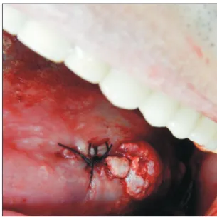
terrupted suture was initiated at the upper region of the lesion for traction (Fig. 4, 5), favoring a circular incision around the lesion.(Fig. 6) Lastly, suturing was performed.(Fig. 7)
Immediately after the operation, an analgesic was pre- scribed for pain. In addition, the patient was required to use topical 0.12% chlorhexidine digluconate mouthwash three times a day for seven days after his oral hygiene routine.
The histopathological exam of the surgical piece showed a squamous cell carcinoma lesion grade I (well differentiated).
Fig. 4. Collection of a lesion sample with needle and suture thread.
Wanessa da Silva Silveira et al: Squamous cell carcinoma from oral lichen planus: a case report of a lesion with 28 years of evolution. J Korean Assoc Oral Maxillofac Surg 2017
Fig. 5. Lesion with suture thread.
Wanessa da Silva Silveira et al: Squamous cell carcinoma from oral lichen planus: a case report of a lesion with 28 years of evolution. J Korean Assoc Oral Maxillofac Surg 2017
Fig. 6. Scaphoid-shaped surgical site immediately after biopsy.
Wanessa da Silva Silveira et al: Squamous cell carcinoma from oral lichen planus: a case report of a lesion with 28 years of evolution. J Korean Assoc Oral Maxillofac Surg 2017
Fig. 7. Suture performed in the incisional biopsy area.
Wanessa da Silva Silveira et al: Squamous cell carcinoma from oral lichen planus: a case report of a lesion with 28 years of evolution. J Korean Assoc Oral Maxillofac Surg 2017
The rate of transformation of LP in squamous cell car- cinoma in individual studies has ranged from 0% to 3.5%.
There might be a higher frequency of malignancy in some sites, such as the tongue, followed by the oral and gingival mucosa7. These findings correspond with those of the present study, considering that the lesion studied was located on the edge of the tongue. The patient described here was a 64-year- old Caucasian man10, consistent with the observation that squamous cell carcinoma is more prevalent in Caucasian men over 50 years. However, the present study disagrees with the conclusion that LP lesions are more common in women11.
In the present case, the lesion initially presented (in 1988) as plaques and white patches, clinical aspects that are char- acteristic of LP lesions. Later (2014), the lesion presented as a vegetation with leukoplakic and erythematous areas on the edge of the tongue, suggestive of a malignant lesion. This was confirmed by the histopathological exam. Such lesion evolution indirectly confirms the evolution of the initial LP diagnosis for squamous cell carcinoma, which potentially presented as vegetation on the edges of the tongue12.
The lesion described in the present work involved the edge of the tongue, whereas squamous cell carcinoma and LP largely affect the tongue itself13-15.
In 2014, the clinical aspect suggesting malignant lesion led to the performance of an incisional biopsy, whose tissue sample was later forwarded for histopathological exam. De- finitive diagnosis of squamous cell carcinoma was performed regression caused by radiotherapy and chemotherapy, the
medical team did not recommend complete excision of the lesion.
III. Discussion
The focus of the present study was to highlight, through a clinical case, the importance of the potential origin of oral squamous cell carcinoma in an LP lesion7-9. In such a lesion, the epithelium covering the LP becomes more susceptible to oncogenic factors.
Fig. 8. Aspect of the lesion after incisional biopsy.
Wanessa da Silva Silveira et al: Squamous cell carcinoma from oral lichen planus: a case report of a lesion with 28 years of evolution. J Korean Assoc Oral Maxillofac Surg 2017
Fig. 9. Aspect of the lesion 2 years after incisional biopsy, 6 che- motherapy sessions, and 35 radiotherapy sessions.
Wanessa da Silva Silveira et al: Squamous cell carcinoma from oral lichen planus: a case report of a lesion with 28 years of evolution. J Korean Assoc Oral Maxillofac Surg 2017
Fig. 10. Grade I squamous cell carcinoma composed of numer- ous atypical pleomorphic cells and keratin pearls (H&E staining, x100).
Wanessa da Silva Silveira et al: Squamous cell carcinoma from oral lichen planus: a case report of a lesion with 28 years of evolution. J Korean Assoc Oral Maxillofac Surg 2017
by biopsy, followed by an anatomopathological exam16. The main etiological factors of oral squamous cell car- cinoma are tobacco, alcohol, and constant stress17. Out of these factors, the patient described here only presented stress, which possibly influenced the occurrence of LP and the later occurrence of squamous cell carcinoma. Stress can act as a triggering agent2.
For the patient described here, the squamous cell carci- noma was treated through surgery, radiotherapy, and che- motherapy18. It is worth noting that, in the present study, the patient quit dental follow-up for 26 years. If the patient had continued dental follow-up via periodic visits, followed by biopsies and routine histopathological exams, the lesion might not have progressed to squamous cell carcinoma. Early detection of premalignant lesions and oral cancer is essential for achieving favorable prognoses19.
In conclusion, considering that oral LP is a relatively com- mon disease with cancerization potential, the present study highlights to the scientific community the need for early treatment and follow-up of precancerous oral lesions. More- over, this work reports the main clinical and histopathological characteristics of oral squamous cell carcinoma, a lesion with high rates of morbidity and mortality.
Conflict of Interest
No potential conflict of interest relevant to this article was reported.
ORCID
Wanessa da Silva Silveira, http://orcid.org/0000-0001- 9848-3917
Ezequiel Gregolin Bottezini, http://orcid.org/0000-0001- 7863-567X
Maria Salete Linden, http://orcid.org/0000-0001-6501-1710 Isadora Rinaldi, http://orcid.org/0000-0003-1864-5985 Luiz Renato Paranhos, http://orcid.org/0000-0002-7599- 0120
João Paulo de Carli, http://orcid.org/0000-0002-4705-6226 Micheline Trentin, http://orcid.org/0000-0001-5040-3578 Pâmela Letícia dos Santos, http://orcid.org/0000-0003- 1734-4187
References
1. Kouhsoltani M, Aghbali A, Shokoohi B, Ahmadzadeh R. Mo- lecular targeting of Her-2/neu protein is not recommended as an adjuvant therapy in oral squamous cell carcinoma and oral lichen planus. Adv Pharm Bull 2015;5(Suppl 1):649-52.
2. Krupaa RJ, Sankari SL, Masthan KM, Rajesh E. Oral lichen pla- nus: an overview. J Pharm Bioallied Sci 2015;7(Suppl 1):S158-61.
3. Santos LCO, Batista OM, Cangussu MCT. Characterization of oral cancer diagnostic delay in the state of Alagoas. Braz J Otorhinolar- yngol 2010;76:416-22.
4. Seoane JM, González-Mosquera A, Velo-Noya J. Oral biopsy in the context of oral cancer and precancer. Av Odontoestomatol 2008;24:89-96.
5. Bharanidharan R, Dineshkumar T, Raghavendhar K, Kumar AR.
Squamous cell carcinoma of the gingiva: a diagnostic enigma. J Oral Maxillofac Pathol 2015;19:267.
6. de Oliveira JMB, Pinto LO, Lima NGM, de Almeida GCM. Oral cancer: assessment of academic dentistry and nursing knowledge as for the risk factors and diagnostic procedures. Rev Bras Can- cerol 2013;59:211-8.
7. Garcia-Pola MJ, Llorente-Pendás S, González-Garcia M, García- Martín JM. The development of proliferative verrucous leukoplakia in oral lichen planus. A preliminary study. Med Oral Patol Oral Cir Bucal 2016;21:e328-34.
8. Budimir V, Richter I, Andabak-Rogulj A, Vučićević-Boras V, Bu- dimir J, Brailo V. Oral lichen planus: retrospective study of 563 Croatian patients. Med Oral Patol Oral Cir Bucal 2014;19:e255-60.
9. Shi W, Yang J, Li S, Shan X, Liu X, Hua H, et al. Potential involve- ment of miR-375 in the premalignant progression of oral squamous cell carcinoma mediated via transcription factor KLF5. Oncotarget 2015;6:40172-85.
10. Cabello BT, Sazo BN, Salgado FA, Martínez RB. Squamous cell carcinoma of the lip survival rate. Rev Med Chil 2015;143:847-55.
11. Mankapure PK, Humbe JG, Mandale MS, Bhavthankar JD. Clini- cal profile of 108 cases of oral lichen planus. J Oral Sci 2016;58:43- 12. Pedron IG, dos Santos ESR, Aburad A, Tortamano IP, Adde CA. 7.
Squamous cell carcinoma: diagnosis and first behavior. J Health Sci Inst 2006;24:237-41.
13. Selvamani M, Yamunadevi A, Basandi PS, Madhushankari GS.
Prevalence of oral squamous cell carcinoma of tongue in and around Davangere, Karnataka, India: a retrospective study over 13 years. J Pharm Bioallied Sci 2015;7(Suppl 2):S491-4.
14. Agha-Hosseini F, Mirzaii-Dizgah I. Serum and saliva collagenase-3 (MMP-13) in patients with oral lichen planus and oral squamous cell carcinoma. Med J Islam Repub Iran 2015;29:218.
15. Oh MS, Kang SH, Kim HJ, Zhenglin Z, Ryu JI, Nam W, et al.
Overall five-year survival rate in squamous cell carcinoma of oral cavity. J Korean Assoc Oral Maxillofac Surg 2009;35:83-8.
16. De Carli JP, Trentin MS, Linden MSS, Bós ÂJG, Pedro REL, Silva SO. Oral squamous cell carcinoma of great extent: protocol diag- nosis. Odonto 2010;18:67-71.
17. Hande AH, Mohite DP, Chaudhary MS, Patel M, Agarwal P, Bohra S. Evidence based demonstration of the concept of ʻfield cancerizationʼ by p53 expression in mirror image biopsies of pa- tients with oral squamous cell carcinoma: an immunohistochemical study. Rom J Morphol Embryol 2015;56:1027-33.
18. Brener S, Jeunon FA, Barbosa AA, Grandinetti HAM. Carcinoma de células escamosas bucal: uma revisão de literatura entre o perfil do paciente, estadiamento clínico e tratamento proposto. Rev Bras Cancerol 2007;53:63-9.
19. Yardimci G, Kutlubay Z, Engin B, Tuzun Y. Precancerous lesions of oral mucosa. World J Clin Cases 2014;2:866-72.