Gene Therapy for Head and Neck Squamous Cell Carcinoma Using KITENIN (KAI1 COOH-Terminal Interacting
Tetraspanin)-Antisense Therapy
Joon Kyoo Lee,
1Dong-Hoon Lee,
1Eun Gene Sun,
2Jeong A Bae,
2Sang Chul Lim,
1Jeong Joon Min,
3Myung-Whun Sung,
4and Kyung Keun Kim
2Departments of 1Otolaryngology-Head and Neck Surgery and 3Nuclear Medicine and 2Medical Research Center for Gene Regulation, Chonnam National University Medical School, Hwasun Hospital, Hwasun;
4Department of Otolaryngology-Head and Neck Surgery, Seoul National University College of Medicine, Seoul, Korea.
Received: July 14, 2010 Revised: August 18, 2010 Accepted: August 30, 2010
Corresponding author: Dr. Joon Kyoo Lee, Department of Otolaryngology-Head and Neck Surgery, Chonnam National University Medical School, Hwasun Hospital, 160 Ilsim-ri, Hwasun-eup, Hwasun 519-763, Korea.
Tel: 82-61-379-7760, Fax: 82-61-379-7761 E-mail: [email protected]
∙ The authors have no financial conflicts of interest.
© Copyright:
Yonsei University College of Medicine 2011 This is an Open Access article distributed under the terms of the Creative Commons Attribution Non- Commercial License (http://creativecommons.org/
licenses/by-nc/3.0) which permits unrestricted non- commercial use, distribution, and reproduction in any medium, provided the original work is properly cited.
Purpose: KAI1 COOH-terminal interacting tetraspanin (KITENIN) has been found to act as a promoter of metastasis in murine models of colon cancer and squamous cell carcinoma (SCC). The suppression of tumor progression and metastasis of es- tablished colon cancer in mice was observed after intravenous delivery of small in- terfering RNA (siRNA) targeting KITENIN. The purpose of this study was to inves- tigate the efficacy of gene therapy targeting KITENIN in human head and neck SCC. Materials and Methods: SNU-1041, a well-established human hypopharyn- geal SCC cell line, was used. KITENIN expression in SNU-1041 was measured by Western blot analysis. The cells were prepared, maintained in culture dishes with media, and divided into two groups: the si-KITENIN group and the scrambled group (control). The siRNA targeting KITENIN (si-KITENIN) and scrambled DNA were transfected into the SNU-1041 cells in each group. The effect of gene therapy was compared by in vitro experiments to evaluate invasion, migration, and proliferation.
Results: KITENIN was strongly expressed in the SNU-1041 cells, and the number of invaded cells was reduced more in the si-KITENIN group than in the scrambled group (p<0.001). The speed for the narrowing gap, made through adherent cells, was lower in the si-KITENIN group (p<0.001), and the number of viable proliferating cells was reduced in the si-KITENIN group compared to the scrambled group (p<
0.001, the third day). KITENIN protein expression was no longer identified in the si- KITENIN group. Conclusion: Gene therapy using an anti-KITENIN strategy might be effective for head and neck squamous carcinoma.
Key Words: Gene therapy, head and neck cancer, squamous cell carcinoma, KIT- ENIN protein, small interfering RNA (siRNA)
INTRODUCTION
Reducing the progression and metastasis of head and neck squamous cell carcino-
ma (SCC) requires an understanding of molecular mechanisms involved in inva-
as gene-coding siRNAs. Recent studies have demonstrated the general application of siRNAs to silence gene expres- sion in a range of cell types and in mammalian models.
16The purpose of this study was to investigate the efficacy of gene therapy (siRNA targeting KITENIN) in human head and neck SCC.
MATERIALS AND METHODS
Cell culture and transfection
SNU-1041 cells,
17-19a well-established human head and neck SCC cell line, were grown in Dulbecco’s modified Eagle’s medium (DMEM, Invitrogen, Carlsbad, CA, USA) supple- mented with 10% fetal bovine serum (Hyclone, Logan, UT, USA) in a humidified atmosphere of 5% CO
2at 37°C. The cells were prepared and maintained in culture dishes with media, and divided into two groups: the si-KITENIN group and the scrambled group (control). The siRNA targeting KIT- ENIN (si-KITENIN) and the scrambled DNA were transfect- ed into the SNU-1041 cells in each group. The sequence of siRNA was as follows: human KITENIN 5’-GCUUG- GACUUCAGCCUCGUAGUCAA-3’. The transfections were performed using the Lipofectamine RNAiMAX (Invit- rogen) according to the manufacturer’s instructions. The cells were maintained and used for further analysis.
Western blot analysis
Western blot analysis was used to investigate KITENIN ex- pression before and after transfection of siRNA (si-KIT- ENIN). The tissues were solubilized in NP40 lysis buffer containing a protease inhibitor mixture (Roche, Indianapo- lis, IN) and 1 mM phenylmethylsulfonyl fluoride. The re- solved proteins (50 µg) were transferred to a nitrocellulose membrane and blotted with the KITENIN antibody and an- tirabbit immunoglobulin-horseradish peroxidase (Amer- sham, Arlington Heights, IL, USA). The blot was reprobed with anti-actin antibody (I-19; Santa Cruz Biotechnology, Santa Cruz, CA, USA) to control loading variations.
Cell invasion assay
Cell migration was measured using a Transwell migration ap- paratus (Costar Inc., Cambridge, UK) with minor modifica- tion as described previously.
9The filters (8-µm pore size) were coated with 1% gelatin solution on both the top and bot- tom surfaces. The cells from each group (si-KITENIN group, scrambled group) were harvested, washed once in serum-free sion and dissemination.
1Gene therapy for tumor metastasis
is a promising because it inhibits the spread of cancer re- gardless of the tumorigenicity.
2,3A large body of evidence suggests that down-regulation of KAI1/CD82 expression occurs with invasive and metastatic disease, and that it is an important step in the progression of many human malignancies.
4-8KAI1, a tumor metastasis suppressor gene, is a transmembrane glycoprotein that is a member of the tetraspanin superfamily. The metastasis sup- pressor function was recently reported to be decreased in a spliced variant of KAI1 at the COOH-terminal region, sug- gesting that the COOH-terminal region of KAI1 is impor- tant for the effects of KAI1 on cell motility.
9Subsequently, a protein that interacted with the COOH-terminal cytoplasm domain of KAI1 was identified via a two-hybrid yeast sys- tem.
10The function of this protein Vang-like 1 (VANGL1
11) during carcinogenesis has not been fully described, and it has been renamed as KAI1 COOH-terminal interacting tet- raspanin (KITENIN).
KITENIN-overexpressing CT-26 mouse colon cancer cells have shown increased tumorigenicity and early hepat- ic metastasis in vivo, as well as increased invasiveness and adhesiveness to fibronectin in vitro, compared to parental cells.
10Moreover, the suppression of progression and me- tastasis of established colon cancer has been observed in mice after intravenous delivery of small interfering RNA (siRNA) targeting KITENIN.
12We recently reported that KITENIN promoted pulmo- nary metastasis in a murine model of SCC by increasing cancer cell invasion and adhesion to fibronectin,
13and that KITENIN represented a more aggressive phenotype in a murine model of oral cavity squamous carcinoma.
14Fur- thermore, we investigated the expression of KITENIN in human laryngeal SCC, and found that KITENIN expres- sion was significantly increased in laryngeal cancer tissues, compared to the adjacent normal tissue mucosa, as well as in metastatic lymph nodes compared to non-metastatic lymph nodes. High KITENIN expression was associated significantly with an advanced disease stage, extent of the tumor, and lymph node metastasis.
15siRNA is a small double-stranded, non-protein coding
RNA (21-31 nucleotides) involved in gene silencing func-
tions, especially RNA interference (RNAi). This ability of
siRNA has provided researchers with a novel tool to block
the expression of disease-causing genes, provided that their
mRNA sequences are known. siRNAs can be delivered to
cells either exogenously as synthetic agents or endogenously
RESULTS
Western blot analysis
Fig. 1 shows that KITENIN was strongly expressed in the SNU-1041 cells and scrambled DNA-transfected SNU-1041 cells (SNU-1041/Scrambled). However, the expression of KITENIN protein was not observed in the si-KITENIN- transfected SNU-1041 cells (SNU-1041/si-KITENIN), dem- onstrating that KITENIN was effectively blocked by siRNA.
Cell invasion assay
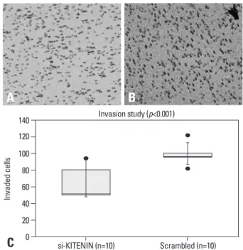
As shown in Fig. 2, the number of invading si-KITENIN- transfected SNU-1041 cells was 64.2±19.2, whereas the number of the scrambled DNA-transfected SNU-1041 cells DMEM/0.2% bovine serum albumin (BSA), and resuspend-
ed at 2×10
6cells/µL DMEM/0.2% BSA. To start the assay, 120 µL (2.4×10
5cells) was loaded onto the upper chamber of the Transwell, whereas 400 µL DMEM/0.2% BSA contain- ing 20 µg/mL human plasma fibronectin (Calbiochem, La Jolla, CA, USA), a chemotactic factor, was loaded onto the lower chamber. The Transwell apparatus was incubated for 24 hours at 37°C. At the end of incubation, the cells attached to the membranes were fixed and stained with Diff-Quick (Inter- national Reagents, Kobe, Japan) by following the manufac- turer’s protocol. The cells on the top surface of the filters were wiped off with cotton balls, and the cells that migrated to the bottom surface were counted in ten random squares of 0.5×0.5 mm
2by microscopic field of view. The results were expressed as mean±SE of the number of cells/field.
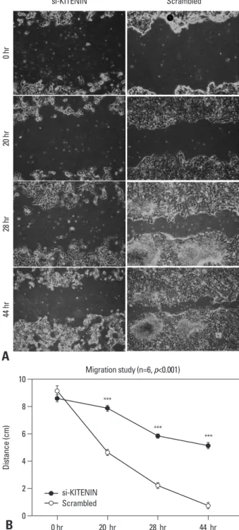
Cell migration assay
si-KITENIN-transfected and scrambled DNA-transfected SNU-1041 cells were seeded in a 6-well plate (1×10
5cells/
well). Twenty-four hours later, the media were changed to serum-free DMEM and incubated for 12 hours. After the media were removed from the wells, a straight transverse line through the adherent cells was drawn using a ruler and a 1000 µL-tip, resulting in a uniform gap. The media were changed to DMEM supplemented with 2% fetal bovine se- rum. At 0, 20, 28, and 44 hours later; the distances between the gaps were measured in centimeters after capture of six random sites by the microscopic field of view.
Cell proliferation assay
The proliferation and viability of the cells were measured using an enhanced cell viability assay kit, EZ CyTox (Daeil Lab Service Co., Seoul, Korea). The cells were seeded at 2
×10
4cells/well in 48-well plates, transfected with si-KIT- ENIN and scrambled DNA, and grown overnight in the presence of serum. Next, EZ CyTox was added according to the manufacturer’s instructions for 2 hours, after which the discolored cells were moved into 96-well plates for analysis. Absorbance at 450 nm was determined using a microplate reader with SOFTmax PRO software (Molecu- lar Devices, Sunnyvale, CA, USA).
Statistical analysis
Experimental differences were tested for statistical signifi- cance with SPSS 16.0 (SPSS Inc., Chicago, IL, USA). The Student’s t-test was used, and p values of less than 0.05 were considered statistically significant.
KITENIN
β-actin
SNU-1041 SNU-1041/si-KITENIN SNU-1041/Scrembled
Fig. 1. KITENIN expression in SNU-1041 cells: KITENIN was strongly ex- pressed in SNU-1041 cells and scrambled DNA-transfected SNU-1041 cells (SNU-1041/Scrambled). The expression of KITENIN protein was not ob- served in si-KITENIN-transfected SNU-1041 cells (SNU-1041/si-KITENIN).
Fig. 2. Cell invasion assay: the number of invading si-KITENIN-transfected SNU-1041 cells (A) was 64.2±19.2, whereas the number was 98.6±10.2 for the scrambled DNA-transfected SNU-1041 cells (B), as measured by 10 random squares of 0.5×0.5 mm2 on the microscopic field of view under con- ditions with 20 µg/mL of fibronectin; the difference between the two groups was statistically significant (p<0.001)(C).
0 20 40 60 80 100 120 140
Invaded cells
si-KITENIN (n=10) Scrambled (n=10) Invasion study (p<0.001)
A B
C
the control group as time passed by 20, 28, 44 hours, com- pared to the si-KITENIN group (p<0.001)(Fig. 3). For the scrambled group, the gap was nearly filled within 44 hours, however, it remained wide open in the si-KITENIN group.
Cell proliferation assay
The number of proliferating cells, as determined by absor- bance, was significantly decreased in the si-KITENIN group compared to the scrambled group (Fig. 4). On the third day, absorbance was 0.4933±0.0484 in the si-KITENIN group, whereas 0.6023±0.0319 in the scrambled group (p<0.001).
DISCUSSION
siRNA, known as RNAi, is a powerful tool for strong and specific suppression of gene expression.
16The use of these molecules can be applied to a wide range of cancers and oth- er proliferative disorders where aberrant gene expression oc- curs. Therefore, oncogenic and mutant tumor suppressor genes might represent potential targets for the RNAi ap- proach. It has been shown that mutated p53 protein, involved in almost half of human malignancies, was eliminated by siRNA and the function of wild-type p53 restored.
20Other examples include: elimination of the Ras oncogene using siRNAs,
21transfection of leukemic cells with siRNAs target- ing a BCR-ABL fusion transcript, thereby inducing apopto- sis,
22,23and targeting of VEGF by siRNAs.
24In this study, siRNA targeting KITENIN was effective.
SNU-1041, an established human head and neck squamous carcinoma cell line,
17-19expressed very high levels of KIT- was 98.6±10.2, as measured by 10 random squares of 0.5×
0.5 mm
2by the microscopic field of view under conditions with 20 µg/mL of fibronectin; the difference between the two groups was statistically significant (p<0.001).
Cell migration assay
The artificial wound gap became significantly narrower in
si-KITENIN
0 hr20 hr28 hr44 hr
Scrambled
A
B
Fig. 3. Cell migration assay: the artificial wound gap became significantly narrower in the control group as time passed by 20, 28, 44 hours, compared to the si-KITENIN group (p<0.001)(B). In the scrambled group, the gap was nearly filled within 44 hours. However, it remained wide open in the si-KIT- ENIN group (A).
Fig. 4. Cell proliferation assay: the proliferating cells, as determined by ab- sorbance, were significantly decreased in the si-KITENIN group compared to the scrambled group. On the third day, absorbance was 0.4933±0.0484 in the si-KITENIN group and 0.6023±0.0319 in the scrambled group (p<0.001).
0 2 4 6 8 10
Distance (cm)
0 hr 20 hr 28 hr 44 hr
Migration study (n=6, p<0.001)
***
***
***
si-KITENIN Scrambled
0.25 0.30 0.35 0.40 0.45 0.50 0.55 0.60 0.65
Absorbance (450 nm, viable cells)
1 day 2 day 3 day 4 day
Proliferation study
***
si-KITENIN (n=6) Scrambled (n=6)
Basic Research Promotion Fund) (KRF-2008-1684).
REFERENCES
1. Howell GM, Grandis JR. Molecular mediators of metastasis in head and neck squamous cell carcinoma. Head Neck 2005;27:710-7.
2. Woodhouse EC, Chuaqui RF, Liotta LA. General mechanisms of metastasis. Cancer 1997;80:1529-37.
3. Yamaguchi H, Wyckoff J, Condeelis J. Cell migration in tumors.
Curr Opin Cell Biol 2005;17:559-64.
4. Jackson P, Marreiros A, Russell PJ. KAI1 tetraspanin and metasta- sis suppressor. Int J Biochem Cell Biol 2005;37:530-4.
5. Takaoka A, Hinoda Y, Satoh S, Adachi Y, Itoh F, Adachi M, et al.
Suppression of invasive properties of colon cancer cells by a me- tastasis suppressor KAI1 gene. Oncogene 1998;16:1443-53.
6. Yang JL, Jackson P, Yu Y, Yoshie O, Ham JM, Russell PJ, et al.
Invasion and metastasis correlate with down-regulation of KAI1 expression in human colon cancer cell lines. GI Cancer 2001;3:
313-22.
7. Yang X, Wei LL, Tang C, Slack R, Mueller S, Lippman ME. Over- expression of KAI1 suppresses in vitro invasiveness and in vivo metastasis in breast cancer cells. Cancer Res 2001;61:5284-8.
8. Jee B, Jin K, Hahn JH, Song HG, Lee H. Metastasis-suppressor KAI1/CD82 induces homotypic aggregation of human prostate cancer cells through Src-dependent pathway. Exp Mol Med 2003;
35:30-7.
9. Lee JH, Seo YW, Park SR, Kim YJ, Kim KK. Expression of a splice variant of KAI1, a tumor metastasis suppressor gene, influ- ences tumor invasion and progression. Cancer Res 2003;63:7247- 10. Lee JH, Park SR, Chay KO, Seo YW, Kook H, Ahn KY, et al. 55.
KAI1 COOH-terminal interacting tetraspanin (KITENIN), a member of the tetraspanin family, interacts with KAI1, a tumor metastasis suppressor, and enhances metastasis of cancer. Cancer Res 2004;64:4235-43.
11. Yagyu R, Hamamoto R, Furukawa Y, Okabe H, Yamamura T, Na- kamura Y. Isolation and characterization of a novel human gene, VANGL1, as a therapeutic target for hepatocellular carcinoma. Int J Oncol 2002;20:1173-8.
12. Lee JH, Cho ES, Kim MY, Seo YW, Kho DH, Chung IJ, et al.
Suppression of progression and metastasis of established colon tu- mors in mice by intravenous delivery of short interfering RNA targeting KITENIN, a metastasis-enhancing protein. Cancer Res 2005;65:8993-9003.
13. Lee JK, Bae JA, Sun EG, Kim HD, Yoon TM, Kim K, et al. KIT- ENIN increases invasion and migration of mouse squamous can- cer cells and promotes pulmonary metastasis in a mouse squa- mous tumor model. FEBS Lett 2009;583:711-7.
14. Lee JK, Lim SC, Kim HD, Yoon TM, Kim K, Nam JH, et al.
KITENIN represents a more aggressive phenotype in a murine model of oral cavity squamous carcinoma. Otolaryngol Head Neck Surg 2010;142:747-52.
15. Lee JK, Yoon TM, Seo DJ, Sun EG, Bae JA, Lim SC, et al. KAI1 COOH-terminal interacting tetraspanin (KITENIN) expression in early and advanced laryngeal cancer. Laryngoscope 2010;120:
953-8.
16. Sioud M. Therapeutic siRNAs. Trends Pharmacol Sci 2004;
25:22-8.
ENIN, which functions as a metastasis-promoting gene.
Our previous data demonstrated that many human head and neck squamous cancer tissues expressed KITENIN, and that the level of KITENIN correlated with the disease stage.
After transfection of si-KITENIN into SNU-1041 cells, KITENIN was no longer identified in the cells. siRNA- transfected SNU-1041 cells showed reduced in vitro inva- sion, migration, and proliferative characteristics compared to the cells in the control group.
siRNAs can be delivered in two ways: exogenous admin- istration of synthetic siRNAs and endogenous expression of siRNA via plasmid or viral vectors.
Generally, synthetic siRNAs are delivered to cells in cul- ture via liposome-based transfection reagents. In this study, we used the lipofectamine trasfection reagent (RNAiMAX, Invitrogen) and the experiments were successful. However, this delivery offers only immediate and/or short-term ef- fects.
25Therefore, siRNA delivery systems which can pro- vide long-term biological effects are needed.
To overcome these problems, several research groups have shown that short hairpin siRNA can be produced from expression plasmids that contain promoters that are depen- dent on either RNA polymerase (pol) II or pol III.
26-29Brum- melkamp, et al.
26developed a new expression vector sys- tem, pSUPER (suppression of endogenous RNA) that directs the synthesis of siRNA-like transcripts in mammali- an cells, and Lee, et al.
12used the pSUPER vector system to deliver KITENIN siRNA for efficient and stable suppres- sion of KITENIN expression in established tumors of syn- geneic mice. The latter observed marked inhibition of the growth of established tumors as well as suppression of dis- tant metastases of colon cancer, following four weekly or semiweekly intravenous injections of pSUPER-KITENIN.
In addition to plasmid vectors, adenoviral and lentiviral- based vectors have also been developed and used success- fully.
30,31For our next in vivo study with animals on anti- KITENIN delivery, plasmid or viral vector systems will be studied.
In conclusion, gene therapy using anti-KITENIN strate- gies may help or delay the progression (invasion, migration and proliferation) of head and neck squamous carcinoma.
ACKNOWLEDGEMENTS
This work was supported by the Korean Research Founda-
tion grant funded by the Korean Government (MOEHRD,
24. Zhang L, Yang N, Mohamed-Hadley A, Rubin SC, Coukos G.
Vector-based RNAi, a novel tool for isoform-specific knock-down of VEGF and anti-angiogenesis gene therapy of cancer. Biochem Biophys Res Commun 2003;303:1169-78.
25. Holen T, Amarzguioui M, Wiiger MT, Babaie E, Prydz H. Posi- tional effects of short interfering RNAs targeting the human coag- ulation trigger Tissue Factor. Nucleic Acids Res 2002;30:1757-66.
26. Brummelkamp TR, Bernards R, Agami R. A system for stable ex- pression of short interfering RNAs in mammalian cells. Science 2002;296:550-3.
27. Miyagishi M, Taira K. U6 promoter-driven siRNAs with four uri- dine 3′ overhangs efficiently suppress targeted gene expression in mammalian cells. Nat Biotechnol 2002;20:497-500.
28. Lee NS, Dohjima T, Bauer G, Li H, Li MJ, Ehsani A, et al. Ex- pression of small interfering RNAs targeted against HIV-1 rev transcripts in human cells. Nat Biotechnol 2002;20:500-5.
29. Xia H, Mao Q, Paulson HL, Davidson BL. siRNA-mediated gene silencing in vitro and in vivo. Nat Biotechnol 2002;20:1006-10.
30. Rubinson DA, Dillon CP, Kwiatkowski AV, Sievers C, Yang L, Kopinja J, et al. A lentivirus-based system to functionally silence genes in primary mammalian cells, stem cells and transgenic mice by RNA interference. Nat Genet 2003;33:401-6.
31. Shen C, Buck AK, Liu X, Winkler M, Reske SN. Gene silencing by adenovirus-delivered siRNA. FEBS Lett 2003;539:111-4.
17. Kim SG, Hong JW, Boo SH, Kim MG, Lee KD, Ahn JC, et al.
Combination treatment of Cetuximab and photodynamic therapy in SNU-1041 squamous cancer cell line. Oncol Rep 2009;22:701-8.
18. Koh TY, Park SW, Park KH, Lee SG, Seol JG, Lee DW, et al. In- hibitory effect of p27KIP1 gene transfer on head and neck squa- mous cell carcinoma cell lines. Head Neck 2003;25:44-9.
19. Sung MW, Roh JL, Park BJ, Park SW, Kwon TK, Lee SJ, et al.
Bile acid induces cyclo-oxygenase-2 expression in cultured hu- man pharyngeal cells: a possible mechanism of carcinogenesis in the upper aerodigestive tract by laryngopharyngeal reflux. Laryn- goscope 2003;113:1059-63.
20. Martinez LA, Naguibneva I, Lehrmann H, Vervisch A, Tchénio T, Lozano G, et al. Synthetic small inhibiting RNAs: efficient tools to inactivate oncogenic mutations and restore p53 pathways. Proc Natl Acad Sci U S A 2002;99:14849-54.
21. Brummelkamp TR, Bernards R, Agami R. Stable suppression of tumorigenicity by virus-mediated RNA interference. Cancer Cell 2002;2:243-7.
22. Scherr M, Battmer K, Winkler T, Heidenreich O, Ganser A, Eder M. Specific inhibition of bcr-abl gene expression by small inter- fering RNA. Blood 2003;101:1566-9.
23. Wilda M, Fuchs U, Wössmann W, Borkhardt A. Killing of leuke- mic cells with a BCR/ABL fusion gene by RNA interference (RNAi). Oncogene 2002;21:5716-24.