∙ Received: June 15, 2012. Accepted: September 27, 2012.
∙ Corresponding author : Yong Ho Do
Department of Nuclear Nedicine, Seoul National University Hospital, 28 Yeongon-dong, Jongno-gu, Seoul, 110-744, Korea
Tel: +82-2-2072-2948, Fax: +82-2-766-9083 E-mail: [email protected]
Original Article
PET/CT 검사 시 IRIS (Iterative Reconstruction in Image Space) 적용에 따른 CT 피폭선량 감소와 PET SUV 비교 연구서울대학교병원 핵의학과
도용호 ․ 송호준 ․ 이형진 ․ 이홍재 ․ 김진의
The Study of Reducing Radiation Exposure Dose and Comparing SUV According to Applied IRIS (Iterative Reconstruction in Image Space) for PET/CT
Yong Ho Do, Ho Jun Song, Hyung Jin Lee, Hong Jae Lee and Jin Eui Kim
Department of Nuclear Medicine, Seoul National University Hospital, Seoul, Korea
Purpose : Presently, hardwares and softwares for reducing radiation exposure are continually developed for PET/CT examination. Purpose of this study is to evaluate effectiveness of reducing radiation exposure dose of CT and SUV changes of PET when applied each kernel to ACCT (Attenuation Correction Computed Tomography) according to adopted IRIS (Iterative Reconstruction in Image Space) software. Materials and Methods : Biograph mCT (Siemens, Germany) was used as a PET/CT scanner. Using AAPM CT performance phantom, from standard (120 kVp, 100 mAs), 7 scans were conducted by reducing 15 mAs each. After image reconstruction by FBP (Filtered Back Projection) and IRIS, noise and spatial resolution were evaluated. The same method was applied to anthropomorphic chest phantom and acquired images were compared. NEMA IEC body phantom was used for SUV evaluation. Injected dose rate for hot sphere (hot) and background cylinder (BKG) were 1:8. CT dose condition (120 kVp, 50 mAs) was the same for each scan and PET scan durations were 1, 2, 3 and 4min. After scanning, each kernel of IRIS was applied to ACCT. And PET images were reconstructed by ACCT adopted IRIS for comparing SUV changes. Results : AAPM phantom test for noise evaluation, SD for FBP 100 mAs, IRIS 55 mAs were 8.8 and 8.9. FBP 85 mAs, IRIS 40 mAs were 9.5 and 9.7. FBP 70 mAs, IRIS 25 mAs were 11.9 and 11.1. Above mAs condition for FBP and IRIS, SD showed similar values. And for spatial resolution test, there was no significant difference. For chest phantom test, when applied the same mAs and kernel to both of FBP and IRIS, every applied kernels showed reduced noise. Lower mAs and higher kernel value showed higher noise reduction. There was no considerable difference only except for I70 very sharp kernel for SUV comparison using NEMA IEC body phantom. Conclusion : In this study, low mAs (55 mAs) applied IRIS and standard mAs (100 mAs) applied FBP showed similar noise. And only except for I70 kernel, there was no significant SUV changes. It is possible to reduce needless radiation exposure and acquire better image quality than FBP's through applying appropriate kernel of IRIS to PET/CT. (Korean J Nucl Med Technol 2012;
16(2):29-34)
Key Words : FBP, IRIS, SUV, kernel
서 론
최근 진단 분야에서 PET/CT는 종양학 분야는 물론 심장, 신경 등 여러 가지 분야에서 널리 활용되고 있다.1) PET/CT 의 비약적인 발달로 인한 검사건수의 증가와 CT 영상의 질
Fig. 1. Siemens Biograph mCT PET/CT scanner.
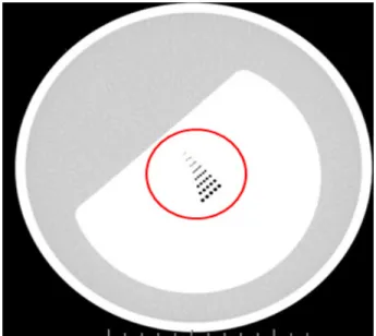
Fig. 2. AAPM CT performance phantom.
Fig. 3. Anthropomorphic chest phantom.
Fig. 4. NEMA IEC body phantom.
향상을 위한 선량 증가에 따라 PET/CT 검사 시 환자와 검사 자의 방사선 피폭 감소를 위한 hardware 및 software의 개발 이 지속적으로 이루어지고 있다.2) CT의 경우 iterative 방식 의 재구성 방법이 적용되기 전까지 FBP (Filtered Back Projection)방식을 통하여 영상이 재구성 되어 왔다.3) FBP 방 식의 경우 재구성 시간이 빠르다는 장점을 가지고 있지만 공 간 분해능과 영상의 노이즈를 분리할 수 없기 때문에 선량 감소 기능을 적용할 수 없다는 단점을 가지고 있다. 반면 Siemens사의 CT 선량 감소 소프트웨어 프로그램인 IRIS (Iterative Reconstruction in Image Space)의 경우 데이터 재 구성 소요 시간은 FBP 방식에 비하여 오래 걸리는 반면 master volume 재구성 이 후의 iterative correction을 통하여 영상의 해상력 감소 없이 노이즈 감소가 가능하다.4-5) 본 연 구에서는 IRIS software의 적용을 통하여 PET/CT 검사 시 선량감소 효과와 각 kernel 적용 시 CT 영상의 비교 그리고 각 kernel의 ACCT (Attenuation Correction computed to-
mography) 적용 시 SUV (Standard Uptake Value) 변화를 비 교하고자 하였다.
실험재료 및 방법
1. 실험 장비 및 재료
실험에 사용된 장비는 Biograph mCT 40 slice (Siemens Medical System, Germany)(Fig. 1)이며, 사용된 phantom은 AAPM (The American Association of Physicists in Medicine) CT performance phantom (Fig. 2), anthropomorphic chest phantom (Canadian Scientific Products, London, Ontario, Canada)(Fig. 3)과 NEMA IEC body phantom (Fig. 4)을 사 용하였다.
Fig. 5. Drawing ROI for noise test. Fig. 6. Visible estimation for spatial resolution.
2. 실험 방법
1) 노이즈 & 해상력 평가
AAPM CT performance에 물을 가득 채운 후 관전압 120 kVp, 관전류 100, 85, 70, 55, 40, 25, 10 mAs, 절편 두께 3 mm, FOV (Field of View) 500 mm 조건으로 설정하고, AEC (Auto Exposure Control)는 적용하지 않았다.
각 관전류를 적용하여 영상 획득 후 FBP kernel B30f, B40f, B50f, B70f와 IRIS kernel I30f, I40f, I50f, I70f을 적용하 여 재구성한 후 각 kernel을 적용한 영상의 6시 방향의 1/4지 점에 16.37 sq.cm의 ROI (Region of Interest)를 그려 노이즈 를 측정하였다(Fig. 5).
AAPM phantom 내의 공간 분해능 측정용 슬라이스를 노 이즈 측정 시와 동일한 방법으로 영상을 획득하여 재구성 후 window width 300, level -100으로 설정 후 1.25 mm 지점이 구분되는지 육안으로 평가하였다(Fig. 6).
2) Chest phantom 영상 평가
Anthropomorphic chest phantom을 이용하여 노이즈, 해 상력 측정 시와 동일한 조건으로 영상 획득하여 FBP, IRIS 각 kernel로 재구성하였다. 노이즈 실험에서 유사한 노이즈 레벨을 나타낸 mAs를 선택하여 FBP와 IRIS 적용에 따른 chest phantom 영상들의 노이즈와 해상력을 평가하였다.
3) SUV 변화 평가
NEMA IEC body phantom 이용하여 55.5 MBq를 back- ground에 주입하고 열소와 배후 방사의 비를 8:1이 되도록
모형을 제작하였다. CT 스캔 조건은 관전압 120 kVp, 관전 류 50 mAs, 절편두께 5 mm로 설정하고 AEC는 적용하지 않 았다. PET 스캔 조건은 스캔 시간 1분, 2분, 3분, 4분으로 하 였으며 TrueX+TOF 알고리즘을 적용하였고 gaussian 5 mm 필터, iteration 2, subset 21로 하였다. 영상 획득 후 FBP B19f, IRIS I26f ASA, I30f, I40f, I50f, I70f kernel을 ACCT에 적용하여 PET 영상을 재구성 후 각 kernel 값 적용에 따른 SUV 변화를 평가하였다.
결 과
1. 노이즈 감소 평가
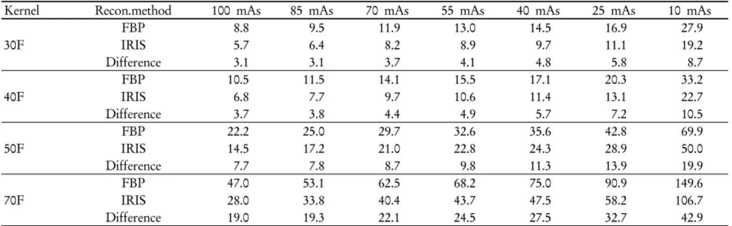
AAPM phantom을 이용한 노이즈 테스트에서 kernel 값이 증가할수록 동일한 mAs에서 FBP와 IRIS방식으로 재구성한 영상의 노이즈 값의 차이가 증가하였고 mAs가 감소할수록 노이즈 값의 차이 역시 증가하였다. 100 mAs에서 FBP 방식 으로 재구성시 노이즈 값 8.8은 55 mAs에서 IRIS 방식으로 재구성한 영상의 노이즈 값 8.9와 유사하였으며 85 mAs FBP 9.5, 40 mAs IRIS 9.7, 70 mAs FBP 11.9, 25 mAs IRIS 11.1로 유사한 노이즈 값이 측정되었다(Table 1). FBP 방식으로 재 구성한 영상에 비하여 IRIS 적용 시 45 mAs 감소 시에도 유 사한 노이즈 값을 나타내었다.
2. 피폭 선량 감소 평가
Table 1에서 FBP 방식과 IRIS 방식으로 재구성 후 유사한
Table 1. The results of noise comparison
Kernel Recon.method 100 mAs 85 mAs 70 mAs 55 mAs 40 mAs 25 mAs 10 mAs
30F
FBP 8.8 9.5 11.9 13.0 14.5 16.9 27.9
IRIS 5.7 6.4 8.2 8.9 9.7 11.1 19.2
Difference 3.1 3.1 3.7 4.1 4.8 5.8 8.7
40F
FBP 10.5 11.5 14.1 15.5 17.1 20.3 33.2
IRIS 6.8 7.7 9.7 10.6 11.4 13.1 22.7
Difference 3.7 3.8 4.4 4.9 5.7 7.2 10.5
50F
FBP 22.2 25.0 29.7 32.6 35.6 42.8 69.9
IRIS 14.5 17.2 21.0 22.8 24.3 28.9 50.0
Difference 7.7 7.8 8.7 9.8 11.3 13.9 19.9
70F
FBP 47.0 53.1 62.5 68.2 75.0 90.9 149.6
IRIS 28.0 33.8 40.4 43.7 47.5 58.2 106.7
Difference 19.0 19.3 22.1 24.5 27.5 32.7 42.9
Table 2. CTDIvol and DLP of each mAs
mAsDose 100 85 70 55 40 25 10
CTDIvol (mGy) 7.19 6.16 5.07 3.97 2.88 1.82 0.73
DLP (mGy*cm) 332 266 236 185 134 85 34
Table 3. The results of resolution comparison
NoRecon 1 2 3 4
FBP 100 mAs (B30f) 85 mAs (B30f) 70 mAs (B30f) 55 mAs (B30f)
IRIS 55 mAs (I30f) 40 mAs (I30f) 25 mAs (I30f) 25 mAs (I30f)
Visible Resolution 1.25 mm
Fig. 7. Chest phantom image comparison (left: FBP 100 mAs, right: IRIS 55 mAs).
노이즈 값을 나타내는 mAs를 선택하여 피폭선량 감소(Table 2)를 확인 한 결과 평균 3.25 mGy가 감소하였다.
3. 해상력 평가
Table 1의 노이즈 값을 기준으로 유사한 노이즈를 나타내
는 mAs를 선택하여 AAPM phantom을 스캔 후 FBP와 IRIS 방식으로 재구성하여 육안적 해상력 측정을 한 결과 모든 영 상에서 1.25 mm의 점이 구분되었다(Table 3).
Fig. 8. SUV comparison of each kernel for scan time 1 minute.
Fig. 9. SUV comparison of each kernel for scan time 2 minutes.
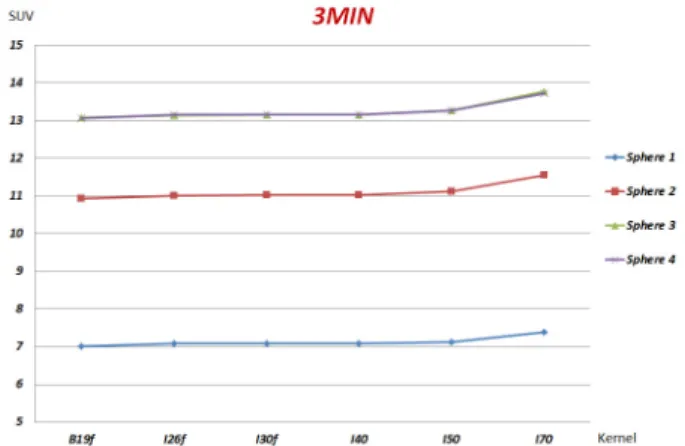
Fig. 10. SUV comparison of each kernel for scan time 3 minutes.
Fig. 11. SUV comparison of each kernel for scan time 4 minutes.
4. Chest phantom 영상 평가
노이즈, 해상력 평가와 동일한 조건으로 스캔 및 재구성한 phantom 영상을 비교한 결과 육안으로도 확인 가능한 노이 즈 감소 효과가 나타났으며 해상력의 저하는 관찰되지 않았 다(Fig. 7).
5. SUV 평가
NEMA IEC body phantom을 이용한 FBP, IRIS 각 kernel 의 ACCT 적용에 따른 SUV 변화는 10 mm, 13 mm, 17 mm, 22 mm sphere 모두 1분(Fig. 8), 2분(Fig. 9), 3분(Fig. 10), 4분 (Fig. 11)에서 동일한 패턴을 보였으며 I70 (IRIS) kernel을 제 외 하고는 ACCT에 기존의 FBP 방식의 kernel을 적용하여 얻은 SUV와 유의한 차이를 보이지 않았다.
결 론
Siemens사의 CT 선량 감소 소프트웨어인 IRIS 적용을 통
하여 AAPM CT performance phantom을 이용한 실험으로 기존 FBP 방식에 비하여 CT 선량이 45% 감소되어도 해상 력 감소 없이 노이즈가 감소되었으며 FBP 방식을 이용한 선 량감소 전 영상과 유사한 영상의 획득이 가능하였다.
Anthropomorphic chest phantom을 이용한 영상 평가 역시 시각적으로 확인 가능한 노이즈 감소 효과가 있었으며 해상 력의 저하는 보이지 않았다. NEMA IEC body phantom의 SUV 평가에서는 ACCT에 기존 FBP 방식을 적용하여 획득 한 SUV에 비교하여 I70f very sharp kernel을 제외하고는 스 캔시간 1분, 2분, 3분, 4분 모두에서 유의할만한 차이가 나타 나지 않았으나 I70 kernel의 ACCT 적용 시에는 SUV 변화에 대한 고려가 필요할 것으로 생각된다. 본 연구를 통하여 IRIS 적용 시 기존 FBP 방식에 비하여 CT 피폭선량 감소와 해상력 저하 없는 노이즈 감소 효과를 입증하였으며 IRIS kernel의 적절한 적용을 통하여 PET/CT 검사 시 환자 피폭 선량 감소는 물론 FBP 방식에 비하여 우수한 영상의 획득이 가능할 것이라 사료된다. 그러나 저 선량을 조사할수록 FBP, IRIS 재구성 방식에 따른 유사한 노이즈를 나타내는 영상의 획득을 위한 선량 감소하는 점과 chest phantom 내부 구조물
의 단순화로 인한 해상력의 정확한 평가가 어려운 한계점이 있 었으며 추후 보다 세분화된 실험이 필요할 것으로 생각된다.
요 약
Siemens사의 CT 선량 감소 소프트웨어인 IRIS의 적용을 통하여 CT 선량 감소 시 노이즈 감소 효과와 해상력의 보존 그리고 ACCT에 IRIS 각 kernel의 적용 시 SUV 변화를 확인 하는데 목적을 두었다.
Biograph mCT 40 slice 스캐너를 이용하여 AAPM CT performance phantom, Anthropomorphic chest phantom을 관전압 120 kVp로 고정하고 100-10 mAs까지 15%감소하여 스캔 후 FBP, IRIS 각 kernel을 적용하여 재구성 하여 영상의 노이즈, 해상력, 영상 평가를 시행하였다. NEMA IEC body phantom을 이용하여 55.5 MBq를 background에 주입하고 열소와 배후 방사의 비를 8:1이 되도록 모형을 제작하였다.
120 kVp, 50 mAs 조건으로 1분, 2분, 3분, 4분 스캔하여 영상 을 획득한 후 ACCT에 IRIS 각 kernel을 적용하여 기존 FBP 방식을 적용한 SUV와의 평가를 시행하였다.
IRIS의 적용 시 기존 FBP 방식에 비하여 45% 선량을 감 소하였음에도 불구하고 해상력 저하 없는 노이즈 감소 효과
가 확인 되었으며 SUV 평가 실험에서 IRIS의 I70f kernel을 제외하고는 기존 FBP 방식을 통하여 획득된 SUV와 유의한 차이가 나타나지 않았다.
본 연구를 통하여 IRIS 적용 시 기존 FBP 방식에 비하여 CT 피폭선량 감소와 해상력 저하 없는 노이즈 감소 효과를 입증하였으며 IRIS kernel의 적절한 적용을 통하여 PET/CT 검사 시 환자 피폭선량 감소는 물론 FBP 방식에 비하여 우 수한 영상의 획득이 가능할 것이라 사료된다.
REFERENCES
1. J E Ortuno, G Kontaxakis, J L Rubio, P Guerra and A Santos.
Efficient methodologies for system matrix modelling in iter- ative image reconstruction for rotating high-resolution PET.
Phys Med Biol 2010;55:1833-1861
2. Yates SJ, Pike LC, Goldstone KE. Effect of multi-slice scanners on patient dose from routine CT examinations in East Anglia.
Br J Radiol 2004;77:472-478
3. 고인호. Textbook of computed tomography. 제1판. 청구.
2003;148-152.
4. Dawson P. Patient dose in multi-slice CT: Why is it increasing and dose it matter? Br J Radiol 2004;77:S10-S13
5. 고창순. 핵의학. 제3판. 고려의학. 2008;60-64.