Antioxidant and Anti-inflammatory Effects of Ethanol Extract of Aster yomena in RAW 264.7 Macrophages
Sung Ok Kim
1, Ji-Suk Jeong
2and Yung Hyun Choi
3,4*
1Department of Food Science and Biotechnology, College of Engineering, Kyungsung University, Busan 48434, Korea
2Gurye-gun Agricultural Center and Gurye Wild Flower Institute, Gurye-gun 57660, Korea
3Anti-Aging Research Center, Dong-eui University, Busan 47340, Korea
4Department of Biochemistry, Dong-eui University College of Korean Medicine, Busan 47227, Korea Received August 31, 2019 /Revised September 20, 2019 /Accepted September 23, 2019
Aster yomena (Kitam.) Honda is an edible vegetable and perennial herb belonging to the Asteraceae
family, and has been used for a long time for the prevention and treatment of various diseases.
Although leaf extracts of A. yomena are known to have antioxidant and anti-inflammatory effects, ac- curate efficacy assessments are still inadequate. In this study, we investigated whether the antioxidant efficacy of ethanol extract of A. yomena leaf (EEAY) is correlated with the anti-inflammatory effect in RAW 264.7 macrophages. The results showed that EEAY significantly inhibited the hydrogen peroxide (H
2O
2)-induced growth inhibition in RAW 264.7 cells, which was associated with increased expression of nuclear factor erythroid 2-related factor-2 (Nrf2) and heme oxygenase-1 (HO-1). EEAY pretreatment also effectively prevented H
2O
2-induced reactive oxygen species generation and apoptosis through in- hibition of caspase-3 activation and poly (ADP-ribose) polymerase degradation. Additionally, EEAY significantly increased the expression and production of interleukin-10, a representative anti-in- flammatory cytokine, which was associated with increased expression of toll-like receptor 4 and mye- loid differentiation factor 88 at transcriptional and translational levels. Furthermore, the increased pro- duction of nitric oxide (NO) by lipopolysaccharide was markedly abolished under the condition of EEAY pretreatment, and the inhibitory effect of NO production by EEAY was further increased by hemin, an HO-1 inducer. Overall, our results suggest that EEAY is able to activate the Nrf2/HO-1 signaling pathway to protect RAW 264.7 macrophages from oxidative and inflammatory stress.
Key words : Aster yomena leaf, ROS, Nrf2/HO-1, IL-10, NO
*Corresponding author
*Tel : +82-51-850-7413, Fax : +82-51-853-4036
*E-mail : [email protected]
This is an Open-Access article distributed under the terms of the Creative Commons Attribution Non-Commercial License (http://creativecommons.org/licenses/by-nc/3.0) which permits unrestricted non-commercial use, distribution, and reproduction in any medium, provided the original work is properly cited.
Journal of Life Science 2019 Vol. 29. No. 9. 977~985 DOI : https://doi.org/10.5352/JLS.2019.29.9.977
서 론
활성산소종(reactive oxygen species, ROS)은 산소의 대사 과정에서 생성되며 세포 내 유전자 발현의 조절을 포함한 주 요 생리적 역할을 조절한다[9, 25]. 비록 적정 수준의 ROS 생성 은 세포의 성장과 증식에 필수적인 신호분자 역할을 하지만 과도한 ROS의 생성은 세포에 산화적 손상을 일으키게 된다 [32, 45]. 미토콘드리아는 세포 내 소기관들 중에서 ROS의 생 성을 담당하는 중요한 기관이지만, 산화적 스트레스에 의한 ROS 과잉 생산을 통한 미토콘드리아 기능 장애는 DNA 손상 과 apoptosis (세포사멸)를 유도하는 중요한 기전 중 하나로 인식되고 있다[34, 37]. 따라서 항산화 방어 시스템은 세포 보
호에 필수적이며, nuclear factor erythroid 2-related factor 2 (Nrf2)는 세포 내 산화-환원 균형을 위한 핵심 전사조절인자 중의 하나이다. 생리학적 조건 하에서, Nrf2는 Kelch-like ECH-associated protein 1 (Keap1)에 결합하여 불활성화된 형 태로 세포질에 존재한다. 그러나 산화적 스트레스 조건 하에 서, Nrf2는 Keap1로 부터 해리되어 핵으로 전위되며, 여기서 다양한 세포보호 효소들의 전사활성을 촉진한다[23, 35]. 최근 여러 연구들에서 다양한 항산화 활성 효능을 가진 천연물들이 항염증 효능을 동시에 나타내고 있음이 보고되고 있다[3, 26].
이러한 항염증 효능은 toll-like receptor (TLR)와 연계된 nu- clear factor kappa B (NF-κB) 신호 경로의 조절을 통한 염증 유발 인자들과 전 염증성 사이토카인(pro-inflammatory cyto- kines)의 생성을 억제하면서 항 염증성 사이토카인(anti-in- flammatory cytokines)의 생성 촉진과 연관되어 있다[20, 28].
따라서 항산화 및 항염증 효능을 동시에 지니는 천연물들은 다양한 질환의 예방과 치료에 효과적일 수 있다.
최근 선행 연구들에서 다양한 질병의 예방과 치료를 위하여
사용되어 온 전통의학 자원은 항산화 및 항염증 효능을 지닌
소재의 발굴을 위한 후보로서 유용성이 매우 높음을 알 수
있다[16, 43]. 쑥부쟁이[Aster yomena (Kitam.) Honda]는 아시
아 전역에 널리 분포하는 국화과(Asteraceae family)에 속하는 다년생 식물로서 기침 및 천식 등을 포함한 여러 질병을 치료 하기 위해 오랫동안 사용되어 왔다[36]. 아울러 쑥부쟁이 추출 물 또는 구성 성분들은 항균[21, 29, 42] 및 항응고[7] 작용과 함께 항산화[14, 27] 및 항염증[15, 18] 효능과 같은 약리학적 활성이 있는 것으로 보고되고 있다. 본 연구실에서도 쑥부쟁 이 추출물이 항염증[17] 작용뿐 만 아니라 항비만 효능[12]이 있음을 보고한 바 있다. 그러나 쑥부쟁이 추출물의 항산화 및 항염증에 관한 복합적인 기전 연구는 여전히 미비한 상황이 다. 따라서 본 연구에서는 쑥부쟁이 추출물의 항산화 및 항염 증 효능을 동시에 평가하기 위하여 쑥부쟁이 에탄올 추출물이 산화적 스트레스에서 의하여 유발된 ROS의 생성에 미치는 영향 및 Nrf2 신호 경로의 연관성과, 항 염증성 사이토카인의 발현에 미치는 영향 및 TLR4 신호 경로의 연관성을 동시에 평가하였다.
재료 및 방법
쑥부쟁이 에탄올 추출물 준비
본 연구에 사용된 쑥부쟁이 잎은 구례야생화연구소(구례 군, 전라남도, 대한민국)에서 제공받았으며, 건조된 잎(50 mg) 을 절단 후 미세 분말로 분쇄한 다음 70% 에탄올(500 ml)에 2일 동안 침전시켰다. Whatman No.3 여과지를 통해 2회 여과 하여 불용성 물질을 제거한 후 rotary evaporator (Rikakikai Co., Ltd., Tokyo, Japan)를 사용하여 농축하였다[17]. 농축된 추출물(ethanol extract of A. yomena leaf, EEAY)은 dime- thylsulfoxide (DMSO, Sigma-Aldrich Chemical Co., St. Louis, MO, USA)에 최종 농도 200 mg/ml로 녹인 후 세포 배양용 배지에 적정 농도로 희석하여 처리하였다.
세포 배양 및 세포 생존율 분석
RAW 264.7 세포는 한국세포주은행(Korean Cell Line Bank, 서울, 대한민국)에서 구입하였으며 10% fetal bovine serum, 100 U/ml penicillin 및 100 mg/ml streptomycin이 함유된 Dulbecco's modified Eagle's medium (DMEM, WelGENE Inc., 대구, 대한민국)을 이용하여 배양하였다. 세포 생존율은 3-[4,5-dimethylthiazol-2-yl]-2,5-diphenyltetrazolium bromide (MTT) 분석법에 준하였다. 이를 위하여 RAW 264.7 세포를 96-well plate (1×10
4cells/well) 분주 후 37℃에서 24시간 동안 배양했다. 이들 세포에 24시간 동안 다양한 농도의 EEAY를 처리하거나 과산화수소(hydrogen peroxide, H
2O
2), lipopoly- saccharide (LPS), N-acetylcysteine (NAC) 또는 hemin (all from Sigma-Aldrich Chemical Co.) 등을 1시간 전처리한 후 EEAY를 24시간 처리하였다. 배양이 끝난 후 배지를 제거하고 MTT 용액(Sigma-Aldrich Chemical Co.; 5 mg/ml in phos- phate-buffered saline, PBS)을 각 well에 넣고 37℃에서 3시간
반응시켜 형성된 formazan을 DMSO로 용해시켰다. 광학 밀 도(Optical density)는 560 nm에서 microplate spectropho- tometer (Molecular Devices, Sunnyvale, CA, USA)를 이용하 여 측정하였다.
Reverse transcription-polymerase chain reaction (RT-PCR)에 의한 mRNA 발현 분석
전사 수준에서 유전자들의 발현 변화를 조사하기 위하여 TRIzol reagent (Invitrogen Co., Carlsbad, CA, USA)를 이용 하여 RNA를 분리하였다. 분리된 RNA를 정량 후, 해당 유전 자의 primer, DEPC water 그리고 ONE-STEP RT-PCR PreMix Kit (Intron, Seoul, Korea)를 넣고 Mastercycler gradient (Eppendorf, Hamburg, Germany)를 이용하여 증폭하였다. 각 PCR 산물들의 양적 차이를 확인하기 위하여 1X TAE buffer로 1.5% agarose gel을 만들고 well 당 각각의 primer에 해당하는 PCR 산물에 DNA gel loading solution을 섞어서 loading 한 후 50 V에서 전기영동을 행하였다. 전기영동으로 DNA 분리 가 끝난 gel을 ethidium bromide (EtBr, Sigma-Aldrich Chem- ical Co.)로 염색한 후 UV 하에서 관찰하였으며, glyceral- dehyde-3-phosphate dehydrogenase (GAPDH)를 internal control로 사용하였다.
단백질의 분리 및 Western blot analysis
번역 수준에서 유전자들의 발현 변화 분석을 위하여 선행 방법에 따라 단백질을 분리 및 정량화 하였다[19]. Western blot analysis를 위해 동량의 단백질들을 sodium dodecyl sul- fate (SDS)-polyacrylamide gel electrophoresis를 이용하여 분 리하고 polyvinylidene difluoride membrane (Schleicher &
Schuell, Keene, NH, USA)으로 전이시켰다. 각각의 mem- brane에 적정 항체 및 enhanced chemiluminescence (ECL, Amersham Corp., Arlington Heights, IL, USA) 용액을 이용 하여 단백질들의 발현 변화를 조사하였다. 본 실험에 사용된 1차 항체들은 Santa Cruz Biotechnology Inc. (Santa Cruz, CA, USA) 및 Calbiochem (Cambridge, MA, USA)에서 구입하였 으며, 2차 항체들은 Amersham Corp.에서 구입하였다.
ROS 생성의 측정
세포 내 ROS 생성의 정도를 확인하기 위하여 준비된 세포들
을 PBS에 수세 후 fluorescent probe인 10 μM의 2’,7’-di-chlor-
odihydrofluorescein diacetate (DCF-DA, Molecular Probes,
Leiden, Netherlands)로 20분간 염색 후 형광현미경(Carl
Zeiss, Oberkochen, Germany) 하에서 ROS의 생성 변화 여부
를 조사하였다. 아울러 동일한 조건에서 준비된 세포들에서
ROS 생성에 대한 정량적인 비교를 위하여 반응이 끝난 세포
들을 flow cytometer (Becton Dickinson, San Jose, CA, USA)
를 적용시켜 ROS 값의 변화를 분석하였다[8].
A
B
EEAY (μg/ml)H2O2 (1 mM) EEAY (μg/ml) NAC (10 mM)
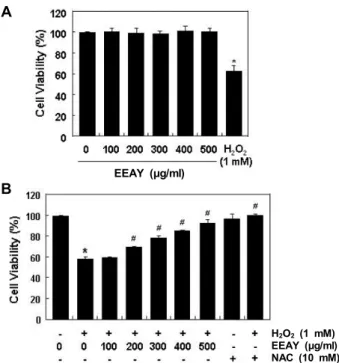
Fig. 1. Protective effects of EEAY against H2O2-induced cytoto- xicity in RAW 264.7 macrophages. The cells were treated with different concentrations of EEAY or 1 mM H2O2 for 24 hr (A) or treated with 1 mM H2O2 for 24 hr, after EEAY or NAC pretreatment with the indicated concen- trations for 1 hr (B). Following treatments, cell viability was examined by MTT assay. The data are shown as mean ± SD obtained from three independent experi- ments (*p<0.05 compared to control; #p<0.05 compared to cells cultured with 1 mM H2O2).
핵의 형태 변화 관찰을 통한 apoptosis 유도 확인 핵의 형태적 변화를 관찰하기 위하여 준비된 세포들을 PBS 로 수세 후 0.1 M phosphate buffer (pH 7.2)에 희석된 2.0%
paraformaldehyde (Sigma-Aldrich Chemical Co.)로 1시간 세 포들을 고정하였다. 고정된 세포들을 PBS로 수세한 다음 0.05 M phosphate buffer (pH 7.2)에 2.5 μg/ml의 농도로 희석된 4,6-diamidino-2-phenylindole (DAPI, Sigma-Aldrich Chemi- cal Co.) 용액으로 상온 암하에서 15분간 염색하였다. 이를 다시 PBS로 수세한 후 형광현미경 하에서 핵의 형태를 비교하였다[8].
Flow cytometry 분석에 의한 apoptosis 유발의 정량화 Apoptosis 정도의 정량적인 분석을 위하여 Cycle TEST PLUS DNA REAGENT Kit (Becton Dickinson, San Jose, CA, USA)을 사용하였다. 이를 위하여 준비된 세포를 protocol에 따라 고정 후 4°C, 암실에서 30분 동안 propidium iodide (PI) 용액에 반응시켰다. 이들 세포를 35-mm mesh를 이용하여 단 일세포로 분리하고 실험군당 최소 10,000개 이상의 세포를 flow cytometer (Becton Dickinson, San Jose, CA, USA)에 적 용시켜 세포 내 DNA 함량에 따른 histogram을 대상으로 sub- G1기에 속하는 세포를 apoptosis가 유발된 세포로 산출하였다.
Interleukin (IL)-10 및 nitric oxide (NO) 생성량의 측정 EEAY의 항염증 효능 평가를 위한 IL-10의 함량은 ELISA kit (R&D Systems, Minneapolis, MN, USA)를 이용하여 제시 된 실험 방법에 준하여 실험을 수행하였으며, NO 생성의 정도 는 Griess reagent를 이용하여 측정하였다[19].
통계 분석
모든 실험결과는 평균±표준편차(mean ± standard devia- tion, SD)로 표시하였고 Sigma Plot (Systat Software Inc., San Jose, CA, USA) Student t-test를 이용하여 통계적 유의성을 얻 었다. 모든 통계처리의 유의성은 p<0.05 수준에서 검증하였다.
결 과
산화적 스트레스에 대한 EEAY의 세포독성 보호 효과 산화적 스트레스에 대한 EEAY의 보호 효능을 조사하기 위 하여 먼저 RAW 264.7 세포의 증식에 미치는 EEAY의 영향을 조사하였다. Fig. 1A에 나타낸 MTT 분석 결과에 의하면, 500 μg/mL의 EEAY 처리군까지 대조군에 비하여 유의적인 세포 독성을 나타내지 않았다. 반면 산화적 스트레스를 유발하여 위하여 처리된 1 mM의 H
2O
2에 노출된 세포에서는 대조군에 비하여 약 60% 정도의 생존율을 나타내었다. Fig. 1B는 세포독 성이 나타내지 않는 500 μg/ml까지 EEAY를 1시간 전처리 후 1 mM의 H
2O
2를 24시간 처리한 후 세포 생존력을 MTT 분석을 수행한 결과이다. 이 결과에서 알 수 있듯이 H
2O
2에
의한 세포독성이 EEAY의 처리 농도 의존적으로 차단되었으 며, 강력한 ROS 생성 억제제인 NAC을 전처리하였을 경우에 도 H
2O
2에 의한 세포생존율 억제가 거의 대조군 수준으로 유 지되었다. 이는 EEAY는 산화적 스트레스에 의한 세포독성을 효과적으로 차단할 수 있었음을 보여주는 결과이다.
Nrf2 및 HO-1의 발현에 미치는 EEAY의 영향
RAW 264.7 세포에서 H
2O
2에 세포독성 유발에 대한 EEAY 의 차단 효과가 Nrf2의 발현과 연관성이 있는지를 조사하기 위하여 Nrf2의 발현에 미치는 EEAY의 영향을 조사하였다.
Fig. 2의 RT-PCR 및 Western blot 분석 결과에 의하면 Nrf2의 발현이 전사 및 번역 수준에서 EEAY의 처리 농도 의존적으로 증가하였음을 알 수 있었다. 이는 또한 대표적인 Nrf2 의존적 세포보호 효소인 HO-1의 발현 증가와도 연관성이 있었으며, 이들 유전자들의 발현 증가가 EEAY의 산화적 스트레스 억제 에 최소한 기여할 가능성을 보여주었다.
H
2O
2에 의한 ROS의 생성에 미치는 EEAY의 영향
EEAY의 산화적 스트레스 유발 세포독성 억제 효능이 ROS
EEAY (μg/ml)
EEAY (μg/ml)
A
B
Fig. 2. Induction of Nrf2 and HO-1 by EEAY in RAW 264.7 macrophages. The cells were treated with the indicated concentrations of EEAY for 24 hr. (A) Total RNAs were prepared for RT-PCR analysis of Nrf2 and HO-1 mRNA expression. (B) Cell lysates were prepared for Western blot analysis with antibodies specific for Nrf2 and HO-1, and an ECL detection system. Experiments were re- peated three times and similar results were obtained.
GAPDH and actin were used as internal controls for the RT-PCR and Western blot analysis, respectively.
A
B
C
H2O2 (1 mM) EEAY (500 μg/ml)
H2O2 (1 mM) EEAY (500 μg/ml) H2O2 (1 mM)
EEAY (500 μg/ml)
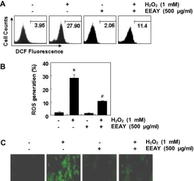
Fig. 3. Inhibition of ROS generation by EEAY in H2O2-treated RAW 264.7 macrophages. The cells were pretreated with 500 μg/ml EEAY for 1 hr and then stimulated with or without 1 mM H2O2 for 1 hr. (A) After stain- ing with DCF-DA florescent dye, DCF fluorescence was monitored using a flow cytometer. (B) The data were shown as mean ± SD obtained from three in- dependent experiments (*p<0.05 compared to control;
#p<0.05 compared to cells cultured with 1 mM H2O2).
(C) The fluorescent images were obtained by a fluo- rescence microscope. Each image is representative of at least three independent experiments.
의 생성 억제와 연관성이 있는지의 여부를 조사하기 위하여 DCF-DA 염색법을 이용하였다. Fig. 3A 및 Fig. 3B의 flow cy- tometry 결과에서 알 수 잇듯이 H
2O
2가 단독 처리된 RAW 264.7 세포에서 ROS의 생성이 매우 증가되었으며 이는 EEAY 의 전처리에 의하여 현저하게 감소되었다. 형광현미경적 관찰 로 이러한 결과를 재확인한 결과에서도 H
2O
2가 처리된 세포
에서 ROS의 생성을 의미하는 형광 강도가 증가되었으나, EEAY가 전처리된 세포에서는 현저하게 억제되었다(Fig. 3C).
즉 EEAY의 산화적 스트레스 억제 효능이 ROS의 생성 차단과 직접 연관성이 있음을 알 수 있었다.
H
2O
2에 의한 apoptosis 유발에 미치는 EEAY의 영향 다음은 H
2O
2에 의한 생존율 감소가 apoptosis 유발과 연관 성이 있는지, 이를 EEAY가 억제할 수 있는지를 조사하였다.
DAPI 염색에 의한 핵의 형태적 변화를 조사한 결과에서, 전형 적인 apoptosis가 유발된 세포에서 관찰되는 DNA의 절단에 의한 염색질의 응축 현상이 H
2O
2가 단독으로 처리된 RAW 264.7 세포에서 현저하게 증가되었으며(Fig. 4A), flow cyto- metry 분석에 의한 apoptosis 유발 세포 집단을 의미하는 sub- G1기에 속하는 세포의 빈도 역시 대조군에 비하여 증가되었 다(Fig. 4B, Fig. 4C). 그러나 H
2O
2에 의한 apoptosis 유발이 EEAY가 전처리된 세포에서는 현저하게 감소되었다. 이러한 EEAY의 apoptosis 억제와 연관된 주요 유전자들의 발현 변화 와의 연관성을 조사한 결과, H
2O
2가 처리된 세포에서 관찰된 caspase-3의 활성형의 발현 증가가 EEAY가 전처리된 조건에 서 감소되었으며, 활성형 caspase-3의 기질 단백질인 poly (ADP- ribose) polymerase (PARP)의 단편화 또한 EEAY에 의 하여 억제되었다(Fig. 4D).
IL-10의 발현 및 생성에 미치는 EEAY의 영향
EEAY의 항산화 효능이 항염증 효능과 연관성이 있는지를
조사하기 위하여 대표적인 항 염증성 사이토카인인 IL-10의
생성에 미치는 EEAY의 영향을 조사하였다. 이를 위하여 다양
한 EEAY가 처리된 세포의 배지에 함유된 IL-10의 양을 조사한
H2O2 (1 mM) EEAY (500 μg/ml)
H2O2 (1 mM) EEAY (500 μg/ml)
H2O2 (1 mM) EEAY (500 μg/ml)
H2O2 (1 mM) EEAY (500 μg/ml)
A
B
C
Fig. 4. Inhibition of apoptosis by EEAY in H2O2-treated RAW 264.7 macrophages. The cells were pre- treated with 500 μg/ml EEAY for 1 hr and then stimulated with or without 1 mM H2O2 for 24 hr.
(A) The stained nuclei stained with DAPI were pictured under a fluorescence microscope. Each image is representative of at least three independ- ent experiments. (B) The cells were stained with PI, and then the percentages of sub-G1 apoptotic cells were determined using flow cytometry. (C) The data were shown as mean ± SD obtained from three independent experiments (*p<0.05 compared to control; #p<0.05 compared to cells cultured with 1 mM H2O2). (D) Cell lysates were prepared for Western blot analysis with the in- dicated antibodies, and an ECL detection system.
Experiments were repeated three times and sim- ilar results were obtained. Actin was used as an internal control for and Western blot analysis.
A
B C
EEAY (μg/ml)
EEAY (μg/ml) EEAY (μg/ml)
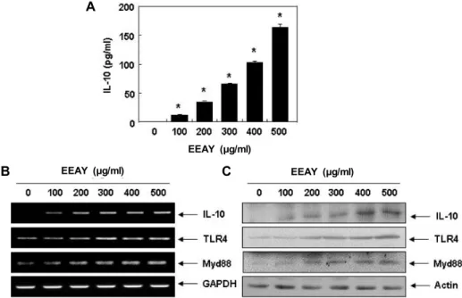
Fig. 5. Increase of IL-10 generation and expression by EEAY in RAW 264.7 macrophages. The cells were treated with the indicated concentrations of EEAY for 24 hr. (A) The levels of IL-10 production were measured by ELISA kit using culture media.
The data are shown as mean ± SD obtained from three independent experiments (*p<0.05 compared to control). (B) Total RNAs were prepared for RT-PCR analysis of IL-10, TLR4 and Myd88 mRNA expression. (C) Cell lysates were prepared for Western blot analysis with antibodies specific for IL-10, TLR4 and Myd88, and an ECL detection system. Experiments were repeated three times and similar results were obtained. GAPDH and actin were used as internal controls for the RT-PCR and Western blot analysis, respectively.
결과, EEAY 처리 농도 의존적으로 IL-10의 생성이 유의적으로 증가하였음을 알 수 있었다(Fig. 5A). 이러한 EEAY에 의한 IL-10의 생성 증가가 IL-10의 발현 증가와 연관성이 있는지를 조사한 결과, EEAY에 노출된 RAW 264.7 세포에서 IL-10의 mRNA 및 단백질의 발현이 EEAY의 처리 농도 증가에 따라 현저하게 증가되었다(Fig. 5B, Fig. 5C). 또한 IL-10의 발현 증
가는 TLR4 및 myeloid differentiation factor 88 (Myd88)의 증가를 동반하여 TLR4 신호계의 활성에 따른 IL-10 유전자의 전사 활성이 증가되었을 것으로 추정된다.
NO의 생성에 미치는 EEAY의 영향
EEAY에 의한 항 염증성 사이토카인인 IL-10의 발현 및 생
A
B
LPS (100 ng/ml) EEAY (500 μg/ml) Hemin (20 mM)
LPS (100 ng/ml) EEAY (500 μg/ml) Hemin (20 mM)
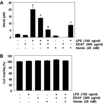
Fig. 6. Inhibition of LPS-induced NO generation by EEAY in RAW 264.7 macrophages. The cell were pretreated with 500 μg/ml EEAY or 20 mM hemin for 1 hr and then stimulated with or without 100 ng/ml LPS for 24 hr.
(A) NO production was measured using the Griess re- agent in culture media. Each value indicates the mean
± SD obtained from three independent experiments (*p<0.05 compared to control; #p<0.05 compared to cells cultured with 100 ng/ml LPS; &p<0.05 compared to cells co-treated with 500 μg/ml EEAY and 100 ng/ml LPS).
(B) Cell viability was gauged by an MTT assay. Data were expressed as percentage of control. Each value in- dicates the mean ± SD obtained from three independent experiments.
성 증가가 항염증 효능과 연관성이 있는지를 확인하기 위하여 대표적인 염증성 매개체인 NO의 생성에 미치는 EEAY의 영 향을 평가하였다. Fig. 6A의 결과에서 알 수 있듯이 LPS가 처 리된 RAW 264.7 세포에서 NO의 생성이 매우 증가되었으나, EEAY를 전처리한 조건에서 배양된 세포에서는 LPS에 의한 NO의 생성이 유의적으로 차단되었다. 아울러 EEAY의 항염 증 효능이 항산화 효능과 관련이 있는지를 조사하기 위하여 HO-1의 유도제인 hemin을 EEAY와 동시에 전처리하였을 경 우 hemin 및 EEAY 단독 전처리군에 비하여 NO의 생성을 더욱 억제하였다. 아울러 이러한 항염증 효능이 세포독성과는 무관하였다(Fig. 6B).
고 찰
산화적 스트레스와 염증성 스트레스는 다양한 질환의 원인 이면서 질환의 이환율(morbidity)을 상승 시키는 요인으로 작 용한다[1, 2, 31]. 비록 이를 억제하기 위한 다양한 약물들이
개발되고 있지만, 이들의 독성을 최소화하기 위한 방법으로 천연물의 활용에 대한 연구가 증폭되고 있다. 본 연구에서도 이러한 천연물의 발굴을 위한 연구의 일환으로 쑥부쟁이 잎의 에탄올 추출물(EEAY)에 대한 항산화 및 항염증 효능을 조사 하였다. 이를 위하여 RAW 264.7 세포 모델을 이용하였으며, 산화적 스트레스를 유발하기 위하여 적정 수준의 세포독성을 지니는 H
2O
2의 농도(1 mM)를 설정하였다. 본 연구의 결과에 의하면 H
2O
2에 대한 세포독성을 EEAY가 효과적으로 차단하 였으며, 이러한 EEAY의 산화적 스트레스 억제 효과가 Nrf2 신호계와 연관성이 있는지를 조사한 결과, 세포독성이 없는 범위에서 EEAY는 Nrf2의 발현뿐만 아니라 대표적인 Nrf2 의 존적 세포 보호 효소인 HO-1의 발현[23, 35]을 전사 및 번역 수준에서 EEAY 처리 농도 의존적으로 증가시켰다. 최근 본 연구의 결과와 유사하게 Qin, et al. [33]은 RAW 264.7 세포에 서 H
2O
2에 의한 산화적 스트레스에 대한 만삼(Codonopsis pilo-
sula) 유래 다당체의 방어 효과가 Nrf2 매개 HO-1의 발현 증가와 연관성이 있음을 제시한 바 있다. 그리고 flavonoid의 일종 인 orientin과 isovitexin이 RAW 264.7 세포에서 H
2O
2및 LPS 에 의한 산화적 스트레스 억제에 Nrf2 및 HO-1의 발현 증가가 관여함이 보고된 바 있다[24, 41]. 비록 Nrf2/Keap1-ARE 신호 전달계에 의하여 조절되는 다른 항산화 유전자의 발현 여부와 전사활성 조절에 대한 EEAY의 기전 연구는 추가적으로 수행 되어야 할 것이지만, H
2O
2에 의한 산화적 스트레스에 대한 EEAY의 억제 효과에는 최소한 Nrf2 신호계의 활성과 연관성 이 있음을 알 수 있었다.
다음은 EEAY의 산화적 스트레스에 대한 보호 효과가 ROS 의 생성 차단과 직접적인 연관성이 있는지를 조사한 결과, RAW 264.7 세포에서 H
2O
2에 의한 ROS의 생성이 EEAY에 의하여 유의적으로 차단되었음을 DCF-DA 염색을 통한 결과 에서 확인하였다. 한편 RAW 264.7 세포에서 H
2O
2에 의한 ROS 의 생성은 mitochondrial membrane potential의 소실을 통한 미토콘드리아의 기능 손상을 초래하여 caspase 의존적 apop- tosis 경로의 활성을 통하여 apoptosis를 유발하는 것으로 알 려져 있다[33, 44]. 미토콘드리아의 기능 손상과 연관된 apop- tosis는 궁극적으로 caspase cascade의 활성을 통하여 effect caspase를 활성화시켜 기질단백질들의 분해를 통하여 apop- tosis를 완성한다[13, 30]. DAPI 염색을 이용한 핵의 형태변화 관찰 및 PI 염색을 통한 apoptosis 빈도 조사 결과를 통한 본 연구의 결과에서도 알 수 있듯이, EEAY는 H
2O
2에 의한 apop- tosis 유도를 효과적으로 억제하였다. 아울러 EEAY의 apopto- sis 억제 효과는 caspase-3의 활성화 억제에 따른 PARP와 같은 기질단백질의 분해 차단과도 연관성이 있었다. 따라서 EEAY 에 의한 ROS의 생성 차단은 미토콘드리아의 기능 손상을 억 제하면서 apoptosis 경로의 활성을 저해하였을 것으로 추정되 며, 이는 EEAY의 항산화 효능에 의한 것으로 사료된다.
한편 염증 반응 조절에 관여하는 다양한 사이토카인 중에서
IL-10은 대표적인 항 염증성 사이토카인이다[20, 28]. 비록 TLR 비의존적 경로에 의하여 IL-10의 발현이 증가될 수도 있 지만, 단핵구에서 대식세포로의 활성화에 의한 IL-10의 생성 이 증가되기 위해서는 NF-κB의 활성을 위한 TLR 경로의 활성 화가 이루어져야 한다[4, 22]. 따라서 EEAY의 항산화 효능이 항염증 효능과 동반되는 현상인지를 확인하기 위하여 IL-10의 생성에 미치는 영향을 조사한 결과, 세포 독성이 없는 범위에 서 EEAY은 IL-10의 생성을 유의적으로 증가시켰으며, 이는 IL-10의 mRNA 및 단백질 수준에서의 발현 증가에 의한 것임 을 알 수 있었다. 또한 이러한 IL-10의 전사 및 번역 활성 증가 에 TLR4-Myd88 신호계가 관여하는지를 조사한 결과, EEAY 처리 농도 의존적으로 TLR4 및 Myd88 발현이 증가되었음을 알 수 있었다. 이는 RAW 264.7 대식세포에서 염증성 반응의 억제를 위한 IL-10의 발현 증가에 TLR4-Myd88 신호계가 관여 한다는 선행 연구들의 결과와 잘 일치된다[11, 38]. RAW 264.7 대식세포에서 TLR4-Myd88 신호계 매개 IL-10의 발현 조절과 함께, 염증 자극에 따른 대표적인 염증성 매개인자인 NO의 생성 증가는 inducible NO synthase의 발현을 증가에 의한 결과이며 이 또한 TLR4-Myd88 신호계가 관여할 수 있다[5, 10, 40]. 따라서 EEAY의 항염증 효능을 재확인하기 위하여 LPS에 의해 유도되는 NO의 생성에 미치는 EEAY의 영향을 조사하였으며, LPS의 처리에 의하여 증가된 NO의 생성이 EEAY의 존재 하에서는 유의적으로 억제되었음을 알 수 있었 다. 이상에서 확인된 EEAY의 항염증 효능이 HO-1의 발현 증 가와 연관된 항산화 효능과 관련성이 있는지의 여부를 추가적 으로 조사하였다. 이를 위하여 HO-1의 활성을 유도하는 heme 의 산화형인 hemin [6, 39]을 사용하여 LPS에 의한 NO의 생성 에 미치는 EEAY의 효능과 비교하였다. 본 연구의 결과에 의하 면, hemin은 세포 독성이 없는 조건에서 LPS에 의한 NO의 생성을 유의적으로 감소시켰으며, EEAY와 hemin을 동시에 전처리한 경우, LPS에 의한 NO의 생성을 더욱 억제시켰다.
본 연구의 결과에 의하면, EEAY의 항산화 효능에는 항염증 효능이 동반되었으며, HO-1의 발현 증가가 관여할 가능성을 보여 주었다. 이는 쑥부쟁이 추출물이 항산화[14, 27] 및 항염 증[15, 17, 18] 효능이 있을 것이라는 선행 연구의 결과를 잘 뒷받침하여 줄 수 있는 결과라고 생각한다. 그러나 이러한 효 능이 쑥부쟁이 잎의 에탄올 추출물에 함유된 어떤 생리활성 성분에 의한 현상인지, HO-1의 전사활성이 Nrf2 의존적인 현 상인지에 대한 추가적인 연구가 수행되어야 할 것이다. 아울러 동물 모델을 이용한 EEAY의 항산화 효능이 항염증 효능과 직 접 연관되어 있는지에 대한 검증 또한 이루어져야 할 것이다.
감사의 글
이 논문은 2019학년도 동의대학교 교내연구비(과제번호:
201902110001) 및 한국연구재단(2018R1A2B2005705) 지원에
의해 연구되었음.
References
1. Ayoub, K. F., Pothineni, N. V. K., Rutland, J., Ding, Z. and Mehta, J. L. 2017. Immunity, inflammation, and oxidative stress in heart failure: Emerging molecular targets. Cardio- vasc. Drugs Ther. 31, 593-608.
2. Bello-Klein, A., Mancardi, D., Araujo, A. S., Schenkel, P. C., Turck, P. and de Lima Seolin, B. G. 2018. Role of redox ho- meostasis and inflammation in the pathogenesis of pulmo- nary arterial hypertension. Curr. Med. Chem. 25, 1340-1351.
3. Cătană, C. S., Atanasov, A. G. and Berindan-Neagoe, I. 2018.
Natural products with anti-aging potential: Affected targets and molecular mechanisms. Biotechnol. Adv. 36, 1649-1656.
4. Chen, S., Lu, Z., Wang, F. and Wang, Y. 2018. Cathelicidin- WA polarizes E. coli K88-induced M1 macrophage to M2- like macrophage in RAW264.7 cells. Int. Immunopharmacol.
54, 52-59.
5. Cheng, X., Gao, D., Chen, B. and Mao, X. 2015. Endotoxin- binding peptides derived from casein glycomacropeptide in- hibit lipopolysaccharide-stimulated inflammatory responses via blockade of NF-
κ
B activation in macrophages. Nutrients 7, 3119-3137.6. Chen-Roetling, J. and Regan, R. F. 2017. Targeting the Nrf2- heme oxygenase-1 axis after intracerebral hemorrhage. Curr.
Pharm. Des. 23, 2226-2237.
7. Choi, J. H., Kim, D. W., Park, S. E., Choi, B. S., Sapkota, K., Kim, S. and Kim, S. J. 2014. Novel thrombolytic protease from edible and medicinal plant Aster yomena (Kitam.) Honda with anticoagulant activity: Purification and partial characterization. J. Biosci. Bioeng. 118, 372-377.
8. Choi, Y. H. 2019. Activation of the Nrf2/HO-1 signaling pathway contributes to the protective effects of coptisine against oxidative stress-induced DNA damage and apopto- sis in HaCaT keratinocytes. Gen. Physiol. Biophys. 38, 281-294.
9. Finkel, T. and Holbrook, N. J. 2000. Oxidants, oxidative stress and the biology of ageing. Nature 408, 239-247.
10. Gao, H., Liu, X., Sun, W., Kang, N., Liu, Y., Yang, S., Xu, Q. M., Wang, C. and Chen, X. 2017. Total tanshinones ex- hibits anti-inflammatory effects through blocking TLR4 di- merization via the MyD88 pathway. Cell Death Dis. 8, e3004.
11. Gupta, P. K., Rajan, M. G. R. and Kulkarni, S. 2017. Activation of murine macrophages by G1-4A, a polysaccharide from Tinospora cordifolia, in TLR4/MyD88 dependent manner. Int.
Immunopharmacol. 50, 168-177.
12. Han, M. H., Jeong, J. S., Jeong, J. W., Choi, S. H., Kim, S.
O., Hong, S. H., Park, C., Kim, B. W. and Choi, Y. H. 2017.
Ethanol extracts of Aster yomena (Kitam.) Honda inhibit adi- pogenesis through the activation of the AMPK signaling pathway in 3T3-L1 preadipocytes. Drug Discov. Ther. 11, 281- 287.
13. Hassan, M., Watari, H., AbuAlmaaty, A., Ohba, Y. and Sakuragi, N. 2014. Apoptosis and molecular targeting ther- apy in cancer. Biomed. Res. Int. 2014, 150845.
14. Heo, B. G., Park, Y. S., Chon, S. U., Lee, S. Y., Cho, J. Y.
and Gorinstein, S. 2007. Antioxidant activity and cytotox- icity of methanol extracts from aerial parts of Korean salad plants. Biofactors 30, 79-89.
15. Hwang, K. A., Hwang, Y. J. and Song, J. 2018. Aster yomena extract ameliorates pro-inflammatory immune response by suppressing NF-κB activation in RAW 264.7 cells. J. Chin.
Med. Assoc. 81, 102-110.
16. Jiao, R., Liu, Y., Gao, H., Xiao, J. and So, K. F. 2016. The anti-oxidant and antitumor properties of plant polysaccha- rides. Am. J. Chin. Med. 44, 463-488.
17. Kang, H. J., Jeong, J. S., Park, N. J., Go, G. B., Kim, S. O., Park, C., Kim, B. W., Hong, S. H. and Choi, Y. H. 2017.
An ethanol extract of Aster yomena (Kitam.) Honda inhibits lipopolysaccharide-induced inflammatory responses in mur- ine RAW 264.7 macrophages. Biosci. Trends 11, 85-94.
18. Kim, A. R., Jin, Q., Jin, H. G., Ko, H. J. and Woo, E. R. 2014.
Phenolic compounds with IL-6 inhibitory activity from Aster yomena. Arch. Pharm. Res. 37, 845-851.
19. Kwon, D. H., Cheon, J. M., Choi, E. O., Jeong, J. W., Lee, K. W., Kim, K. Y., Kim, S. G., Kim, S., Hong, S. H., Park, C., Hwang, H. J. and Choi, Y. H. 2016. The immun- omodulatory activity of Mori folium, the leaf of Morus alba L., in RAW 264.7 macrophages in vitro. J. Cancer Prev. 21, 144-151.
20. Lai, C. Y., Su, Y. W., Lin, K. I., Hsu, L. C. and Chuang, T. H. 2017. Natural modulators of endosomal toll-like re- ceptor-mediated psoriatic skin inflammation. J. Immunol.
Res. 2017, 7807313.
21. Lee, W., Woo, E. R. and Lee, D. G. 2016. Phytol has anti- bacterial property by inducing oxidative stress response in Pseudomonas aeruginosa. Free Radic. Res. 50, 1309-1318.
22. Liu, D., Cao, G., Han, L., Ye, Y., SiMa, Y. and Ge, W. 2016.
Flavonoids from Radix Tetrastigmae inhibit TLR4/MD-2 mediated JNK and NF-κB pathway with anti-inflammatory properties. Cytokine 84, 29-36.
23. Loboda, A., Damulewicz, M., Pyza, E., Jozkowicz, A. and Dulak, J. 2016. Role of Nrf2/HO-1 system in development, oxidative stress response and diseases: an evolutionarily conserved mechanism. Cell. Mol. Life Sci. 73, 3221-3247.
24. Lv, H., Yu, Z., Zheng, Y., Wang, L., Qin, X., Cheng, G. and Ci, X. 2016. Isovitexin exerts anti-inflammatory and anti-oxi- dant activities on lipopolysaccharide-induced acute lung in- jury by inhibiting MAPK and NF-κB and activating HO-1/
Nrf2 pathways. Int. J. Biol. Sci. 12, 72-86.
25. Moloney, J. N. and Cotter, T. G. 2018. ROS signalling in the biology of cancer. Semin. Cell Dev. Biol. 80, 50-64.
26. Neha, K., Haider, M. R., Pathak, A. and Yar, M. S. 2019.
Medicinal prospects of antioxidants: A review. Eur. J. Med.
Chem. 178, 687-704.
27. Nugroho, A., Kim, K. H., Lee, K. R., Alam, M. B., Choi, J. S., Kim, W. B. and Park, H. J. 2009. Qualitative and quanti- tative determination of the caffeoylquinic acids on the Korean mountainous vegetables used for chwinamul and their peroxynitrite-scavenging effect. Arch. Pharm. Res. 32, 1361-1367.
28. Ore, A. and Akinloye, O. A. 2019. Oxidative stress and anti- oxidant biomarkers in clinical and experimental models of non-alcoholic fatty liver disease. Medicina (Kaunas) 55, E26.
29. Park, S. H. and Eom, A. H. 2007. Effects of mycorrhizal and endophytic fungi on plant community: A microcosm study.
Mycobiology 35, 186-190.
30. Pfeffer, C. M. and Singh, A. T. K. 2018. Apoptosis: A target for anticancer therapy. Int. J. Mol. Sci. 19, 448.
31. Pickering, R. J., Rosado, C. J., Sharma, A., Buksh, S., Tate, M. and de Haan, J. B. 2018. Recent novel approaches to limit oxidative stress and inflammation in diabetic complications.
Clin. Transl. Immunology 7, e1016.
32. Pizzino, G., Irrera, N., Cucinotta, M., Pallio, G., Mannino, F., Arcoraci, V., Squadrito, F., Altavilla, D. and Bitto, A. 2017.
Oxidative stress: Harms and benefits for human health.
Oxid. Med. Cell. Longev. 2017, 8416763.
33. Qin, T., Ren, Z., Liu, X., Luo, Y., Long, Y., Peng, S., Chen, S., Zhang, J., Ma, Y., Li, J. and Huang, Y. 2019. Study of the selenizing Codonopsis pilosula polysaccharides protects RAW264.7 cells from hydrogen peroxide-induced injury.
Int. J. Biol. Macromol. 125, 534-543.
34. Rigoulet, M., Yoboue, E. D. and Devin, A. 2011. Mitochon- drial ROS generation and its regulation: mechanisms in- volved in H2O2 signaling. Antioxid. Redox Signal. 14, 459-468.
35. Robledinos-Antón, N., Fernández-Ginés, R., Manda, G. and Cuadrado, A. 2019. Activators and inhibitors of NRF2: A review of their potential for clinical development. Oxid. Med.
Cell. Longev. 2019, 9372182.
36. Sim, J. H., Lee, H. S., Lee, S., Park, D. E., Oh, K., Hwang, K. A., Kang, H. R., Ye, S. K. and Kim, H. R. 2014. Anti-asth- matic activities of an ethanol extract of Aster yomena in an ovalbumin-induced murine asthma model. J. Med. Food 17, 606-611.
37. Sosa, V., Moline, T., Somoza, R., Paciucci, R., Kondoh, H.
and Leonart, M. E. 2013. Oxidative stress and cancer: an overview. Ageing Res. Rev. 12, 376-390.
38. Tian, H., Liu, Z., Pu, Y. and Bao, Y. 2019. Immunomodulatory effects exerted by Poria cocos polysaccharides via TLR4/
TRAF6/NF-κB signaling in vitro and in vivo. Biomed. Phar- macother. 112, 108709.
39. Tsiftsoglou, A. S., Tsamadou, A. I. and Papadopoulou, L.
C. 2006. Heme as key regulator of major mammalian cellular functions: molecular, cellular, and pharmacological aspects.
Pharmacol. Ther. 111, 327-345.
40. Wang, Y., Cui, Y., Cao, F., Qin, Y., Li, W. and Zhang, J. 2015.
Ganglioside GD1a suppresses LPS-induced pro-inflamma- tory cytokines in RAW264.7 macrophages by reducing MAPKs and NF-κB signaling pathways through TLR4. Int.
Immunopharmacol. 28, 136-145.
41. Xiao, Q., Piao, R., Wang, H., Li, C. and Song, L. 2018.
Orientin-mediated Nrf2/HO-1 signal alleviates H2O2-induced oxidative damage via induction of JNK and PI3K/AKT activation. Int. J. Biol. Macromol. 118, 747-755.
42. Yun, J., Lee, H., Ko, H. J., Woo, E. R. and Lee, D. G. 2015.
Fungicidal effect of isoquercitrin via inducing membrane disturbance. Biochim. Biophys. Acta. 1848, 695-701.
초록:RAW 264.7 대식세포에서 쑥부쟁이 추출물의 항산화 및 항염증 효능에 관한 연구
김성옥
1․정지숙
2․최영현
3,4*
(1경성대학교 공과대학 식품응용공학부, 2구례야생화연구소, 3동의대학교 항노화연구소, 4동의대학교 한의과대학
생화학교실)
쑥부쟁이는 국화과에 속하는 다년생 식물로서 다양한 질병의 예방 및 치료에 오랫동안 사용되어 왔다. 최근 연구에서 쑥부쟁이 잎 추출물이 항산화 및 항염증 효과가 있는 것으로 알려져 있지만, 정확한 효능 평가에 관한 연구는 여전히 미비한 실정이다. 본 연구에서는 RAW 264.7 대식세포에서 쑥부쟁이 잎 에탄올 추출물(EEAY)의 항산화 효능이 항염증 효능과 연관이 있는지의 여부를 조사하였다. 본 연구의 결과에 의하면, EEAY는 H
2O
2처리 에 의한 RAW 264.7 세포의 세포 독성을 유의적으로 억제시켰으며, 이는 Nrf2 및 HO-1의 발현 증가와 관련이 있음을 보여 주었다. 또한 EEAY는 H
2O
2에 의한 apoptosis를 유의적으로 억제하였으며, 이는 caspase-3의 활성 억제에 따른 PARP의 분해 차단과 연관성이 있었다. 그리고 EEAY는 대표적인 항 염증성 사이토카인인 IL-10의 발현 및 생산을 증가시켰으며, 이는 전사 및 번역 수준에서의 TLR-4 및 Myd88 발현 증가와 관련이 있었다. 아울 러 EEAY는 LPS에 의한 염증성 매개인자인 NO의 생성 증가를 현저히 억제하였으며, EEAY에 의한 NO 생성의 억제 효과는 HO-1 유도제인 hemin에 의해 더욱 증가되었다. 따라서 본 연구의 결과는 EEAY에 의한 산화적 및 염증성 스트레스에 대한 RAW 264.7 대식세포의 보호 효과에 최소한 Nrf2/HO-1 신호 경로의 활성화가 관여할 가능성을 보여주었다.
43. Zhang, C., Wang, N., Xu, Y., Tan, H. Y., Li, S. and Feng, Y. 2018. Molecular mechanisms involved in oxidative stress-associated liver injury induced by Chinese herbal medicine: An experimental evidence-based literature review and network pharmacology study. Int. J. Mol. Sci. 19, E2745.
44. Zhang, X. T., Sun, X. Q., Wu, C., Chen, J. L., Yuan, J. J., Pang, Q. F. and Wang, Z. P. 2018. Heme oxygnease-1 in-
duction by methylene blue protects RAW264.7 cells from hydrogen peroxide-induced injury. Biochem. Pharmacol. 148, 265-277.
45. Zimmermann, M. and Reichert, A. S. 2017. How to get rid of mitochondria: crosstalk and regulation of multiple mi- tophagy pathways. Biol. Chem. 399, 29-45.