LPS로 자극한 RAW 264.7 세포에서 중국 연변에 자생하는 약용 식물 에탄올 추출물의 항염증 효과 연구
박예진1#, 서종환1#, 길태영1, 천세윤1, 박인철2, 이상우3, 차윤엽4*, 안효진1*
1 : 상지대학교 한의과대학 약리학교실, 2 : 옌벤대학교 농과대학
3 : 한국생명공학연구원 해외생물소재센터, 4 : 상지대학교 한의과대학 재활의학교실
Anti-inflammatory Effects of Ethanol Extract of Chinese Medicinal Plants in Yanjin on LPS-stimulated RAW 264.7 Macrophages
Yea-Jin Park
1#, Jong-Hwan Seo
1#, Tae-Young Gil
1, Se-Yun Cheon
1, Ren-Zhe Piao
2Sang-Woo Lee
3, Yun-Yeop Cha
4*and Hyo-Jin An
1*1 : Department of Pharmacology, College of Korean Medicine, Sangji University, 83 Sangjidae-gil, Wonju-si, Gangwon-do 26339, Republic of Korea
2 : College of Agriculture, Yanbian University, Jilin, Yanji 133002, China
3 : International Biological Material Research Center, Korea Research Institute of Bioscience and Biotechnology, Daejeon 34141, Korea
4 : Department of Rehabilitative Medicine of Korean Medicine and Neuropsychiatry, College of Korean Medicine, Sangji University, Wonju, Gangwon-do 26339, Republic of Korea
ABSTRACT
Objectives : This study was fulfilled to investigate nominee materials as anti-inflammatory agent from ethanol extract of Chinese medicinal plants in Yanjin. Among the 20 candidates, we selected most effective one, the ethanol extract of
Cicuta virosaL. (CVL). The mechanism underlying the anti-inflammatory effects of CVL is not clearly identified as yet. Accordingly, we clarified the anti-inflammatory effects of CVL and its underlying molecular mechanisms in LPS-stimulated RAW 264.7 macrophages.
Methods : RAW264.7 macrophages were incubated with CVL (12.5, 25, or 50 M) and/or lipopolysaccharide (LPS) (1 ㎍/㎖). Cytotoxicity was determined using a 3-(4,5-dimethylthiazol-2-yl)-2,5-diphenyl-tetrazolium bromide assay and the level of nitric oxide (NO) production was measured with Griess reagent. The prostaglandin E
2(PGE
2) production was measured with enzyme immunoassay kits and the protein expression of inducible nitric oxide synthase (iNOS) was determined using Western blot analysis.
1)Results : Among the 20 ethanol extract of Chinese medicinal plants of Yanjin tested, CVL significantly reduced the production of NO in a dose-dependent manner via inhibition the protein expressions of iNOS without cytotoxicity on the LPS-stimulated RAW 264.7 macrophages. In addition, CVL also effectively declined the production of PGE
2in LPS-simulated RAW 264.7 macrophages.
Conclusions : Taken together, these data presented in this study demonstrate that CVL possesses anti- inflammatory activity by suppressing the production of pro-inflammatory mediators NO and PGE
2, and pro- inflammatory protein iNOS expression in LPS-stimulated RAW 264.7 macrophages.
Key words :
Cicuta virosaL., lipopolysaccharide, nitric oxide, prostaglandin E
2, inducible nitric oxide synthase
*Corresponding author : Hyo-Jin An, Department of Pharmacology, College of Korean Medicine, Sangji University, 83 Sangjidae-gil, Wonju-si, Gangwon-do, 26339, Republic of Korea.
·Tel : +82-33-738-7503 ·Fax : +82-33-730-0679 ·E-mail : [email protected]
*Co-coresponding author : Yun-Yeop Cha, Department of Rehabilitative Medicine of Korean Medicine and Neuropsychiatry, College of Korean Medicine, Sangji University, Wonju, Gangwon-do 26339, Republic of Korea.
#First author : Yea-Jin Park, Department of Pharmacology, College of Korean Medicine, Sangji University, 83 Sangjidae-gil, Wonju-si, Gangwon-do, 26339, Republic of Korea.
·Tel : +82-33-738-7503 ·Fax : +82-33-730-0679 ·E-mail : [email protected]
Jong-Hwan Seo, Department of Pharmacology, College of Korean Medicine, Sangji University, 83 Sangjidae-gil, Wonju-si, Gangwon-do, 26339, Republic of Korea.
·Tel : +82-33-738-7503 ·Fax: +82-33-730-0679 ·E-mail : [email protected]
·Received : 10 October 2018 ·Revised : 07 November 2018 ·Accepted : 25 November 2018
Ⅰ. Introduction
Inflammation is part of a multiple biological response of the body to noxious stimuli (e.g., damaged cells, pathogens, or irritants)
1). Despite its necessary roles, the excessive or aberrant inflammation can lead to various acute and chronic human diseases, such as rheumatic disease, atherosclerotic lesions, and type Ⅱ diabetes
2). The activation of pro-inflammatory cells, mainly macrophages and monocytes, plays a crucial function in inflammatory response
3). In addition, macrophages serve as host defenses about harmful substances and are concerned in diverse disease processes including infections, autoimmune diseases, and inflammatory disorders
4). Macrophages sense and react to some pathogen through pattern-recognition receptors (PRRs) including toll-like receptors (TLRs) and consequently regulate the inflammatory response
5).
Lipopolysaccharide (LPS), which represents an outer membrane structure and an vital virulence factor of the cell wall of Gram-negative bacteria, acts an essential role in metabolic processes via linkage to the pathogen- sensing system, inducing release of a large number of inflammatory cytokines
6). LPS activates macrophages by binding to TLR4
1)and activated macrophages produce various pro-inflammatory molecules, including nitric oxide (NO), prostaglandin E
2(PGE
2), tumor necrosis factor-α (TNF-α ), and interleukines
7). For that reason, many researchers have been used the LPS-stimulated macrophages on experiments of inflammation.
Korean is an absolute poor country for biological resources. There are about 1.5 million species of biological resources worldwide, but in Korea, there are about 30,000 species of biological resources. Therefore, we conducted this study using the Chinese plants, which have not yet been studied a lot. Among the ethanol extract of Chinese plants,
Cicuta virosaL.
(CVL) is a member of the
Apiaceaefamily and the study on anti-inflammatory effect of CVL is not clearly identified as yet. Therefore, in this current study, we explored the anti-inflammatory effects of CVL in RAW 264.7 macrophages, which can be activated with LPS to mimic the situation of inflammation.
Ⅱ. Materials and Methods
1. Chemicals and Reagents
Ethanol extract of Chinese plants was supplied by Foreign Plant Extract Bank (Daejeon, Republic of Korea).
Dulbecco’s modified Eagle’s medium (DMEM), fetal bovine serum (FBS), penicillin, and streptomycin were purchased from Life Technologies Inc. (Grand Island, NY, USA). LPS (
Escherichia coli, serotype 055:B5), 3-(4,5-dimethylthiazol-2-yl)-2,5-diphenyltetrazoliu m bromide (MTT), N6-(1-Iminoethyl)lysine (NIL), NS- 398, and Griess reagent were purchased from Sigma Chemical Co. (St. Louis, MO, USA). The enzyme immunoassay (EIA) kits for prostaglandin E
2(PGE
2) were obtained from RD Systems. Dimethyl sulfoxide (DMSO) was purchased from Junsei Chemical Co., Ltd.
(Tokyo, Japan). iNOS and -actin monoclonal antibodies were purchased Santa Cruz Biotechnology (Santa Cruz, CA, USA).
2. Cell Culture and Sample Preparation
The RAW 264.7 macrophage cell line was obtained from Korea Cell Line Bank (KCLB, Seoul, Republic of Korea). The cells were cultured in DMEM supplemented with 10% FBS, penicillin (100 U/ml), and streptomycin (100 ㎍/㎖) in a 37℃ and humidified atmosphere of 5%
CO
2. The plants were collected in Antu fengxingcun (安 图), China in 2012 and authenticated by Yanbian University (YU), Renbo-An. Briefly, the dried and refined Chinese plants were extracted with 1000 ㎖ of 95% (v/v) ethanol for 2 h, twice. The extract was percolated with filter paper (3 ㎜; Whatman PLC, Kent, UK), condensed using a rotary evaporator (Buchi, Swiss), and lypophilized using a freeze dryer (Christ, Germany). The powder was dissolved in DMSO and filtered using Acrodisc®
Syringe Filters 0.2 ㎛ Supor® Membrane (Rall Life Sciences, MI, USA).
3. MTT Assay for Cell Viability
Cell viability was assessed using the MTT assay.
Briefly, RAW 264.7 cells were seeded into a 96-well plate at a density of 1 × 10
4cells per well, and then treated with various concentrations (0, 15.63, 31.25, 62.5, 125, 250, or 500 ㎍/㎖) of ethanol extract of plants for 24 h. After treatment, the extract-treated cells were incubated with MTT solution (5 ㎎/㎖) for 4 h at 37℃. After discarding the supernatant, the formazan product that formed in the cell was dissolved in DMSO.
Cell viability was measured at 570 nm using an Epoch microplate spectrometer (Biotek, Winooski, VT, USA).
4. NO Assay
NO content was analyzed indirectly by measuring
the supernatants of cultured RAW 264.7 cells for nitrite
using the Griess reagent (1% sulfanilamide in 5%
phosphoric acid, 1% α-naphthylamide in H
2O). RAW 264.7 cells were seeded into a 24-well plate at a density of 5 × 10
5cells per well, and then treated with various concentrations of ethanol extract of plants for 1 h. After pre-incubation of the extract, the cells were stimulated with LPS (1 ㎍/㎖) for 48 h. A 50
㎕ amount of cell culture media was mixed with 50 ㎕ of Griess reagent in a 96-well plate, incubated at room temperature for 15 min, and then measured at 540 nm using an Epoch microplate spectrometer (Biotek, Winooski, VT, USA).
5. PGE
2Assay
RAW 264.7 cells were seeded into a 24-well plate at a density of 5 × 10
5cells per well, and then treated with various concentrations of CVL for 1 h. After pre- incubation of CVL, the cells were stimulated with LPS (1 ㎍/㎖) for 24 h. The release of PGE
2in the cultured media was measured by ELISA kit according to the manufacturer’s instructions.
6. Western Blot Analysis
The cells were suspended in PRO-PREP
TMprotein extraction solution (Intron Biotechnology, Seoul, Republic of Korea). The suspension was incubated on the ice for 20 min and then centrifuged 11,000
´gfor 30 min. The protein concentration was determined using the Bio- Rad protein assay reagent according to the manufacturer’s instructions (Bio-Rad, Hercules, CA, USA). Equal amounts (30 ㎍) of protein sample were separated on a sodium dodecyl sulfate (SDS) polyacrylamide gel, followed by transferred onto a polyvinylidene fluoride (PVDF) membrane. Membranes were incubated for 1 h with 5%
skim milk at room temperature and then incubated overnight with a 1:1000 dilution of primary antibody at 4℃. The blots were washed three times with Tween 20/Tris-buffered saline (T/TBS) and incubated with a 1:2500 dilution of horseradish peroxidase- conjugated secondary antibody for 2 h at room temperature. The blots were again washed three times with T/TBS and then visualized by enhanced chemiluminescence (GE Healthcare, Waukesha, WI, USA).
7. Statistical Analysis
Each result is expressed as the mean ± standard deviation (SD) of triplicate experiments. Statistical analysis was fulfilled using SPSS statistical analysis software (version 19.0; International Business Machines,
Armonk, NY, USA). Statistically significant differences were determined using analysis of variance and Dunnett’s post hoc test, and P-values of less than 0.05 were considered statistically significant.
Ⅲ. Results
1. Anti-inflammatory Effect of Ethanol Extract of Chinese Medicinal Plants in Yanjin
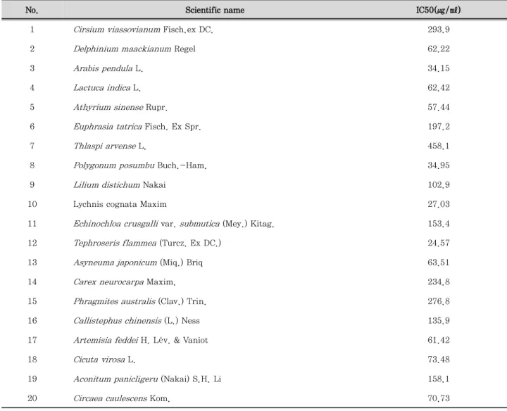
To select candidate of anti-inflammation agents, we investigated the effect of ethanol extract of Chinese plants on cell viability and LPS-induced NO production in RAW264.7 macrophages. As shown in Table 1, among the ethanol extract of 20 plants, 8 plants (
Delphinium maackianumRegel; DMR,
Arabis pendulaL.; APL,
Lactuca indicaL.; LIL,
Athyrium sinenseRupr.; ASR,
Polygonum posumbuBuch.-Ham.; PPBH,
Lychnis cognataMaxim; LCM,
Tephroseris flammea(Turcz. Ex DC.); TF, and
Artemisia feddeiH. Lév. &
Vaniot; AFH) had cytotoxicity at concentrations of less than 62.5 ㎍/㎖ as determined by the MTT assay.
Thus, there were only 12 non-cytotoxicity plants at concentrations of more than 62.5 ㎍/㎖, and we selected these 12 plants for NO screening. In table 2,
‘IC50’ is the concentration of the individual plants required to inhibit 50% of NO production and the
‘inhibitory ratio’ is measured to assess the inhibition effects in maximum concentration of individual plants.
The data showed that only 4 plants (
Cirsium viassovianumFisch.ex DC.; CVF,
Carex neurocarpaMaxim.; CNM,
Cicuta virosaL.; CVL, and
Circaea caulescensKom.; CCK) had the inhibitory ratio of more than 30% on NO production. In addition,
Cicuta virosaL. (CVL) provoked inhibitory effect of NO production about 70%. Based on these data, we selected CVL for the subsequent experiments.
2. Effect of CVL on the viability of RAW 264.7 macrophages
Cytotoxicity of CVL in RAW 264.7 cells was performed using MTT assay. As shown in Figure 1, RAW 264.7 cells were treated with different concentrations of CVL (15.63, 31.25, 62.5, 125, 250, or 500 ㎍/㎖) for 24 h.
The cell viability was not affected by CVL up to 62.5
㎍/㎖. Accordingly, we investigated the anti-inflammatory effects of CVL with concentration 12.5, 25, and 50 ㎍/
㎖ in LPS-stimulated RAW 264.7 macrophages.
No. Scientific name IC50(㎍/㎖)
1 Cirsium viassovianum Fisch.ex DC. 293.9
2 Delphinium maackianum Regel 62.22
3 Arabis pendula L. 34.15
4 Lactuca indica L. 62.42
5 Athyrium sinense Rupr. 57.44
6 Euphrasia tatrica Fisch. Ex Spr. 197.2
7 Thlaspi arvense L. 458.1
8 Polygonum posumbu Buch.-Ham. 34.95
9 Lilium distichum Nakai 102.9
10 Lychnis cognata Maxim 27.03
11 Echinochloa crusgalli var. submutica (Mey.) Kitag. 153.4
12 Tephroseris flammea (Turcz. Ex DC.) 24.57
13 Asyneuma japonicum (Miq.) Briq 63.51
14 Carex neurocarpa Maxim. 234.8
15 Phragmites australis (Clav.) Trin. 276.8
16 Callistephus chinensis (L.) Ness 135.9
17 Artemisia feddei H. Lév. & Vaniot 61.42
18 Cicuta virosa L. 73.48
19 Aconitum panicligeru (Nakai) S.H. Li 158.1
20 Circaea caulescens Kom. 70.73
Each value represents the mean ± SD (n=3).
Table 1. Effect of Ethanol extract of Chinese medicinal plants in Yanjin on the cell viability in RAW 264.7 macrophages.
No. Scientific name Inhibition ratio(%) IC50(㎍/㎖)
1 Cirsium viassovianum Fisch.ex DC. 39.40 ± 0.80 126.02
2 Euphrasia tatrica Fisch. Ex Spr. 12.77 ± 1.58 97.77
3 Thlaspi arvense L. 26.92 ± 0.26 193.05
4 Lilium distichum Nakai 17.31 ± 0.44 102.53
5 Echinochloa crusgalli var. submutica (Mey.) Kitag. 13.93 ± 0.71 171.43
6 Asyneuma japonicum (Miq.) Briq 7.76 ± 1.63 >500
7 Carex neurocarpa Maxim. 40.96 ± 0.97 63.26
8 Phragmites australis (Clav.) Trin. 22.66 ± 0.90 249.56
9 Callistephus chinensis (L.) Ness 19.17 ± 0.37 167.68
10 Cicuta virosa L. 71.63 ± 0.29 36.55
11 Aconitum panicligeru (Nakai) S.H. Li 7.63 ± 0.93 117.14
12 Circaea caulescens Kom. 38.75 ± 0.62 33.75
Each value represents the mean ± SD (n=3).
Table 2. Effect of Ethanol extract of Chinese medicinal plants in Yanjin on LPS-stimulated NO production level in RAW 264.7 macrophages.
Figure 1. Effect of CVL on the viability of RAW 264.7 cells. The cells were treated with different concentrations of CVL for 24 h, and their viability were determined using MTT assay. Values represent mean ± SD of three independent experiments. The values are represented as mean ± S.D. (n = 4). **P < 0.01, ***P < 0.001 compared to DMSO.
3. Effect of CVL on LPS-stimulated NO production in RAW 264.7 macrophages
To further estimate the inhibition of CVL on LPS- stimulated NO production in RAW 264.7 macrophages, the cells were pre-treated with or without CVL (1.5, 3.1, 6.2, 12.5, 25, or 50 ㎍/㎖) for 1 h before LPS stimulation (1 ㎍/㎖). The LPS-stimulated NO production was significantly decreased by CVL in a dose-dependent manner. In addition, high concentration of CVL (50 ㎍/
㎖) showed elevated inhibitory effect on NO production than NIL used as a positive control.
Figure 2. Effect of CVL on production of NO in LPS-stimulated RAW 264.7 cells. RAW 264.7 cells were pretreated indicated concentrations of CVL (1.5, 3.1, 6.2, 12.5, 25, or 50 ㎍/㎖) for 1 h, and followed by LPS stimulation (1 ㎍/㎖) for 48 h. The nitric oxide (NO) production was determined using Griess reagent.
The values are represented as mean ± SD (n = 6). ###P<0.001, compared to CON; *P<0.05 and ***P<0.001 compared to LPS.
4. Effect of CVL on LPS-stimulated PGE
2production in RAW 264.7 macrophages
Next, we examined the effect of CVL on LPS-stimulated PGE
2production in RAW 264.7 macrophages. The cells were treated with or without CVL (12.5, 25, or 50 ㎍/㎖) for 1 h and then treated with LPS (1 ㎍/㎖) for 24 h.
As shown in Figure 3, LPS-stimulated PGE
2production
was significantly diminished by CVL. Our data revealed that CVL exhibits suppressive activity on PGE
2production as well as NO production in LPS- stimulated RAW 264.7 macrophages.
Figure 3. Effect of CVL on production of PGE2 in LPS-stimulated RAW 264.7 cells. RAW 264.7 cells were pretreated indicated concentrations of CVL (12.5, 25, or 50 ㎍/㎖) for 1 h, and followed by LPS stimulation (1 ㎍/㎖) for 24 h. The prostaglandin-E2
(PGE2) levels in the culture media was measured by ELISA kit.
The values are represented as mean ± SD (n=6). ###P<0.001, compared to CON; ***P<0.001 compared to LPS.
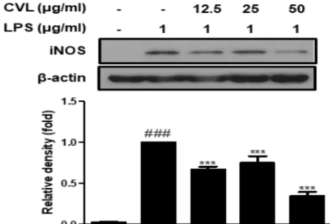
5. Effect of CVL on LPS-stimulated iNOS protein expression in RAW 264.7
macrophages
Previous reports suggested that LPS effectively activated iNOS transcription leading to the overproductions of NO in RAW 264.7 macrophages
8). We explored whether CVL could inhibit the iNOS protein expression using western blot analysis. The expression of iNOS protein was strongly increased after stimulation with LPS in RAW 264.7 macrophages. However, pre-treated cells with CVL were significantly abolished the expression of iNOS protein (Figure 4). We suggest that inhibition of iNOS expression may be related to the inactivation of NO production by CVL in LPS-stimulated RAW 264.7 cells.
Figure 4. Effect of CVL on iNOS protein expression in LPS-stimulated RAW 264.7 cells. RAW 264.7 cells were pretreated indicated concentrations of CVL (12.5, 25, or 50 ㎍/㎖) for 1 h, and followed by LPS stimulation (1 ㎍/㎖) for 24 h. The values are represented as mean ± SD (n = 6). ###P < 0.001, compared to CON; ***P <
0.001 compared to LPS.