Anti-inflammatory Effects of Ethanol Extract of Various Korean Compositae Herbs in LPS-induced RAW 264.7 Macrophages
Min-gyu Seo
1#, Yun-Mi Kang
1#, Kyung-Sook Chung
2, Se-Yun Cheon
1, Jong Hyuk Park
3, Young-Cheol Lee
4and Hyo-Jin An
1*1 : Department of Pharmacology, College of Korean Medicine, Sangji University, 83 Sangjidae-gil, Wonju-si, Gangwon-do 26339, Republic of Korea
2 : Catholic Precision Medicine Research Center, College of Medicine, The Catholic University of Korea, 222, Banpo-daero, Seocho-gu, Seoul, 06591, Republic of Korea
3 : Institute of Natural Cosmetic Industry for Namwon, Jeollabuk-do, 55801, Republic of Korea 4 : Department of Herbology, College of Korean Medicine, Sangji University, Wonju-si, Republic of Korea
ABSTRACT
Objective : This study was designed to evaluate candidate materials as anti-inflammation agent from extracts of various Korean Compositae herbs in Hwaak mountain. Among Korea medicinal herbs, Ainsliaea acerifolia (AA) belongs to the Compositae family, has been used for the treatment of rheumatic arthritis. However, AA has not been previously reported to have an anti-inflammatory effect. Therefore, we investigated the anti-inflammatory effects of AA and its underlying molecular mechanisms in lipopolysaccharide (LPS)-induced RAW 264.7 macrophages.
Methods : Cell viability was determined by 3-(4,5-dimethylthiazol-2-yl)-2,5-diphenyltetrazolium bromide (MTT) assay in RAW 264.7 macrophages. Nitric oxide (NO) was measured with Griess reagent and pro-inflammatory cytokines were detected by enzyme immunoassay (EIA) kits in LPS-stimulated RAW 264.7 macrophages. Protein expressions of inducible nitric oxide synthase, and cyclooxygenase-2 (COX-2) and p65 subunit of nuclear factor-κ B (NF-κ B) were determined by Western blot analysis.
Results : Among 8 extracts of Korean Compositae herbs tested, AA showed the inhibition of NO production without cytotoxicity. Consistent with the observation, AA reduced the expression levels of iNOS and COX-2 proteins in LPS- simulated RAW 264.7 macrophages in dose-dependent manner. In addition, AA inhibited the productions of TNF-α and IL-6 in LPS-simulated RAW 264.7 macrophages. However, AA did not inhibit activation of p65 NF-κ B in LPS- simulated RAW 264.7 macrophages.
Conclusion : These results suggest that down-regulation of iNOS, COX-2 protein expression and TNF-α and IL-6 production by AA are responsible for its anti-inflammatory effects.
1)
Key words : Ainsliaea acerifolia (AA), anti-inflammation, lipopolysaccharide (LPS), nitric oxide (NO), inducible nitric oxide synthase (iNOS), cyclooxygenase-2 (COX-2)
Ⅰ. INTRODUCTION
Inflammation is a defensive mechanism of a host to
various stimuli including cellular injury or infection with biological, chemical, or physical stimuli
1). Macrophages are key mediators of the early stages during the
*Corresponding author : Hyo-Jin An, Department of Pharmacology, College of Korean Medicine, Sangji University, 83 Sangjidae-gil, Wonju-si, Gangwon-do, 26339, Republic of Korea.
·Tel:+82-33-738-7503 ·Fax: +82-33-730-0679 ·E-mail : [email protected]
#First author : Min-gyu Seo, Department of Pharmacology, College of Korean Medicine, Sangji University, 83 Sangjidae-gil, Wonju-si, Gangwon-do, 26339, Republic of Korea.
·Tel:+82-33-738-7503 ·Fax: +82-33-730-0679 ·E-mail : [email protected]#Co-first author : Min-gyu Seo, Yun-Mi Kang, Department of Pharmacology, College of Korean Medicine, Sangji University, 83 Sangjidae-gil, Wonju-si, Gangwon-do, 26339, Republic of Korea.
·Tel:+82-33-738-7503 ·Fax: +82-33-730-0679 ·E-mail : [email protected] ·Received:25 January 2017 ·Revised:24 February 2017 ·Accepted:15 March 2017
inflammatory process and their activation by bacterial products such as lipopolysaccharide (LPS) promotes the synthesis and release of nitric oxide (NO), pro- inflammatory cytokines, eicosanoids and bioactive lipids, all of them intermediaries involved in the inflammatory onset
2).
LPS is the main outer membrane component of gram- negative bacteria and plays a key role during severe gram-negative infection, sepsis, and shock. The most prominent LPS-sensitive cell population are cells of the monocyte/macrophages lineage
3). RAW 264.7 was found to be uniquely efficient for measuring infection-related pro-inflammatory mediators. It is a good model to study inflammatory responses, which can be activated by LPS and trigger the production of inflammatory mediators, such as leukotrienes, tumor necrosis factor alpha (TNF- α ), interleukins (ILs), inducible nitric oxide synthase (iNOS), and cyclooxygenase-2 (COX-2)
4). These inflammatory mediators can be regulated by activation of the transcription factors
5).
Medicinal herbs have been used as therapeutics for centuries throughout the world. Phytochemicals derived from these medicinal plants are used to treat various neurological and immunological disorders
6). Korean medicinal herbs for alternative or complementary medicine are an important part of the culture and traditions of Korean populations. Most of the population in Korea have been reliant on herbal medicines for their health care needs. Many studies reported that Korean medicinal herbs possess various pharmacological effects, including immune-enhancing
7), anti-diabetic
8)and anti-oxidant activities
9). Among Korea medicinal herbs, Ainsliaea acerifolia (AA) Sch.Bip. belongs to the Compositae family, has been used for the treatment of rheumatic arthritis and enteritis in the oriental folk medicine
10). It has been reported that sesquiterpenoids which are characteristic constituents of the Ainsliaea genus possess diverse biological activities, including cytotoxic, antiviral, antibacterial, and anti-inflammatory activities
11). Nonetheless, AA has not been previously reported to have an anti-inflammatory effect. In the present study, as a part of our screening project to evaluate the anti- inflammatory potentials of various Korean Compositae herbs from Hwaak mountain, we investigated the anti- inflammatory effect of AA in RAW 264.7 macrophage cell line, which can be stimulated with LPS to mimic the condition of inflammation.
Ⅱ. MATERIALS AND METHODS
1. Chemicals and Reagents
Dulbecco’s modified Eagle’s medium (DMEM), fetal
bovine serum (FBS), penicillin, and streptomycin were purchased from Life Technologies Inc. (Grand Island, NY, USA). LPS ( Escherichia coli , serotype 055:B5), 3-(4,5-dimethylthiazol-2-yl)-2,5-diphenyltetrazolium bromide (MTT), N6-(1-Iminoethyl)lysine (NIL), NS-398, and Griess reagent were purchased from Sigma Chemical Co. (St. Louis, MO, USA). Dimethyl sulfoxide (DMSO) was purchased from Junsei Chemical Co., Ltd. (Tokyo, Japan). iNOS, COX-2 and β -actin monoclonal antibodies were purchased Santa Cruz Biotechnology (Santa Cruz, CA, USA). NF-κ B and PARP antibodies were purchased from Cell Signaling Technology (Danvers, MA, USA).
The enzyme immunoassay kits for TNF-α and IL-6 were obtained from R&D Systems (Minneapolis, MN, USA).
2. Preparation of Ethanol Extracts of Various Korean Compositae Herbs
Extracts of various Korean Compositae herbs in Hwaak mountain were obtained from Institute of Natural Cosmetic Industry for Namwon (Namwon, Jeollabuk-do, Republic of Korea). The dried and powdered 8 plant materials were extracted with 95% EtOH three times by maceration.
The extracts were evaporated in vacuo at 40℃ and were filtered, freeze-dried in vaccum . The freeze- dried samples were dissolved in DMSO with the final concentration of 50 ㎎/㎖ for bioassays.
3. Cell Culture
The RAW 264.7 macrophage cell line was obtained from Korea Cell Line Bank (KCLB, Seoul, Republic of Korea).
The cells were cultured in DMEM supplemented with 10%
FBS, penicillin (100 U/㎖), and streptomycin (100 ㎍/㎖) in a 37 ℃ and 5% CO
2incubator.
4. MTT Assay for Cell Viability
Cell viability was assessed using the MTT assay.
Briefly, AA-treated cells were incubated for 24 h, and then cells were incubated with MTT solution (5 ㎎/㎖) for 4 h at 37 ℃. After washing out the supernatant, the insoluble formazan product was dissolved in DMSO.
Cell viability was measured at 570 ㎚ using an automatic microplate reader (Titertek Multiskan, Huntsville, AL, USA).
5. NO Assay
NO content was determined indirectly by assaying
the culture supernatants for nitrite using the Griess
reagent (1% sulfanilamide in 5% phosphoric acid, 1% α-
naphthylamide in H
2O). NO production from RAW 264.7 macrophages was a form of NO
2that exists in culture media. A 50 ㎕ amount of cell culture media was mixed with 50 ㎕ of Griess reagent in a 96-well plate, incubated at room temperature for 15 min, and then measured at 540 ㎚ using an automatic microplate reader (Titertek Multiskan).
6. TNF-α and IL-6 Assays
Culture media were collected about 24 h after treatment with AA and then stored at −70℃. The levels of TNF- α and IL-6 were measured by enzyme immunoassay (EIA) kits according to the manufacturer’s instructions.
7. Preparation of Nuclear Protein Extraction
The cells were treated with AA for 1 h prior to the addition of LPS (1 ㎍/㎖) for 1 h and washed with cold PBS. Nuclear extracts of the cells were prepared as described previously
12).
8. Western Blot Analysis
The cells were resuspended in a commercial lysis buffer (PRO-PREP, Intron Biotechnology, Seoul, Republic of Korea) and incubated for 20 min at 4℃. Cell debris was removed by microcentrifugation, followed by quick freezing of the supernatants. The protein concentration was determined using the Bio-Rad protein assay reagent according to the manufacturer’s instructions (Bio-Rad, Hercules, CA, USA). Aliquots of each protein sample (30 ㎍) were separated on a sodium dodecyl sulfate (SDS) polyacrylamide gel and transferred onto a polyvinylidene fluoride (PVDF) membrane. Membranes were incubated for 1 h with 5% skim milk at room temperature, followed by incubation overnight with a 1:1000 dilution of primary antibody at 4℃. Blots were washed three times with Tween 20/Tris-buffered saline (T/TBS) and incubated with a 1:2500 dilution of horseradish peroxidase-conjugated secondary antibody for 2 h at room temperature. Blots were again washed three times with T/TBS and then developed by enhanced chemiluminescence (GE Healthcare, Waukesha, WI, USA).
9. Statistical Analysis
Data are expressed as the mean ± SD of triplicate experiments. Statistically significant values were compared using ANOVA and Dunnett’s post hoc test, and p -values less than 0.05 were considered statistically significant.
Ⅲ. RESULTS
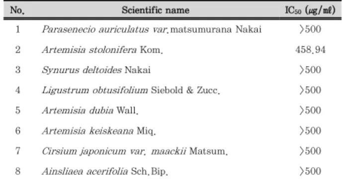
1. Anti-inflammatory Effects of Ethanol Extracts from Various Korean Compositae Herbs from Hwaak Mountain
To select candidate of anti-inflammation agents, we investigated the effect of various Korean Compositae herbs from Hwaak mountain on cell viability and LPS- induced NO production in RAW 264.7 macrophages. As shown in Table 1, most of the ethanol extract of herbs had no effect on the cell viability as determined by MTT assay at 500 ㎍/㎖ except A. stolonifera Kom.
These results suggested that ethanol extract of other 7 plants indicated nonspecific cytotoxicity in RAW 264.7 macrophages. In addition, as shown in Table 2, our data revealed that 5 plants ( S. deltoides Nakai;SD, L.
obtusifolium Siebold & Zucc.;LO, A. keiskeana Miq.;AK, C. japonicum var. maackii Matsum.;CJ, Ainsliaea acerifolia Sch.Bip.;AA) represent the IC
50value in less than 200
㎍/㎖ and AA showed the best inhibitory effect on NO production. Considering all of these, AA was selected in Korean Compositae herbs from Hwaak mountain, and we investigated the anti-inflammatory effect of AA underlying molecular mechanisms in RAW 264.7 macrophages.
No. Scientific name IC50 (㎍/㎖)
1 Parasenecio auriculatus var.matsumurana Nakai >500
2 Artemisia stolonifera Kom. 458.94
3 Synurus deltoides Nakai >500
4 Ligustrum obtusifolium Siebold & Zucc. >500
5 Artemisia dubia Wall. >500
6 Artemisia keiskeana Miq. >500
7 Cirsium japonicum var. maackii Matsum. >500 8 Ainsliaea acerifolia Sch.Bip. >500 Each value represents the mean ± SD (n = 3).
Table 1. Effect of Ethanol Extracts of Various Korea Compositae Herbs on the Cell Viability in RAW 264.7 Macrophages.
No. Scientific name Inhibition ratio(%) IC50
(㎍/㎖) 1 P. auriculatus var. matsumurana Nakai 17.59 ± 6.76** >500 2 A. stolonifera Kom. 18.62 ± 3.23** >500
3 S. deltoides Nakai 26.52 ± 4.18*** 196.62
4 L. obtusifolium Siebold & Zucc. 19.43 ± 11.06** 172.02
5 A. dubia Wall. 16.81 ± 3.35** >500
6 A. keiskeana Miq. 26.32 ± 3.80*** 137.31
7 C. japonicum var. maackii Matsum. 32.68 ± 0.99*** 195.42 8 A. acerifolia Sch.Bip. 38.93 ± 0.95*** 59.42 Inhibition ratio evaluated at dose of 31.25㎍/㎖. Each value represents the mean ± SD (n=3).**p < 0.01, ***p <0.001
Table 2. Effect of Ethanol Extracts of Various Korea Compositae Herbs on the LPS-induced NO Production Level in RAW 264.7 Macrophages.
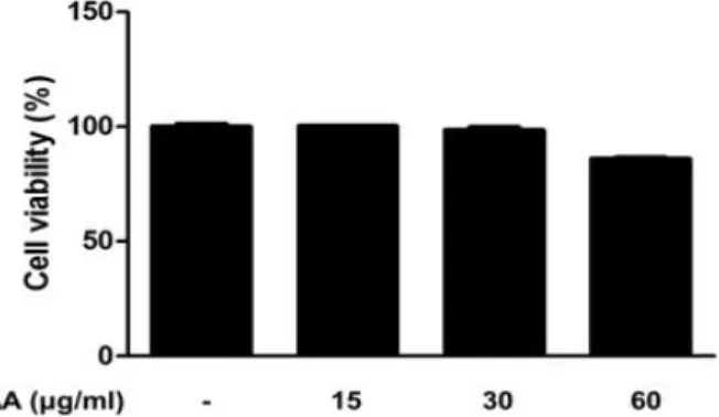
2. Effect of AA on Cell Viability in RAW 264.7 Macrophages
The cell viability of AA was examined by MTT assay in RAW 264.7 macrophages. As shown in Figure 1, treatment with AA (15, 30 and 60 ㎍/㎖) for 24h had nonspecific cytotoxicity in RAW 264.7 macrophages (cell viability at the highest concentration was 84.96%).
Accordingly, we investigated the anti-inflammatory effects of AA with concentration 15, 30 and 60 ㎍/㎖ in LPS-stimulated RAW 264.7 macrophages.
Figure 1. Effect of AA on the cell viability in RAW 264.7 macrophages.
RAW 264.7 macrophages were treated with different concentrations of AA for 24 h, and their viability was determined using MTT assay.
Values represent mean ± S.D. of three independent experiments.
3. Effect of AA on LPS-induced NO Production in RAW 264.7 Macrophages
To determine the inhibitory effects of AA on LPS- induced NO production in RAW 264.7 macrophages, cells were treated with or without AA (15, 30 and 60
㎍/㎖) for 1h and then treated with LPS (1 ㎍/㎖) for 24h. As shown in Figure 2, AA inhibited significantly the LPS-induced NO production in a concentration- dependent manner and its effect at the highest concentration is similar to NIL used as a positive control inhibitor.
Figure 2. Effect of AA on LPS-induced NO production in RAW 264.7 macrophages. Cells were treated with different concentrations of AA for 1 h prior to the addition of LPS (1 ㎍/㎖), and the cells were further incubated for 24 h. NO levels were determined with Griess reagent. NIL (20 μM) was used as a positive control inhibitor.
The data shown represent the mean ± SD of three independent experiments. ###p < 0.001 vs the control group; ***p < 0.001 vs the LPS-treated group.
4. Effects of AA on the LPS-induced iNOS and COX-2 Protein Expression in RAW 264.7 Macrophages
In macrophages exposed to LPS, the expression of iNOS and COX-2 is modulated, which ultimately results in overproduction of NO
13). Therefore, we investigated the effect of AA on the levels of iNOS and COX-2 protein expressions by Western blot analysis.
In unstimulated RAW 264.7 macrophages, the levels of iNOS and COX-2 protein expressions were a little.
However, the expression levels of iNOS and COX-2 proteins were significantly up-regulated in response to LPS (1 ㎍/㎖), while pretreatment with AA inhibited the expression levels of iNOS and COX-2 proteins in a dose-dependent manner.
Figure 3. Effects of AA on the LPS-induced iNOS and COX-2 protein expression in RAW 264.7 macrophages. Cells were treated with 15, 30, and 60 ㎍/㎖ AA for 1 h prior to the addition of LPS (1 ㎍/㎖), and the cells were further incubated for 1 h. The protein levels of iNOS and COX-2 were determined by Western blot analysis using specific antibodies. Densitometric analysis was performed using Bio-Rad Quantity One software. The data shown represent the mean ± SD of three independent experiments. #p <
0.05 vs the control group; ***p < 0.001 vs the LPS-treated group.
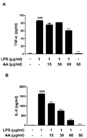
5. Effects of AA on the LPS-induced Production of Pro-inflammatory Cytokines in RAW 264.7 Macrophages
Cytokines are local protein mediators, known to be involved in biological processes, including cell growth and activation, inflammation, immunity, and differentiation.
Thus, they have a role in an autoimmune disease, in which
chronic inflammation
14). To examine the inhibitory effects
of AA on pro-inflammatory cytokine production induced
by LPS, we investigated its effects on LPS-induced
TNF-α and IL-6 productions by using EIA kits. As shown in Figure 4, LPS significantly increased TNF-α and IL-6 productions (P < 0.001), while pretreatment with AA reduced the LPS-induced these cytokines productions (P < 0.001).
Figure 4. Effects of AA on the LPS-induced production of pro- inflammatory cytokines in RAW 264.7 macrophages. Cells were treated with 15, 30, or 60 ㎍/㎖ AA for 1 h prior to the addition of LPS (1 ㎍/㎖), and the cells were further incubated for 24 h.
TNF-α and IL-6 levels were determined by using EIA kits. The data shown represent mean ± SD of three independent experiments.
###p < 0.001 vs the control group; ***p < 0.001 vs the LPS-treated group.
6. Effects of AA on the LPS-induced NF-κ B Activation and the Nuclear Translocation
Independent of the initial pro-inflammatory stimulus, cellular responses are related to gene modulation mediated by changes in the activation of transcription factors
15). Because NF-κ B is an important transcriptional factor in inflammatory response
16), we examined the effect of AA on the nuclear translocation of the p65 subunit of NF-κB in RAW 264.7 macrophages. As shown in Figure 5, LPS induced the translocation of p65 NF-κ B to the nucleus. However, pretreatment with AA could not suppress this process in LPS-simulated RAW 264.7 macrophages. These data suggested that the concerted action of multiple distinct transcription factors might be strongly associated with the expression
of inflammatory-related genes in AA-treated RAW 264.7 macrophages.
Figure 5. Effect of AA on LPS-induced NF-κB activation in RAW 264.7 macrophages. Cells were pretreated with AA for 1 h prior to the addition of LPS (1 ㎍/㎖) for 1 h. Nuclear (N) and cytosolic (C) extracts were isolated, and the levels of p65 in each fraction were determined by Western blot analysis. PARP and α-tubulin were used as internal controls.