Available at http://www.formulastudy.com
HFS
Original Article / 원저
RAW 264.7 대식세포에서 淸腦明神丸에 의한 염증성 및 산화적 스트레스 반응 억제 효능
손변우, 이명화, 황원덕* 동의대학교 한의과대학 내과학교실
Anti-inflammatory and Antioxidant Effects of Cheongnoimyungshin-hwan in RAW 264.7 Macrophages
Byun Woo Son, Myeong Hwa Lee, Won Deok Hwang*
Department of Korean Internal Medicine, College Of Korean Medicine, Dong-Eui University
ABSTRACT
Objectives : Cheongnoimyungshin-hwan (CNMSH) is a Herbal compound prescription that is composed mainly of herbal medicines such as Ginseng Radix Alba, Angelicae Gigantis Radix, Dioscoreae Rhizoma, Longan Arillus and cornus cervi parvum, and for the purpose of improving memory and preventing dementia.
Methods : In this study, it was investigated whether CNMSH could suppress inflammatory response and oxidative stress in the lipopolysaccharide (LPS)-stimulated RAW 264.7 macrophage cells. As a result, CNMSH decreased expression of inducible nitric oxide (NO) synthase and cyclooxygenase-2, and also inhibited production of NO, prostaglandin E2.
Results : This effect was associated with the suppression of the expression of p65, one of the nuclear factor-kappaB (NF-kB) subunits, and increased expression of IκB-α, inhibit the NF-κB transcription factor. In addition, CNMSH significantly blocked intracellular reactive oxygen species accumulation in response to LPS stimulation. Furthermore, CNMSH increased expression of nuclear factor erythroid 2-related factor (Nrf)-2 activation and heme oxygenase (HO)-1.
Conclusions : Therefore, it has been shown anti-inflammatory and antioxidant effects by inhibiting the expression and production of inflammatory mediators in LPS-stimulated macrophages, and is associated with ROS generation and is activated by Nrf2/HO-1 signaling pathway.
ⓒ 2018 The Korean Medicine Society For The Herbal Formula Study
This paper is available at http://www.formulastudy.com which permits unrestricted non-commercial use, distribution, and reproduction in any medium, provided
the original work is properly cited.
Key words : Cheongnoimyungshin-hwan (CNMSH), Anti-inflammatory and Antioxidant Effects, RAW 264.7 Macrophages
Ⅰ. 서론
1)한약재는 동물, 식물 또는 광물에서 채취된 것으로, 옛 선조들이 오랫동안 임상적인 경험을 토대로 시행착 오를 거쳐 약리활성이 있는 것만을 사용한 것이다1. 식 품과 한약재 내의 유효성분이 서로 공존하여 면역시스 템 중 보체계의 활성화, 여러 가지 cytokine의 활성화 등 질병에 대한 생체방어, 시스템 보강 등의 시너지 효 과를 나타내며, 환경호르몬의 영향을 상쇄시키는 등 인 체 항상성에도 영향을 미치는 것으로 알려져 있다2. 최 근 이러한 한약재의 유효성분 동정 및 특성에 대해 연 구하여 신규물질 개발 원료로써 가치가 높아지고 있다1. 청뇌명신환(淸腦明神丸)은 동의대학교 부속 한방병원 6내과에서 기억력 증진 및 치매 예방을 목적으로 개발 된 환약으로 침향(沈香), 원지(遠志)와 인삼(人蔘), 당 귀(當歸), 산약(山藥), 녹용(鹿茸) 등의 한약재가 주재 료로서, 원대(元代) 위역림(危亦林)이 황제께 진상한 공 진단(拱辰丹)을 근간한 처방이다3. 현재까지 청뇌명신 환에 대한 연구로는, 인체 신경세포인 SH-SY5Y 세포에 서 H2O2로 유도된 산화적 스트레스에 대해 Nrf2/HO-1 신호전달경로를 경유하여 ROS 생성과 이와 연계된 미 토콘드리아의 기능 손상을 억제하여 세포를 보호하는 효과4와 미세아교세포인 BV2 cell에서 inflammatory mediator인 NO, PGE2와 IL-1β의 생성을 저해하여 항염증 효과5를 나타낸다는 연구결과가 있다. 또한
in vivo
model에서 흰쥐에 양측 총경동맥을 영구 폐쇄하 여 만성뇌혈류저하를 유발하였을 때, Morris water maze와 면역조직화학염색 등의 결과를 토대로 해마 신 경세포의 자연사와 β-amyloid 침착을 억제하여 학습 과 기억 장애를 개선한다6는 연구와 ICR 마우스를 이용 하여 급성 독성 실험을 진행한 결과 체중, 장기, 생화학 적 지표 등에 유의한 변화가 관찰되지 않아 독성을 나 타내지 않는다7는 연구 결과가 있다. 청뇌명신환에 포 함되는 각각의 재료에 대한 염증효능은 이미 많이 알려 져 있지만8,9,10,11, 이것을 환으로 만든 청뇌명신환에 대한 연구는 아직 미비한 실정이다.
한편, 대식세포(macrophage)는 염증반응이 일어나 게 되면 tumor necrosis factor (TNF)-α, interleukin (IL) 등의 분비를 통해 외부물질에 대해 인지하여 생체 방어에 중요한 역할을 한다12,13. 그람음성세균(Gram negative bacteria)의 세포벽 구성성분인 lipopolysaccharide (LPS)는 국소 및 전신의 염증반응을 유발하며, 대식세 포(macrophages)에서 toll-like receptor (TLR) 4 수 용체를 자극하여 염증매개인자(pro-inflammatory mediators)인 nitric oxide (NO)와 prostaglandin (PG) E2, 그리고 pro-inflammatory cytokine인 TNF-α, IL-1β 등을 생성시키므로 염증 실험 모델에서 주로 이 용되고 있다14,15,16,17. 또한 RAW 264.7 세포는 LPS 자 극에 의해 inducible nitric oxide synthase (iNOS) 및 cyclooxygenase (COX)-2의 발현을 유도하여 NO와 PGE2 등을 생성하므로 염증반응의 대표적인 세포 실험 모델로 이용되고 있다18,19.
따라서 본 연구에서는 마우스 유래 대식세포주인 RAW 264.7 세포에서 pro-inflammatory mediator의 발현을 확인하였고, 더 나아가서 염증과 관련된 신호전 달경로인 nuclear factor-kappaB (NF-kB) 신호계와 강력한 항산화 활성 신호계로 잘 알려진 nuclear factor erythroid 2-related factor (Nrf)-2 activation/heme oxygenase (HO)-1 신호계에 미치는 청뇌명신환의 영 향을 추가로 조사하였다.
Ⅱ. 재료 및 방법
1. 시료준비
본 연구에서는 청뇌명신환(淸腦明神丸, Cheongnoimyung shin-hwan, CNMSH)을 이용하여 항염증 실험을 진행 하였다. 청뇌명신환은 인삼, 당귀, 산약, 용안육, 육계, 원지, 사향, 침향과 용뇌(중국)를 세말하고, 여기에 녹 용과 부자를 5시간 정도 끊인 교(膠)와 꿀을 첨가한 후, 이를 4 g씩 오자대 크기로 75 개 환을 만들어 금박으로
* Corresponding author : Won Deok Hwang, Department of Korean Internal Medicine, College Of Korean Medicine, Dong-Eui University, 52-57, Yangjeong-ro, Busanjin-gu, Busan, 47227, Republic of Korea.
Tel: +82-51-850-8625, Fax: +82-51-853-4063, E-mail: [email protected]
∙Received : January 12, 2018 / Revised : January 23, 2018 / Accepted : January 25, 2018
입힌 것으로 각 구성성분의 중량은 Table 1.에서 나타 내었다. 이 중에서 무작위로 1개의 환을 선정하여, 이 를 멸균된 3차 증류수를 이용하여 100 mg/ml의 농도 로 만든 다음 0.2 μm 필터로 여과하여 실험에 이용하 였다.
2. 세포 배양
실험에 사용된 마우스 유래 대식세포주인 RAW 264.7 세포는 한국세포주은행(Korea Cell Line Bank, Seoul, Korea)에서 분양받아 사용하였으며, 10% fetal bovine serum (FBS, WELGENE Inc., Daegu, Korea)과 L-glutamine (2 mM), penicillin (100 U/ml) 및 streptomycin (100 U/ml, WelGENE Inc.)이 첨가된 Dulbecco's Modified Eagle's Medium (DMEM, WELGENE Inc.)을 사용하여 37℃, 5% CO2 incubator 에서 배양하였다.
3. 세포독성 평가
RAW 264.7 대식세포에서 세포 증식에 미치는 청뇌명신 환의 영향을 조사하기 위하여 3-(4,5-dimethylthiazol-2-yl)-2, 5-diphenyltetrazolium bromide (MTT) assay를 실시하였다. 먼저 6 well plate에 배양된 RAW 264.7 세포에 농도별 청뇌명신환을 1시간 전처리한 후 LPS (100 ng/ml,
Escherichia coli
055:B5, Sigma-Aldrich Chemical Co., St. Louis, MO, USA)를 처리하여 24 시간 배양하였다. 배지를 제거하고, 0.5 mg/ml 농도의 MTT (Sigma-Aldrich Chemical Co.) 시약을 분주하 여 37℃, 5% CO2 배양기에서 2시간 반응시켰다. 배양 이 끝난 다음 배지를 제거하고, dimethyl sulfoxide (DMSO, Sigma-Aldrich Chemical Co.))로 생성된 formazan을 모두 녹인 후 96 well plate에 200 μl씩 옮겨서 enzyme-linked immunosorbent assay (ELISA) reader (Molecular Devices, Sunnyvale, CA, USA)로 540 nm에서 흡광도 변화를 측정하였다. 각 세포에 대 한 세포 독성은 대조군 값을 기준으로 백분율로 계산하 여 나타내었다. 또한 RAW 264.7 세포의 형태학적인 변형은 위상차 현미경(Carl Zeiss, Oberkochen, Germany)을 이용하여 관찰하였다.4. NO 및 PGE2 생산량 측정
RAW 264.7 세포에서 염증성 지표인 NO 및 PGE2 함 량을 측정하기 위하여 청뇌명신환을 농도별로 1시간 처
리하였고, LPS (100 ng/ml)를 처리하여 24시간 배양 한 후 세포 배양액을 수집하였다. LPS 자극에 의한 NO 생성에 미치는 청뇌명신환의 영향을 조사하기 위해서 세포 배양액과 동량의 Griess reagent (Sigma-Aldrich Chemical Co.)를 반응시켰으며, 540 nm에서 ELISA reader를 이용하여 흡광도 값을 측정한 후, sodium nitrite (NaNO2) 표준곡선을 이용하여 나타내었다.
LPS 자극에 의한 PGE2 생성에 미치는 청뇌명신환의 영향은 commercial ELISA kit (Cayman Chemical Co., Ann Arbor, Michigan, USA)를 이용하여 측정하 였으며, 제조사의 protocol에 따라 실험을 진행하였다.
5. ROS 함량 측정
RAW 264.7 세포에서 청뇌명신환이 LPS 자극에 의한 ROS 생성에 미치는 영향을 조사하기 위해 2’,7’-dichlorodihydrofluorescein diacetate (DCF-DA) 염색 법을 이용하였다. 이를 위하여 RAW 264.7 세포에 청 뇌명신환을 1시간 처리한 후 LPS (100 ng/ml)를 처리 하여 6시간 동안 배양하였다. 그후 10 μM의 DCF-DA 용액(Molecular Probes Inc., Leiden, Netherlands) 을 처리하여 37℃, 5% CO2 배양기에서 20분 염색하였 으며, flow cytometer (Becton-Dickinson, San Jose, CA, USA)를 이용하여 ROS 생성 변화 여부를 측정하였 다. 또한 동일한 조건에서 mounting medium을 이용 하여 chamber slide에 봉입한 후 형광현미경(Carl Zeiss, Oberkochen, Germany)을 이용하여 ROS 생성 변화 여부를 관찰하였다. LPS에 의한 ROS 과다 생성 이 산화적 스트레스와 연관성이 있는지 확인하기 위하 여 청뇌명신환과 동일한 조건에서 N-acetylcysteine (NAC, Sigma-Aldrich Chemical Co.)을 처리하여 그 효과를 비교하였다.
6. 단백질 분리, 전기영동 및 Western blot analysis RAW 264.7 세포에서 청뇌명신환이 항염증 및 산화 적인 스트레스 억제 관련 유전자들의 발현에 미치는 영 향을 조사하기 위하여 실험조건에 따라 배양된 세포들 의 단백질을 수집하였다. 각 세포들의 단백질을 분리하 기 위하여 lysis buffer [25 mM Tris-Cl (pH 7.5), 250 mM NaCl, 5 mM EDTA, 1% Nonidet-P40 (NP-40), 1 mM phenymethylsulfonyl fluoride (PMSF), 5 mM dithiothreitol (DTT)]를 첨가하여 4℃
에서 1시간 이상 반응시킨 후, 14,000 rpm으로 30분간
원심 분리하여 상층액을 수거하였다. 또한 NE-PER nuclear and cytosolic extraction reagents (Pierce Biotechnology, Rockford, IL, USA)를 사용하여 제조 사의 protocol에 따라 핵과 세포질에서 단백질을 분리 하였다. Bio-Rad 단백질 정량시약(Bio-Rad, Hercules, CA, USA)을 이용하여 그 사용방법에 따라 정량하여 단 백질 농도를 결정하였고, 동량의 Laemilni sample buffer (Bio-Rad)와 혼합하여 sodium dodecyl sulphate (SDS)-polyacrylamide gel을 이용하여 전기영동을 실 시하였다. 이를 다시 polyvinylidene difluoride membrane (Schleicher and Schuell, Keene, NH, USA)에 전이 시키고, 5% skim milk를 1시간 처리하여 비 특이적인 단백질들에 대한 blocking을 실시하였다. 적정 1차 항 체를 4℃에서 over night 반응시킨 다음 PBS-T (PBS with Tween 20)로 세척하였으며, 1차 항체에 맞는 2차 항체를 처리하여 상온에서 1시간 반응시켰다. 반응이 끝난 후 enhanced chemiluminoesence (ECL) solution (Thermo scientific, Meridian Rd., Rockford, USA) 을 적용시킨 다음 X-ray film에 감광시켜 특정 단백질 의 발현 변화를 분석하였다. 실험에 사용된 1차 항체는 iNOS, COX-2, NF-κB p65, IκBα, Nrf2, HO-1, Kelch Like ECH Associated Protein 1 (Keap1), actin과 lamin B이며, Santa Cruz Biotechnology, Inc. (Santa Cruz, CA, USA) 및 Cell Signalling Technology Inc. (Danvers, MA, USA)에서 구입하였 다. 2차 항체로 사용된 horseradish peroxidase (HRP)-conjugated anti-mouse, anti-rabbit 및 anti-goat 항체는 Santa Cruz Biotechnology, Inc.에 서 구입하여 사용하였다.
7. Reverse transcription-polymerase chain reaction (RT-PCR)
RAW 264.7 세포에서 청뇌명신환이 mRNA 수준에서 항염증 관련 유전자들의 발현에 미치는 영향을 조사하 기 위하여 RT-PCR을 수행하였다. 이를 위하여 청뇌명 신환을 농도별로 1시간 처리 한 후, LPS (100 ng/ml) 를 처리하여 24시간동안 배양하였고, TRIzol reagent (Invitrogen Life Technologies, Carlsbad, CA, USA) 를 이용하여 total RNA를 추출하였다. iNOS primer (forward, 5‘-CGT GTT TAC CAT GAG GCT GA-3' and reverse, 5'-GCT TCA GGT TCC TGA TCC AA-3'), COX-2 primer (forward, 5'-CAA AGG CCT
CCA TTG ACC AGA-3' and reverse, 5'-TGG ACG AGG TTT TTC CAC CAG-3')와 One-step RT-PCR PreMix kit (iNtRON Biotechnology, Gyeonggi, Korea)를 이용하여 PCR을 수행하였으며, 증폭된 DNA 산물은 ethidium bromide (EtBR, Sigma-Aldrich Chemical Co.)를 사용하여 1.5% agarose gel (Affymetrix, Inc., Cleveland, Phio, USA)에 전기 영 동하여 UV 하에서 관찰하였다. 아울러 loading control 로 Glyceraldehyde-3-phosphate dehydrogenase (GAPDH, forward, 5'-GTC ATC ATC TCC GCC CCT TCT GC-3' and reverse, 5'-GAT GCC TGC TTC ACC ACC TTC TTG-3')를 사용하였다.
8. 통계학적인 분석
모든 실험 결과는 Statistical Package for the Social Sciences (SPSS) 통계 프로그램을 이용하여 평균
± 표준편차 (mean ± SD)로 나타내었으며, p<0.05 수준에서 Student’s t-test와 Duncan’s multiple range test를 이용하여 각 결과에 대한 유의성을 검증 하였다.
Ⅲ. 결과
1. RAW 264.7 세포의 증식에 미치는 청뇌명신환의 영향 모든 농도에서 세포독성을 나타내지 않았으며, 본 실 험에서는 2 mg/ml 농도까지 실험 조건으로 선정하여 추후 실험을 진행하였다(Fig. 1A). 또한 광학현미경을 이용해 청뇌명신환 처리에 따른 형태적 변화에 대해 확 인해 본 결과, 청뇌명신환만 처리하였을 때 독성이 없 는 조건에서 큰 변화를 확인하기가 어려웠지만, LPS를 처리하였을 때 증가하였던 대식세포의 활성이 농도 의 존적으로 감소되는 결과를 확인하였다(Fig. 1B).
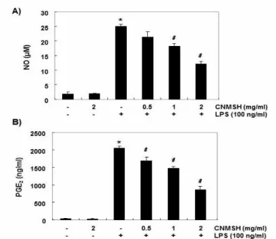
2. LPS 자극에 따른 염증성 매개인자 생성에 미치는 청 뇌명신환의 영향
LPS 자극을 주었을 때 NO와 PGE2의 생성이 대조군 에 비하여 매우 증가되었고, 청뇌명신환을 동시 처리 조건에서는 농도 증가에 따라 유의적으로 감소되었다 (Fig. 2A 및 B). 또한, NO와 PGE2의 생성에 관여하는 iNOS와 COX-2의 단백질 및 mRNA 발현에 미치는 청 뇌명신환의 영향을 Western blotting과 RT-PCR을 통 해 조사한 결과, LPS 자극에 의하여 증가된 두 유전자
의 발현이 추출물 전처리된 조건에서 농도 의존적으로 감소되는 것을 확인할 수 있었다(Fig. 3A 및 B). 따라 서 청뇌명신환은 iNOS와 COX-2의 발현을 차단함으로 서 NO와 PGE2의 생성을 억제함을 알 수 있었다.
3. NF-κB 신호계의 활성에 미치는 청뇌명신환의 영향 Fig. 4에 나타난 바와 같이 LPS 처리에 의해 핵에서 의 NF-κB p65의 발현이 증가하였고 세포질 κB-α 의 발현이 억제되는 것을 확인 할 수 있었으며, 청뇌명 신환을 동시 처리한 조건에서 회복되는 것을 관찰 할 수 있었다. 따라서 청뇌명신환이 나타내는 염증성 매개 인자 생성 억제가 NF-κB 활성 억제를 통하여 일어남 을 알 수 있었다.
4. LPS 자극에 따른 ROS 생성에 미치는 청뇌명신환의 영향
LPS만 처리했을 때 ROS 생성이 확연히 증가하였으 며 추출물을 전처리하였을 때 ROS scavenger인 NAC 전처리 군과 유사하게 ROS 생성이 억제되는 것을 flow cytometer와 형광현미경을 이용하여 확인하였다(Fig. 5).
따라서 청뇌명신환이 항염증 효능 뿐 만 아니라 산화적 인 스트레스를 억제하는 능력도 있음을 알 수 있었다.
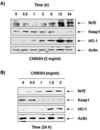
5. Nrf2/HO-1 신호전달 경로에 미치는 청뇌명신환의 영향
Fig. 6에 나타낸 바와 같이, 청뇌명신환의 처리 농도 및 시간 증가에 따라 Nrf2 QNs만 아니라 HO-1의 단백 질 발현이 증가하였으며, Nrf2의 억제 인자인 Keap1의 발현은 감소되었다. 이러한 결과는 청뇌명신환의 산화 적인 스트레스 억제효능이 Nrf2/HO-1 신호계 활성화 와 연관성이 있음을 의미하는 것이다.
Ⅳ. 고찰
본 연구에서는 LPS 자극에 의해 증가된 RAW 264.7 대식세포 모델에서 염증성 매개인자들의 생성 및 그들 조절 유전자들의 발현 변화를 측정하여 청뇌명신환의 항염증 효능을 조사하였다. 그람음성 세균의 세포벽 구 성성분인 LPS는 대식세포에서 TLR4 수용체를 자극하 여 염증성 매개인자와 염증성 사이토카인들의 생성시 키는 것으로 알려져 있다14,15,16,17. LPS 자극에 의해 증 가된 염증성 매개인자와 사이토카인의 분비는 대식세
포에서 외부물질에 대해 인지하여 염증반응 등을 나타 내며, 이를 통해 생체 방어에 중요한 역할을 한다12,13. 따라서 염증성 매개인자의 발현을 조절하는 것만으로 도 염증을 억제할 수 있는 핵심 방안으로 이용될 수 있 다. 본 실험에서는 iNOS와 COX-2의 분비에 의해 유도 되는 NO와 PGE2의 생성18,19에 미치는 청뇌명신환의 영 향을 조사한 결과, 청뇌명신환이 LPS 자극에 의해 증 가하였던 NO와 PGE2 생성량을 농도 의존적으로 감소 시켰다. 또한 청뇌명신환은 동일 조건에서 iNOS와 COX2의 발현을 감소시켰으며, 이는 청뇌명신환이 LPS 에 의한 iNOS와 COX-2의 발현 증가를 차단함으로서 NO와 PGE2의 생성을 억제하였음을 의미한다.
iNOS 및 COX-2와 같은 유전자들의 전사활성을 조 절하는 핵심 전사조절 인자로 가장 대표적인 단백질이 NF-κB이다20. 대식세포의 경우 LPS와 같은 병원체가 세포외막에 존재하는 toll-like receptor (TLR)에 결합 되는 신호에 의하여 IκB 인산화효소 복합체가 NF-κ B 이합체에 결합하여 인산화와 유비퀴틴 작용을 통해 NF-κB 이합체를 IκB로부터 분리시킨다. 분리된 NF-κB는 핵막을 통해 핵으로 들어가서 다양한 염증 성 인자들의 전사활성을 촉진시킴으로서 염증 반응을 일으킨다21.
따라서 청뇌명신환이 RAW 264.7 세포에서 LPS 자 극에 의한 NF-κB 신호계의 활성을 차단할 수 있는지 의 여부를 조사한 결과, LPS가 처리된 배지에서 배양 된 RAW 264.7 세포의 핵에서 NF-κB subunit인 p65 의 발현이 증가한 반면, 세포질에서는 NF-κB의 발현 감소와 함께 IκB-α의 발현 또한 억제되었다. 그러나 청뇌명신환이 처리된 조건에서 LPS의 자극을 받은 RAW 264.7 세포에서는 핵으로의 NF-κB 전위뿐 만 아니라 IκB-α의 발현 감소도 억제되었다. 이는 청뇌 명신환이 IκB의 분해를 차단함으로서 NF-κB의 핵 내 이동을 억제하였음을 의미한다. 따라서 청뇌명신환 이 나타내는 항염증 효능인 NO와 PGE2의 생성 억제는 NF-κB의 활성 차단에 의한 iNOS와 COX-2의 발현이 억제되었기 때문에 나타나는 현상이라고 할 수 있다.
한편 항산화 방어 시스템과 활성산소종(ROS)의 불균 형으로 증가된 산화적인 스트레스는 생체막의 지질과 산화, 단백질 변성 및 DNA 손상을 유도하여 세포와 조 직의 정상적인 기능을 억제하고, 만성염증을 유도하여 체내에서 비가역적인 병변을 유도한다22,23. 또한 산화 적인 스트레스에 동반된 ROS 생성은 호중구와 대식세
포를 염증부위로 이주시켜 염증 유발인자들의 생성을 추가적으로 촉진시킨다24,25. 따라서 LPS가 자극된 RAW 264.7 세포에서 청뇌명신환의 산화적 스트레스를 억제할 수 있는지의 여부를 ROD 생성 억제 효능으로 조사하였다. 본 연구의 결과에 의하면 LPS가 처리된 RAW 264.7 세포에서 ROS의 생성이 매우 증가하였음 을 flow cytometry 및 형광현미경학적 분석을 통하여 확인하였으며, 이러한 산화적 스트레스의 유발이 청뇌 명신환이 존재하는 조건에서 배양된 RAW 264.7 세포 에서는 유의적으로 감소되었다. 이러한 현상은 양성 대 조군으로 사용된 NAC 처리군에서도 유사하게 관찰되 어, 청뇌명신환은 염증성 반응에 동반되는 산화적인 스 트레스도 억제할 수 있음을 알 수 있었다.
Nrf2의 전사활성에 의하여 조절되는 HO-1은 heme 으로부터 CO, biliverdin 그리고 free iron으로 분해되 는 반응에서 염증성 인자들의 생성을 효과적으로 차단 하는 표적인 항산화 효소이다. 또한 heme의 분해산물 중 CO와 biliverdin은 iNOS와 COX-2의 발현을 감소 시켜 NO와 PGE2의 생성을 억제할 수도 있다26,27,28. 따 라서 이러한 항염증 및 항산화 효능을 동시에 가질 수 있는 HO-1의 발현을 조절하는 전사인자인 Nrf2는 세 포의 염증성 및 산화적 자극에 대한 항상성 유지에 중 요한 역할을 담당한다7,29. 일반적으로 Nrf2는 세포질에 서 억제인자인 Keap1에 결합되어 있지만, 스트레스 등 의 자극에 노출하게 되면 Keap1에서 떨어져 핵 내로 이 동하여 세포를 보호하기 위하여 항산화 유전자들의 전 사활성을 촉진시킨다30,31. 이에 근거하여 RAW 264.7 대식세포 모델에서 청뇌명신환이 Nrf2/HO-1 신호계 의 활성을 향상시킬 수 있는지의 여부를 조사한 결과, 청뇌명신환의 처리 시간 및 농도 의존적으로 Nrf2와 HO-1의 발현이 증가하였으며, Keap1의 발현 억제가 동반되었다. 이러한 결과는 청뇌명신환의 항산화 활성 이 최소한 Nrf2/HO-1 신호계의 활성을 통하여 이루어 지고 있음을 보여주는 결과이다.
결론적으로, 청뇌명신환은 염증성 매개인자의 생성 및 발현과 ROS 생성을 억제함으로써 항염증 효능 및 산화적인 스트레스 억제 효능을 나타냈으며, 이 과정에 서 NF-κB와 Nrf2/HO-1 신호전달 경로가 관여하고 있음을 알 수 있었다. 비록 두 신호전달경로에 대한 추 가적인 규명과 동물 실험을 통한 항염증 및 힝산화 효 능에 대한 연구가 더 필요하지만, 청뇌명신환이 기억력 증진 및 치매예방 뿐만 아니라 항염증 및 항산화 효능
소재로서의 개발 가능성이 높음을 보여주었다.
Ⅴ. 결론
청뇌명신환(淸腦明神丸)은 인삼(人蔘), 당귀(當歸), 산약(山藥), 용안육(龍眼肉)과 녹용(鹿茸) 등의 한약재 를 주재료로 하여 기억력 증진 및 치매 예방을 목적으 로 만들어진 환이다. 본 실험에서는 LPS 자극에 의해 유도된 RAW 264.7 대식세포 모델에서 청뇌명신환이 염증반응과 산화적인 스트레스를 억제할 수 있는지의 여부를 조사하였다. 본 연구의 결과에 의하면, 청뇌명 신환은 LPS 자극에 의해 증가된 iNOS 및 COX-2의 발 현을 농도 의존적으로 감소시켰고, 이들로 유도된 염증 매개인자인 NO와 PGE2의 생성 또한 유의적으로 억제 하였다. 이러한 효과는 청뇌명신환의 NF-κB subunit 중 하나인 p65의 발현 억제 및 NF-κB 전사요소 억제 제인 IκB-α 발현 증가와 연관이 있었다. 또한 청뇌명 신환은 LPS 자극에 대한 RAW 264.7 세포 내 ROS 축 적을 차단시켰으며, 이는 Nrf2/HO-1 신호전달경로의 활성화와 연관성이 있었다. 따라서 청뇌명신환은 LPS 자극으로 인해 증가된 대식세포에서 염증매개인자의 발현 및 생성을 억제하여 항염증 효능을 나타냈으며, ROS 생성과 연관하여 Nrf2/HO-1 신호전달 경로의 활성 화를 통해 산화적인 반응을 억제하였음을 알 수 있었다.
감사의 글
이 논문은 2017년도 동의대학교 교내연구비지원에 의해 연구되었슴(연구과제번호:201702600001)
References
1. Lee SH, Jung BH, Kim SY, Chung BC.
Determination of phytoestrogens in traditional medicinal herbs using gas chromatography–
mass spectrometry. J Nutr Biochem. 2004; 15:
452-460.
2. Wasser SP, Weis AL. Therapeutic effects of substances occurring in higher basidiomycetes mushrooms: a modern perspective. Crit Rev Immunol. 1999; 19: 65-96.
3. Heo J. The Dongui Bogam. Yeo-Gang Publisher.
2001:1750
4. Pi GH, and Hwang WD. Neuroprotective effects of Cheongnoemyeongsin-hwan against hydrogen peroxide-induced DNA damage and apoptosis in human neuronal-derived SH-SY5Y cells.
Herbal Formula Science. 2017; 25(1): 51-68.
5. Im YG, Choi YH, Hwang WD. Anti-inflammatory Effects of Cheongnoimyungshin-hwan in Microglia Cells. The Korean Society of Oriental Neuropsychiatry. 2014; 25(4): 423-433.
6. Chang SH, Hwang WD. Ameliorating Effects of Cheongnoemyeongsin-hwan on Learning and Memory Impairment Induced by Cerebral Hypoperfusion in Rats The Korean Medicine Society for The Herbal Formula Study 2017;
25(1): 69-87.
7. Lee DH, Park JS, Lee YS, Sung SH, Lee YH, and Bae SH. The hypertension drug, verapamil, activates Nrf2 by promoting p62-dependent autophagic Keap1 degradation and prevents acetaminophen-induced cytotoxicity. BMB Rep. 2017; 50: 91-96.
8. Kong YH, Lee YC, Choi SY. Neuroprotective and Anti-inflammatory effects of phenolic compounds in Panax ginseng C.A. Meyer. J Ginseng Res. 2009; 33: 111-114.
9. Jang SI, Kim HJ, Hwang KM, Pae HO, Yun YG, Chung HT, Kim YC. Anti-Inflammatory Effect of Ethanol Extract of Angelica uchiyamana in Activated Murine RAW 264.7 macrophages.
Korean J Oriental Medical Prescription. 2002;
10: 189-197.
10. Choi GY, Kim BW. Experimental study on the antioxidant and antimicrobial properties of Dioscoreae Rhizoma. Korean J Orient Int Med, 2010; 31: 290-297.
11. Kim MR, Lim EM. Effects of Longanae arillus water extract on inflammatory response and cytokines in mouse macrophage cells. J Korean Obstet Gynecol. 2014; 27: 1-11.
12. Willoughby DA, Heberden O. Human arthritis applied to animal models. Towards a better
therapy. Ann Rheum Dis. 1975; 34: 471-478.
13. Sirisinha S. Insight into the mechanisms regulating immune homeostasis in health and disease. Asian Pac J Allergy Immunol. 2001;
29: 1-14.
14. Kook YB. Effect of Hwangryunhaedok-tang on blood pressure and renal functions in spontaneously hypertensive rats. The Korean Journal of Oriental Medical Prescription. 2002;
10: 113-129.
15. Mann JR, Backlund MG, DuBois RN. Mechanism of disease: Inflammatory mediators and cancer prevention. Nat Clin Pract Oncol. 2005; 2:
202-210.
16. Cho BO, Ryu HW, So Y, Lee CW, Jin CH, Yook HS, et al. Anti-inflammatory effect of mangostenone F in lipopolysaccharide-stimulated RAW 264.7 macrophages by suppressing NF- κB and MAPK activation. Biomol Ther. 2014;
22: 288-294.
17. Higuchi M, Hisgahi N, Taki H, Osawa T.
Cytolytic mechanisms of activated macrophages.
Tumor necrosis factor and L-arginine-dependent mechanisms act synergistically as the major cytolytic mechanisms of activated macrophages. J Immunol. 1990; 144: 1425-1431.
18. Jin KS, Park JY, Cho MK, Jang JH, Jeong JH., Ok S, Bak MJ, Song YS, Kim MJ, Cho CW, Jeong WS. Modulation of Nrf2/ARE and inflammatory signaling pathways by Hericium erinaceus mycelia extract. Food Sci Biotechnol.
2009; 18: 1204-1211.
19. Park CM, Jing KS, Lee YW, Song YS. Luteolin and chicoric acid synergistically inhibited inflammatory responses via inactivation of PI3K-Akt pathway and impairment of NF-κB translocation in LPS stimulated RAW 264.7 cells. Eur J Pharmacol. 2011; 660: 454-459.
20. Karin M, Ben-Neriah Y. Phosphorylation meets ubiquitination: The control of NF-κB activity. Annu Rev Immunol. 2000; 18: 621-663.
21. Elewaut D, DiDonato JA, Kim JM, Truong F,
Eckmann L, Kagnoff MF. NF-κB is a central regulator of the intestinal epithelial cell innate immune response induced by infection with enteroinvasive bacteria. J Immunol.
1999; 163: 1457-1466.
22. Halliwell B. Antioxidants in human health and disease. Annu Rev Nutr. 1996; 16: 33-50.
23. McCord JM, Wong K, Stokes SH, Petrone WF, English D. Superoxide and inflammation: a mechanism for the anti-inflammatory activity of superoxide dismutase. Acta Physiol Scand Suppl. 1980; 492: 25-30.
24. Mittal M, Siddiqui MR, Tran K, Reddy SP, Malik AB. Reactive oxygen species in inflammation and tissue injury. Antioxid Redox Signal.
2014; 20: 1126-1167.
25. Kasahara E, Sekiyama A, Hori M, Hara K, Takahashi N, Konishi M, et al. Mitochondrial density contributes to the immune response of macrophages to lipopolysaccharide via the MAPK pathway. FEBS Lett. 2011; 585:
2263-2268.
26. Maines MD. The heme oxygenase system : a regulator of second messenger gases. Annu Rev Pharmacol Toxicol. 1997; 37: 517-554.
27. Lee TS, Chau LY. Heme oxygenase-1 mediates the anti-inflammatory effecy of interleukin-10 in mice. Nat Med. 2002; 8: 240-246.
28. Nakao A, Otterbein LE, Overhaus M, Sarady JK, Tsung A, Kimizuka K, et al. Bilicerdin protects the functional integrity of a transplanted syngeneic small bowel. Gastroenterology. 2004;
127: 595-606.
29. Huang BP, Lin CH, Chen HM, Lin JT, Cheng YF, Kao SH. AMPK activation inhibits expression of proinflammatory mediators through downregulation of PI3K/p38 MAPK and NF-κB signaling in murine macrophages. DNA Cell Biol. 2015; 34:
133-141.
30. Huang Y, Li W, Su ZY, Kong AN. The complexity of the Nrf2 pathway: beyond the antioxidant response. J Nutr Biochem. 2015; 26: 1401-1413.
31. Kang KA, Hyun JW. Oxidative stress, Nrf2, and epigenetic modification contribute to anticancer drug resistance. Toxicol Res. 2017;
33: 1-5.
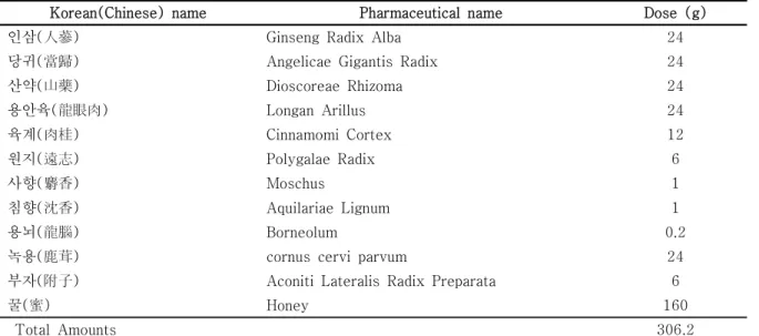
Table 1. Herbal Constitution of Cheongnoimyungshin-hwan. (CNMSH)
Korean(Chinese) name Pharmaceutical name Dose (g)
인삼(人蔘) Ginseng Radix Alba 24
당귀(當歸) Angelicae Gigantis Radix 24
산약(山藥) Dioscoreae Rhizoma 24
용안육(龍眼肉) Longan Arillus 24
육계(肉桂) Cinnamomi Cortex 12
원지(遠志) Polygalae Radix 6
사향(麝香) Moschus 1
침향(沈香) Aquilariae Lignum 1
용뇌(龍腦) Borneolum 0.2
녹용(鹿茸) cornus cervi parvum 24
부자(附子) Aconiti Lateralis Radix Preparata 6
꿀(蜜) Honey 160
Total Amounts 306.2
Fig. 1. Effects of CNMSH on the cell viability in RAW 264.7 macrophages.
Cultured RAW 264.7 cells was pretreated with the different concentrations of CNMSH for 1 h and then incubated with 100 ng/ml LPS for 24 h. (A) Cell viability was gauged by an MTT assay. Data were expressed as percentage of control. Each value indicates the mean ± SD and is representative of results obtained from three independent experiments. (B) Cell morphology was visualized by light microscopy (Magnification, × 200).
Fig. 2. Effects of CNMSH on LPS-induced NO and PGE2 production in RAW 264.7 macrophages.
Cell were pretreated with the indicated concentrations of CNMSH for 1 h and then stimulated with 100 ng/ml LPS for 24 h. (A) NO production was measured using the Griess reagent in culture media and (B) the amounts of PGE2 production was measured by ELISA kit. Each value indicates the mean ± SD and is representative of results obtained from three independent experiments (*P<0.05 compared with the untreated control group; #P<0.05 compared with LPS‑treated group).
Fig. 3. Effects of CNMSH on LPS-induced iNOS and COX-2 protein and mRNA expressions in RAW 264.7 macrophages.
Cell were pretreated with the indicated concentrations of CNMSH for 1 h and then stimulated with 100 ng/ml LPS for 24 h. (A) Cell lysates were prepared for Western blot analysis with antibodies specific for murine iNOS and COX-2, and an ECL detection system. (B) The total RNAs were prepared for RT-PCR analysis of the iNOS and COX-2 mRNA expression using the indicated primers. The experiment was repeated three times, and similar results were obtained. Actin and GAPDH were used as the internal controls for the Western blot analysis and RT-PCR, respectively.
Fig. 4. Effects of CNMSH on LPS-induced NF-κB nuclear translocation in RAW 264.7 macrophages.
Cell were pretreated with the CNMSH (2 mg/ml) for 1 h and then stimulated with 100 ng/ml LPS for 1 h. The nuclear (A) and cytosolic proteins (B) were prepared for Western blot analysis using anti-NF-κB p65 and anti-IκB-α antibodies, and an ECL detection system. Lamin B and actin were used as the internal controls for the nuclear and cytosolic fractions, respectively.
Fig. 5. Effects of CNMSH on intracellular ROS production in LPS-stimulated RAW 264.7 macrophages.
RAW 264.7 cells were pretreated with 2 mg/ml CNMSH, and 10 mM NAC, and then treated with 100 ng/ml LPS for 6 h. (A) The cells were incubated with 10 μM DCF-DA for 30 min at 37°C.
Cells were collected, and DCF fluorescence was measured by a flow cytometry. Values represent the means ±r SD of two independent experiments. (B) Images were obtained using a fluorescence microscope and presented are from one experiment and are representative of at least 3 independent experiments.
Fig. 6. Effects of CNMSH on Nrf2 and HO-1 expression in RAW 264.7 macrophages.
(A) RAW 264.7 cells were incubated with 2 mg/ml of CNMSH for the indicated times, or (B) various concentrations of CHMSH for 24 h. Expression of Nrf2, Keap1 and HO-1 at the protein level was determined by Western blot analysis with cell lysates. Actin was used as an internal control