Socheongja and Socheong 2 Extracts Suppress Lipopolysaccharide-induced Inflammation and Oxidative Stress in RAW 264.7 Macrophages through Activating Nrf2/HO-1 Signaling and Suppressing MAPKs Pathway
Da Hye Kwon1,2, Eun Ok Choi1,2, Hye-Jin Hwang3, Kook Jin Kim4, Su Hyun Hong1,2, Dong Hee Lee4 and Yung Hyun Choi1,2*
1Open Laboratory for Muscular & Skeletal Disease Control and Department of Biochemistry, Dongeui University College of Korean Medicine, Busan 47227, Korea
2Anti-Aging Research Center and Blue Bio Industry RIC, Dongeui University, Busan 47340, Korea
3Department of Food and Nutrition, College of Nursing, Healthcare Sciences & Human Ecology, Dongeui University, Busan 47340, Korea
4Genomine Advanced Biotechnology Research Institute, Genomine Inc., Pohang 37668, Korea Received October 10, 2017 /Revised February 7, 2018 /Accepted February 13, 2018
Inflammatory response and oxidative stress play critical roles in the development and progression of many human diseases. Therefore, a great deal of attention has been focused on finding functional ma- terials that can control inflammation and oxidative stress simultaneously. The purpose of this study was to investigate the effects of Socheongja and Socheong 2, Korean black seed coat soybean varieties, on the inflammatory and oxidative stress induced by lipopolysaccharide (LPS) in RAW 264.7 macro- phages. Our data indicated that the extracts of Socheongja (SCJ) and Socheong 2 (SC2) significantly suppressed LPS-induced production of nitrite oxide (NO) and prostaglandin E2, key pro-inflammatory mediators, by suppressing the expression of inducible NO synthase and cyclooxygenase-2. It was also found that SCJ and SC2 reduced the LPS-induced secretion of pro-inflammatory cytokines, such as tumor necrosis factor-α and interleukin-1β, which was concomitant with a decrease in the protein levels. In addition, SCJ and SC2 markedly diminished LPS-stimulated intracellular reactive oxygen species accumulation, and effectively enhanced nuclear factor erythroid 2-related factor 2 (Nrf2) and heme oxygenase (HO)-1 expression. Furthermore, LPS-induced activation of mitogen-activated protein kinases (MAPKs) was abrogated by SCJ and SC2. Taken together, these data suggest that SCJ and SC2 may offer protective roles against LPS-induced inflammatory and oxidative responses in RAW 264.7 macrophages through attenuating MAPKs pathway, and these effects are mediated, at least in part, through activating Nrf2/HO-1 pathway. Given these results, we propose that SCJ and SC2 have ther- apeutic potential in the treatment of inflammatory and oxidative disorders caused by over-activation of macrophages.
Key words : Anti-inflammation, antioxidant, RAW 264.7 macrophages, Socheongja, Socheong 2
*Corresponding author
*Tel : +82-51-850-7413, Fax : +82-51-853-4036
*E-mail : [email protected]
This is an Open-Access article distributed under the terms of the Creative Commons Attribution Non-Commercial License (http://creativecommons.org/licenses/by-nc/3.0) which permits unrestricted non-commercial use, distribution, and reproduction in any medium, provided the original work is properly cited.
Journal of Life Science 2018 Vol. 28. No. 2. 207~215 DOI : https://doi.org/10.5352/JLS.2018.28.2.207
서 론
염증반응은 물리적인 상처나 생체에 박테리아 독소, 바이러 스 등의 외부 물질에 감염되었을 때 면역세포가 이를 인지하 여 염증 유도 인자들을 분비함으로써 나타내는 생체 방어 기 전 중 하나이다[34, 40]. 특히 대식세포(macrophage)는 그람음 성세균(Gram negative bacteria) 표층의 peptide glycan을 둘 러싸는 외막의 중요 구성성분 중의 하나인 지질다당류(lipo-
polysaccharide, LPS)등에 의해 활성화되어 염증반응을 일으 키는데, 이 때 염증매개인자(pro-inflammatory mediators)인 nitric oxide (NO)나 prostaglandin E2 (PGE2)와 pro-inflam- matory cytokine인 tumor necrosis factor-α (TNF-α), inter- leukin-1β (IL-1β) 등을 분비하여 생체방어에 대비한다[3, 9, 21]. 생체 내에서의 NO의 일반적인 기능과는 달리 inducible nitric oxide synthase (iNOS)의 발현 증가에 의한 NO의 과잉 생산은 혈관투과성과 부종 등의 염증반응을 촉진시킬 뿐만 아니라, DNA 손상과 돌연변이를 일으킬 수 있다. 또한 cyclo- oxygenase (COX-2)에 의해 PGE2가 과잉 생산하게 되면 통증, 발열, 과도한 염증 반응 및 암 발생 촉진 등에도 관여하게 된다 [6, 8, 18, 22, 35, 41]. 한편 세포 내 강력한 항산화 신호계에 속하는 nuclear factor E2-related factor 2 (Nrf2)의 표적 인자 인 heme oxygenase-1 (HO-1)은 heme으로부터 carbon mon- oxide (CO), biliverdin, free iron으로 분해되는 반응을 촉진하
는 효소로, pro-inflammatory cytokine의 생성을 줄이고, an- ti-inflammatory cytokine의 생성을 촉진시키는 기능을 한다 [17, 20]. 또한 HO-1은 염증성 및 산화적 스트레스로부터 세포 를 보호하는 기전에서 중요하게 작용하는 것으로 알려져 있지 만[27], 활성화된 대식세포에서 NO의 생성을 조절한다고도 알려져 있다[26].
검정콩은 민간에서 약콩이라고도 불리며, 검은 빛의 콩을 통칭한다[7]. 일반적으로 대두보다 다량의 isoflavone을 함유 하고 있는데, 그 중에서도 genistein은 유해한 활성산소종 (reactive oxygen species, ROS)을 제거하는 항산화 효과[31]와 heat shock protein (HSP), glucose related protein (GRPs)과 같은 단백질의 발현을 억제하여 항암효과를 나타내는 것으로 알려져 있다[39]. 또한 콩의 기능성 성분인 anthocyanin, iso- flavone, 올리고당, saponin 등은 항암 활성뿐 만 아니라, 골다 공증, 심장질환, 신부전증 예방 등의 효과가 큰 것으로 보고되 어 있다[1, 4, 37]. 검은콩의 품종 중 하나인 녹색자엽이며 검정 소립인 소청자(밀양251호)는 조지방, isoflavone 함량과 an- thocyanin 중에서도 Cy3glc의 비율이 다른 콩에 비해 높게 나 타나며, 떡소용 및 제과용으로 널리 이용된다. 또한, 녹색자엽 이며 검정소립인 소청2호(밀양78호)는 수분흡수율과 isoflavone 함량이 높은 것으로 알려져 있으며, 청국장용으로 많이 이용 된다[15, 32]. 현재까지 일반적인 검정콩의 항염증, 항산화, 항 암 효과 등에 대해서는 이미 많이 알려져 있지만, 소청자와 소청2호에 대한 연구는 아직 미비한 실정이다. 본 연구에서는 검은콩 품종 중에서 이들 두 품종을 선정하여 이들 추출물의 항염증 및 항산화 효능을 검증하였다. 이를 위하여 LPS로 자 극된 RAW 264.7 대식세포에서 염증성 매개인자(NO 및 PGE2)와 염증성 cytokine (TNF-α 및 IL-1β)의 생성 억제 정도 와, 염증성 반응 매개 ROS의 생성 억제 여부 및 강력한 항산화 활성 신호계로 알려진 Nrf2/HO-1 신호계에 미치는 이들 추출 물의 영향을 조사하였다. 아울러 이러한 항염증 및 항산화 효 능과 연관된 세포 내 주요 신호 전달계인 mitogen-activated protein kinases (MAPKs)의 관련성 여부를 추가로 조사하였다.
재료 및 방법
시료
본 연구에서는 국립식량과학원(National Institute of Crop Science, Rural Development Administration, Korea)에서 분 양받은 소청자 및 소청2호를 이용하여 실험을 진행하였으며, 유효성분을 보다 많이 함유할 수 있는 저온용출 방식과 25 kg의 대량생산을 위한 추출법을 표준화하여 추출물을 제조하 였다. 소청자 및 소청2호 추출물은 2시간 동안 저온 처리된 종자를 발아시켜 콩나물 제조기를 이용하여 실온, 암실 조건 에서 6일 동안 재배하여 시료로 사용하였으며, 이를 분쇄하여 정제수와 1:1 비율로 희석하였다. 이 후 저온(4℃)에서 2~12시
간 정도 200 rpm으로 교반하였고, 여과하여 여액을 분리하였 다. 마지막으로 5,000 rpm 원심 분리하여 상층액을 수집하여, 저온상태로 저장하여 실험에 이용하였다.
세포 배양
실험을 진행하기 위해 사용된 RAW 264.7 세포는 한국세포 주은행(Korea Cell Line Bank, Seoul, Korea)에서 분양받아 사 용하였으며, 10% fetal bovine serum (FBS, WELGENE Inc., Daegu, Korea)과 L-glutamine (2 mM), penicillin (100 U/ml) 및 streptomycin (100 U/ml, WelGENE Inc.)이 첨가된 Dulbecco's Modified Eagle's Medium (DMEM, WELGENE Inc.)를 사용하여 37℃, 5% CO2 incubator에서 배양하였다.
세포독성 평가
RAW 264.7 대식세포의 증식에 미치는 소청자 및 소청2호 추출물의 영향을 조사하기 위하여 3-(4,5-dimethylthiazol-2- yl)-2,5-diphenyltetrazolium bromide (MTT) assay를 실시하 였다. 이를 위하여 6 wells plate에 RAW 264.7 세포를 seeding 하여 24시간 동안 안정화시켰다. 이들 세포에 농도별 추출물 을 1시간 처리한 후 100 ng/ml 농도로 LPS (Escherichia coli 055:B5, Sigma-Aldrich Chemical Co., St. Louis, MO, USA)를 처리하였다. 24시간 배양 후 배지를 제거하고, 0.5 mg/ml 농도 의 MTT (Sigma-Aldrich Chemical Co.)를 넣은 후 37℃, 5%
CO2 incubator에서 반응시켰다. 2시간 후 배지를 제거하고 di- methyl sulfoxide (DMSO)로 생성된 formazan을 모두 녹인 후 96 wells plate에 200 μl씩 옮겨서 enzyme-linked im- munosorbent assay (ELISA) reader (Molecular Devices, Sunnyvale, CA, USA)로 540 nm에서 흡광도 변화를 측정하였 다. 각 세포에 대한 세포 독성은 대조군 값을 기준으로 백분율 로 계산하여 나타내었다.
NO 및 PGE2 생산량의 측정
염증성 지표인 NO 및 PGE2 함량의 측정을 위하여 소청자 및 소청2호 추출물을 적정 농도로 1시간 동안 처리한 후, LPS (100 ng/ml)를 처리하여 24시간 동안 배양하였다. LPS 자극에 의한 NO 생성에 미치는 소청자 및 소청2호 추출물의 영향을 조사하기 위해서는 세포 배양액을 수집하여 Griess reagent (Sigma-Aldrich Chemical Co.)와 반응시켰으며, ELISA read- er로 540 nm에서 흡광도 값을 측정하였다. NO 함량은 so- dium nitrite (NaNO2)로 표준곡선을 작성한 후, 계산하여 나 타내었다. PGE2의 생성 정도는 동일 조건에서 배양된 세포 배양액을 대상으로 PGE2 ELISA kit (Cayman Chemical Co., Ann Arbor, Michigan, USA)를 이용하여 측정하였다.
염증성 cytokine 함량의 측정
대표적인 염증성 cytokine인 TNF-α와 IL-1β의 생성에 미치
Fig. 1. Effects of Socheongja and Socheong 2 on the cell viability in RAW 264.7 macrophages. Cultured RAW 264.7 cells was pretreated with the different concentrations of ex- tracts of Socheongja (SCJ) and Socheong 2 (SC2) for 1 hr and then incubated with 100 ng/ml LPS for 24 hr.
Cell viability was gauged by an MTT assay. Data were expressed as percentage of control. Each value indicates the mean ± SD and is representative of results obtained from three independent experiments.
는 소청자 및 소청2호 추출물의 영향을 조사하기 위하여 상기 와 동일한 조건에서 수집된 상층액을 대상으로 R&D system (Minneapolis, USA)에서 구입한 cytokine sandwich ELISA kit를 이용하여 제조사의 protocol에 따라 측정하였다.
ROS 함량의 측정
LPS 자극에 의한 ROS 생성에 미치는 각 추출물의 억제 효 과를 조사하기 위하여 2’,7’-dichlorodihydrofluorescein diac- etate (DCF-DA) 염색법을 이용하였다. 이를 위하여 소청자 및 소청2호 추출물을 1시간 전처리한 후 LPS (100 ng/ml)를 처리 하여 6시간 동안 배양하였다. 준비된 세포들을 모아 10 μM의 DCF-DA 용액(Molecular Probes Inc., Leiden, Netherlands)을 이용하여 37℃, 5% CO2 incubator에서 20분 염색시키고 flow cytometer (Becton-Dickinson, San Jose, CA, USA)를 이용하 여 ROS 생성 변화 여부를 조사하였다. 또한 동일한 조건에서 배양된 세포를 mounting medium을 이용하여 chamber slide 에 mounting 후 형광현미경(Carl Zeiss, Oberkochen, Ger- many) 하에서 ROS 생성 변화 여부를 관찰하였다. 아울러 LPS 에 의한 ROS의 과다 생성이 산화적 스트레스와 연관성이 있 음을 확인하기 위하여 LPS를 처리하기 전에 1시간 동안 N- acetylcysteine (NAC, Sigma-Aldrich Chemical Co.)을 전처리 하여 소청자 및 소청2호 추출물과의 효과를 비교하였다.
단백질의 분리, 전기영동 및 Western blot analysis 항산화 및 항염증 관련 유전자들의 발현 변화에 미치는 소 청자 및 소청2호 추출물의 영향을 조사하기 위하여 다양한 조건에서 배양된 세포들의 단백질을 분리하였다. 이를 위하여 준비된 세포에 적당량의 lysis buffer [25 mM Tris-Cl (pH 7.5), 250 mM NaCl, 5 mM EDTA, 1% Nonidet-P40 (NP-40), 1 mM phenymethylsulfonyl fluoride (PMSF), 5 mM dithiothreitol (DTT)]를 첨가하여 4℃에서 1시간 이상 반응시킨 후, 14,000 rpm으로 30분간 원심 분리하여 상층액을 수거하였다. 상층액 의 단백질 농도는 Bio-Rad 단백질 정량 시약(Bio-Rad, Her- cules, CA, USA)과 그 사용방법에 따라 정량 한 다음 동량의 Laemilni sample buffer (Bio-Rad)와 혼합하여 sodium do- decyl sulphate (SDS)-polyacrylamide gel을 이용하여 전기영 동을 실시하였다. 이를 다시 polyvinylidene difluoride mem- brane (Schleicher and Schuell, Keene, NH, USA)에 electro- blotting에 의해 전이시키고 5% skim milk를 1시간 처리하여 비특이적인 단백질들에 대한 blocking을 실시하였다. 그리고 적정 1차 항체를 처리하여 상온에서 2시간 이상 또는 4℃에서 over night 반응시킨 다음 PBS-T (PBS with Tween 20)로 세척 하고 1차 항체에 맞는 2차 항체를 사용하여 상온에서 1시간 정도 반응시켰다. 반응이 끝난 후 enhanced chemiluminoe- sence (ECL) solution (Thermo scientific, Meridian Rd., Rock- ford, USA)을 적용시킨 다음 X-ray film에 감광시켜 특정 단백
질의 발현 변화를 분석하였다. 실험에 사용된 1차 항체는 iNOS, COX-2, TNF-α, IL-1β, Nrf2, HO-1, Kelch Like ECH Associated Protein 1 (Keap1), extracellular signal-regulated kinase (ERK), c-Jun N-terminal kinases (JNK), p38 MAPK, p-ERK, p-JNK, p-p38 MAPK 및 actin이며, Santa Cruz Biotechnology, Inc. (Santa Cruz, CA, USA) 및 Cell Signalling Technology Inc. (Danvers, MA, USA)에서 구입하였다. 2차 항체로 사용된 horseradish peroxidase (HRP)-conjugated an- ti-mouse, anti-rabbit 및 anti-goat 항체는 Santa Cruz Biotech- nology, Inc.에서 구입하였다.
통계학적인 분석
모든 실험 결과는 Statistical Package for the Social Sciences (SPSS) 통계 프로그램을 이용하여 평균 ± 표준편차(mean ± SD)로 나타내었으며, p<0.05 수준에서 Student’s t-test와 Dun- can’s multiple range test를 이용하여 각 결과에 대한 유의성 을 검증하였다.
결 과
RAW 264.7 세포의 증식에 미치는 소청자 및 소청2호 추 출물의 영향
소청자 및 소청2호 추출물의 세포독성 여부를 확인하기 위 하여 RAW 264.7 세포에 다양한 농도의 추출물을 단독 또는 LPS와 동시 처리한 후에 MTT assay를 실시하였다. 그 결과 소청자 추출물이 소청2호 추출물에 비하여 최고 처리 농도인 1 mg/ml 처리군에서 약간의 세포독성을 보였으나, 큰 차이는 없었다(Fig. 1). 따라서 유의적인 세포독성을 보이지 않는 0.5 mg/ml 를 최고 농도로 선정하여 추후 실험을 진행하였다.
A B
C D
Fig. 2. Effects of Socheongja and Socheong 2 on LPS-induced NO and PGE2 production, and iNOS and COX-2 protein expression in RAW 264.7 macrophages. Cell were pretreated with the indicated concentrations of SCJ and SC2 for 1 hr and then stimulated with 100 ng/ml LPS for 24 hr. (A) NO production was measured using the Griess reagent in culture media and (B) the amounts of PGE2 production was measured by ELISA kit. Each value indicates the mean ± SD and is representative of results obtained from three independent experiments. (C and D) Expression of iNOS and COX-2 at protein levels was de- termined by Western blot analysis with cell lysates. Actin was used as an internal control.
LPS 자극에 의한 염증성 매개인자의 생성에 미치는 소청 자 및 소청2호 추출물의 영향
소청자 및 소청2호 추출물의 항염증 효과를 알아보기 위하 여 염증성 매개인자의 생성에 미치는 영향을 조사하였다. 이 를 위하여 RAW 264.7 세포에서 LPS 자극에 의하여 증가된 NO와 PGE2의 생성 억제 여부를 조사한 결과, LPS 자극에 의 하여 증가된 NO와 PGE2의 분비양이 소청자 및 소청2호 추출 물의 농도 증가에 따라 유의적으로 감소되었다(Fig. 2A 및 Fig.
2B). 또한 NO와 PGE2의 생성에 관여하는 iNOS와 COX-2의 발현에 미치는 소청자 및 소청2호 추출물의 영향을 조사한 결과, LPS 자극에 의하여 증가된 두 유전자의 발현이 소청자 및 소청2호 추출물의 전처리에 의하여 농도 의존적으로 감소 되었다(Fig. 2C 및 Fig. 2D). 따라서 소청자 및 소청2호 추출물 에 의한 NO와 PGE2의 생성 억제 효과는 iNOS 및 COX-2의 발현 억제와 연관성이 있음을 알 수 있었다.
LPS 자극에 의한 염증성 cytokine의 생성에 미치는 소청 자 및 소청2호 추출물의 영향
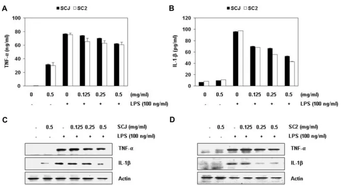
다음은 염증성 cytokine의 생성에 미치는 소청자 및 소청2 호 추출물의 영향을 조사하기 위하여 TNF-α와 IL-1β의 생성 및 발현에 미치는 영향을 조사하였다. Fig. 3의 결과에서 알 수 있듯이, RAW 264.7 세포에서 LPS 자극에 의하여 증가된 TNF-α와 IL-1β의 생성 및 그들의 발현 모두가 소청자 및 소청
2호 추출물 처리에 의하여 감소되어, 소청자 및 소청2호 추출 물에 의한 염증성 cytokine의 생성 억제 또한 해당 유전자들의 발현 감소에 의한 것임을 알 수 있었다.
LPS 자극에 의한 ROS 생성에 미치는 소청자 및 소청2호 추출물의 영향
이상에서 관찰된 소청자 및 소청2호 추출물의 항염증 효능 이 산화적 스트레스 억제와 연관성이 있는지의 여부를 조사하 기 위하여 LPS 자극에 의하여 증가된 ROS의 생성에 미치는 소청자 및 소청2호 추출물의 영향을 조사하였다. Fig. 4A에 나타낸 결과에서 알 수 있듯이, LPS가 단독 처리된 RAW 264.7 세포에서의 증가된 ROS 생성이 소청자 및 소청2호 추출물이 전처리된 조건에서는 거의 완벽하게 차단되었다. 이러한 현상 은 대표적인 항산화제인 NAC의 전처리군에서도 유사하게 관 찰되었으며, 형광현미경을 이용한 관찰에서도 유사한 결과를 얻었다(Fig. 4B). 따라서 소청자 및 소청2호 추출물은 항염증효 과 뿐 만 아니라 항산화력도 매우 우수함을 알 수 있었다.
Nrf2/HO-1 신호전달 경로에 미치는 소청자 및 소청2호 추출물의 영향
소청자 및 소청2호 추출물 항산화 효과가 Nrf2/HO-1 신호 전달 경로의 활성화와 연관성이 있는지의 여부를 조사하기 위하여 두 추출물이 처리된 RAW 264.7 세포를 대상으로 Nrf2
A B
C D
Fig. 3. Effects of Socheongja and Socheong 2 on LPS-induced TNF-α and IL-1β production and protein expression in RAW 264.7 macrophages. Cell were pretreated with the indicated concentrations of SCJ and SC2 for 1 hr and then stimulated with 100 ng/ml LPS for 24 hr. (A and B) The levels of TNF-α and IL-1β production were measured by ELISA kits using culture media. Each value indicates the mean ± SD and is representative of results obtained from three independent experiments.
(C and D) Expression of TNF-α and IL-1β at the protein level was determined by Western blot analysis with cell lysates.
Actin was used as an internal control.
A
B
Fig. 4. Effects of Socheongja and Socheong 2 on intra- cellular ROS production in LPS-stimulated RAW 264.7 macrophages. RAW 264.7 cells were pre- treated with 0.5 mg/ ml SCJ and SC2, and 10 mM NAC, and then treated with 100 ng/ml LPS for 6 hr. (A) In order to monitor ROS production, the cells were incubated at 37˚C in the dark for 20 min with culture medium containing DCF- DA (10 μM).
ROS generation was measured by flow cytometry.
Each point represents the mean of two independent experiments. (B) Images were obtained using a flu- orescence microscope, and those presented are from one experiment, and are representative of at least three independent experiments.
및 HO-1의 발현 변화 여부를 관찰하였다. Fig. 5A 및 Fig. 5B의 결과에서 알 수 있듯이, 소청자 추출물의 처리 농도 및 처리 시간의 증가에 따라 Nrf2와 HO-1의 발현이 점차 증가하였으 며, Keap1의 발현은 억제되었다. 이러한 결과는 소청2호 추출
물의 처리군에서도 유사한 경향성을 나타내어(Fig. 5C 및 Fig.
5D), 두 추출물의 항산화 효능이 Nrf2/HO-1 신호전달계의 활 성화와 최소한의 연관성이 있음을 알 수 있었다.
A B
C D
Fig. 5. Effects of Socheongja and Socheong 2 on Nrf2 and HO-1 expression in LPS-stimulated RAW 264.7 macrophages. RAW 264.7 cells were in- cubated with 0.5 mg/ml of SCJ (A) and SC2 (C) for the indicated times, or various concen- trations of SCJ (B) and SC2 (D) for 24 hr. Total cellular proteins were separated on SDS-poly- acrylamide gels and then transferred onto membranes. The membranes were probed with the indicated antibodies. Proteins were visualized using an ECL detection system.
Actin was used as an internal control.
A B
Fig. 6. Effects of Socheongja and Socheong 2 on MAPKs expression in LPS-stimulated RAW 264.7 macrophages. The cells were pretreated with 0.5 mg/ml SCJ (A) and SC2 (B) for 1 hr and then stimulated with 100 ng/ml LPS for 30 min. The cells were lysed, and the ali- quots containing the total proteins were sub- jected to SDS-polyacrylamide gels followed by immunoblot analysis with the indicated antibodies.
MAPKs 신호전달 경로에 미치는 소청자 및 소청2호 추출 물의 영향
이상에서 관찰된 소청자 및 소청2호 추출물의 항염증 및 항산화 효능과 연관된 세포 내 신호 전달계에 미치는 영향을 조사하기 위하여 3가지 MAPKs의 활성에 미치는 영향을 조사 하였다. 이를 위하여 RAW 264.7 세포에서 LPS에 의한 MAPKs 의 인산화에 미치는 두 추출물의 영향을 조사한 결과, LPS에 의하여 증가된 ERK, JNK 및 p38 MAPK의 활성 증가가 소청 자 및 소청2호 추출물이 전처리된 조건에서는 모두 억제되었 음을 알 수 있었다(Fig. 6A 및 Fig. 6B). 따라서 두 추출물의 항염증 및 항산화 효능은 MAPKs의 활성 억제와 관련이 있음 을 알 수 있었다.
고 찰
본 연구에서는 LPS에 의하여 자극된 RAW 264.7 대식세포 모델을 대상으로 검은콩 품종인 소청자와 소청2호의 항산화
및 항염증 효능을 평가하였다. 이를 위하여 LPS 자극에 의한 염증성 매개인자 및 cytokine의 생성 증가에 미치는 소청자 및 소청2호 추출물의 억제 효과와, ROS의 생성 및 Nrf2/HO-1 신호계에 미치는 두 추출물의 영향을 조사하였다.
체내에서 대식세포는 면역 방어 기전에 중요한 역할을 하는 세포로서, 정상적인 상황에서 iNOS와 COX-2 효소에 의해 생 성되는 NO와 PGE2 및 cytokine을 포함한 염증 관련 매개체는 숙주 생존과 조직 복구에 필수적인 역할을 담당한다[34, 40].
그러나 이러한 염증성 매개인자와 cytokine이 특정 염증성 자 극에 반응하여 과잉 생성되면 염증과 관련된 다양한 질환의 발병과 진행을 촉진한다[3, 9, 21]. 더욱이 TNF-α와 IL-1β를 포함한 염증성 cytokine은 iNOS와 COX-2의 발현을 증가시키 고 NO와 PGE2 생산을 현저히 증가시킨다[5, 38]. 따라서 염증 성 cytokine의 생성 억제와 iNOS와 COX-2의 저해를 통한 NO 와 PGE2의 생성 억제에 대한 연구는 항염증제의 개발에 매우 중요하다. 본 연구의 결과에 의하면, LPS가 처리된 RAW 264.7 대식세포에서 NO 및 PGE2의 생성이 유의적으로 증가되었으
며, 세포 독성이 없는 범위의 소청자 및 소청2호 추출물이 전 처리된 조건에서는 이들의 방출이 현저히 저해되었다. 또한, Western blot 분석의 결과에 따르면 소청자 및 소청2호 추출물 의 전처리는 LPS로 유도된 iNOS 및 COX-2 발현을 감소시켰 으며, ELISA 결과에서 소청자 및 소청2호 추출물이 LPS로 유 도된 TNF-α 및 IL-1β 생성 또한 억제시켰음을 알 수 있었다.
이러한 결과와 일치되게 소청자 및 소청2호 추출물이 TNF-α 및 IL-1β의 단백질 수준을 유의적으로 억제하였음을 확인하였 다. 이러한 결과는 소청자 및 소청2호 추출물이 염증 유발 유 전자들의 발현 감소를 통하여 항염증 효과를 보여줌을 시사한 다.
비록 적정량의 ROS가 세포 내 항상성 유지에 기본적으로 필요하지만, 비정상적인 ROS의 과다 생산으로 규정되는 산화 적 스트레스는 염증을 포함한 다른 많은 병리학적 상태와 밀 접하게 연관되어있다[2, 23]. 특히 만성 염증 상태에서의 지속 적인 ROS 생산은 염증 관련 유전자의 과다 발현을 통해 대식 세포의 염증 신호를 증폭시킨다[14, 36]. 또한 염증반응에 동반 된 산화적 스트레스에 의한 과도한 ROS의 생산은 염증 부위 로의 추가적인 호중구(neutrophils)와 대식세포의 이주를 자 극하여 염증성 매개인자와 cytokine의 추가적인 분비를 촉진 시킨다[13, 24]. 따라서 LPS에 의한 ROS 생성에 대한 소청자 및 소청2호 추출물의 억제 효과를 조사한 결과, LPS에 자극받 은 RAW 264.7 대식세포에서 두 추출물은 ROS 축적을 유의적 으로 감소시켰다. 이러한 소청자 및 소청2호 추출물의 ROS 생성 억제는 염증 동반 산화적 스트레스를 동시에 억제할 수 있는 가능성을 시사하여 준다.
전사인자 Nrf2는 다양한 염증성 및 산화적 자극에 대한 세 포 내 항상성 유지에 중요한 역할을 담당한다[11, 16]. 정상적 인 산화-환원 조건 하에서, Nrf2는 세포질에서 Nrf2의 대표적 인 억제인자인 Keap1에 결합하고, ubiquitin-proteasome 경로 를 통해 쉽게 분해된다. 그러나 스트레스 또는 산화방어 유도 물질에 노출되면 Nrf2는 Keap1에서 해리되고 핵으로 이동하 며 세포 보호 및 항산화 효소 관련 유전자들의 전사 활성을 촉진시킨다[10, 12]. 본 연구의 결과에 따르면, Nrf2의 발현은 소청자 및 소청2호 추출물 처리 농도와 시간이 증가함에 따라 크게 증가했으며, Keap1의 감소가 이 과정에서 동반했다. 또 한, Nrf2의 대표적인 표적 유전자인 HO-1의 발현이 두 추출물 의 처리에 의해 유의하게 증가되었다. HO-1에 의한 heme의 분해산물 중의 하나인 biliverdin은 biliverdin reductase에 의 해 bilirubin으로 전환된다[29, 33]. 특히 이 효소 반응의 생성 물들은 ROS 제거를 통해 항산화 및 세포 보호 기능에서 중요 한 생물학적 역할을 담당한다[19, 25]. ROS 생산의 저해와 Nrf2/HO-1 신호 전달 활성화 사이의 관계에 대한 추가 연구 가 필요하지만, 본 연구의 결과는 소청자 및 소청2호 추출물에 의한 Nrf2와 HO-1의 유도는 산화적 스트레스의 감소를 통하 여 LPS에 의한 염증성 반응의 차단에 기여할 것으로 추정된
다.
한편 MAPKs 신호전달경로는 대식세포에서 염증성 매개인 자 및 cytokine의 생성을 유도하는 경로뿐 만 아니라 ROS 생 성 자극 경로의 활성화에 핵심적인 역할을 한다[30]. 따라서 MAPKs 신호전달경로 표적 치료제는 다양한 염증성 자극 신 호 차단을 유도할 수 있기 때문에 염증 질환 치료에 효과적일 수 있다[28]. 소청자 및 소청2호 추출물의 항염증 효과가 MAPKs 경로를 통해 매개되는지 여부를 조사하기 위해, LPS 에 의하여 유도된 MAPKs 계열 단백질의 활성에 미치는 두 추출물의 영향을 조사하였다. 본 연구의 결과에 의하면, 소청 자 및 소청2호 추출물의 전처리는 LPS에 의하여 유발된 3가지 MAPKs (ERK, JNK 및 p38 MAPK)의 인산화를 완벽하게 억제 하였다. 비록 nuclear factor-κB를 포함한 염증성 반응에 중요 한 역할을 하는 또 다른 세포 내 신호전달계에 대한 평가도 추가적으로 이루어져야겠지만, 본 연구의 결과는 소청자 및 소청2호 추출물에 의한 항염증 효능에 MAPKs 활성의 차단이 최소한 관여하고 있음을 의미한다.
결론적으로, 본 연구의 결과에서는 소청자 및 소청2호 추출 물이 염증성 매개인자와 cytokine의 생성과 ROS의 생성을 억 제함으로서 항염증 및 항산화 효과를 나타냄을 보여주었다.
아울러 이 과정에서 Nrf2/HO-1 신호전달 활성 증대 및 MAPKs 신호경로의 활성 감소가 동반되었다. 두 추출물에 의한 항염 증성 및 항산화적 신호 경로 연결에 대한 추가적인 연구가 필요하지만, 소청자 및 소청2호 추출물이 다중 신호 전달 경로 조절을 통하여 세포독성에 대한 차단효과를 지닐 수 있는 유 용한 식의약 소재로서의 개발 가능성이 높음을 보여주었다.
감사의 글
본 연구는 산업통상자원부와 한국산업기술진흥원의 지역 특화산업육성사업(KIAT, R0005567) 및 한국연구재단(2015R1 A2A2A01004633)의 지원을 받아 수행된 연구임.
References
1. Barnes, S. 1998. Evolution of the health benefits of soy isoflavone. Proc. Soc. Exp. Biol. Med. 217, 386-392.
2. Brüe, B., Dehne, N., Grossmann, N., Jung, M., Namgaladze, D., Schmid, T., von Knethen, A. and Weigert, A. 2013. Redox control of inflammation in macrophages. Antioxid. Redox.
Signal. 19, 595-637.
3. Cho, B. O., Ryu, H. W., So, Y., Lee, C. W., Jin, C. H., Yook, H. S., Jeong, Y. W., Park, J. C. and Jeong, I. Y. 2014. Anti-in- flammatory effect of mangostenone F in lipopolysaccharide- stimulated RAW 264.7 macrophages by suppressing NF-κB and MAPK activation. Biomol. Ther. 22, 288-294.
4. Choung, M. G., Baek, I. Y., Kang, S. T., Han, W. Y., Shin, D. C., Moon, H. P. and Kang, K. H. 2001. Isolation and de- termination of anthocyanins in seed coats of black soybean
(Glycine max (L.) Merr.). J. Agric. Food Chem. 49, 5848-5851.
5. Dinarell, C. A. 2000. Proinflammatory cytokines. Chest 118, 503-508.
6. Golden, B. D. and Abramson, S. B. 1999. Selective cyclo- oxygenase-2 inhibitors. Rheum. Dis. Clin. North. Am. 25, 359- 378.
7. Gong, G. L. 2001. Studies on the standardization of manu- facture and cancer protection and anti-obesity effects of Kochujang, Master Degree dissertation, Pusan National University, Busan, Korea.
8. Guo, S., Qiu, P., Xu, G., Wu, X., Dong, P., Yang, G., Zheng, J., McClements, D. J. and Xiao, H. 2012. Synergistic anti-in- flammatory effects of nobiletin and sulforaphane in lip- opolysaccharide-stimulated RAW 264.7 cells. J. Agric. Food Chem. 60, 2157-2164.
9. Higuchi, M., Hisgahi, N., Taki, H. and Osawa, T. 1990.
Cytolytic mechanisms of activated macrophages: Tumor ne- crosis factor and L-arginine-dependent mechanisms act syn- ergistically as the major cytolytic mechanisms of activated macrophages. J. Immunol. 144, 1425-1431.
10. Huang, B. P., Lin, C. H., Chen, H. M., Lin, J. T., Cheng, Y.
F. and Kao, S. H. 2015. AMPK activation inhibits expression of proinflammatory mediators through downregulation of PI3K/p38 MAPK and NF-κB signaling in murine macro- phages. DNA Cell Biol. 34, 133-141.
11. Huang, Y., Li, W., Su, Z. Y. and Kong, A. N. 2015. The com- plexity of the Nrf2 pathway: beyond the antioxidant response. J. Nutr. Biochem. 26, 1401-1413.
12. Kang, K. A. and Hyun, J. W. 2017. Oxidative stress, Nrf2, and epigenetic modification contribute to anticancer drug resistance. Toxicol. Res. 33, 1-5.
13. Kasahara, E., Sekiyama, A., Hori, M., Hara, K., Takahashi, N., Konishi, M., Sato, E. F., Matsumoto, S., Okamura, H.
and Inoue, M. 2011. Mitochondrial density contributes to the immune response of macrophages to lipopolysaccharide via the MAPK pathway. FEBS Lett. 585, 2263-2268.
14. Kauppinen, A., Suuronen, T., Ojala, J., Kaarniranta, K. and Salminen, A. 2013. Antagonistic crosstalk between NF-κB and SIRT1 in the regulation of inflammation and metabolic disorders. Cell Signal. 25, 1939-1948.
15. Korea seed & Variety service, https://www.seed.go.kr 16. Lee, D. H., Park, J. S., Lee, Y. S., Sung, S. H., Lee, Y. H.
and Bae, S. H. 2017. The hypertension drug, verapamil, acti- vates Nrf2 by promoting p62-dependent autophagic Keap1 degradation and prevents acetaminophen-induced cytotoxicity.
BMB Rep. 50, 91-96.
17. Lee, T. S. and Chau, L. Y. 2002. Heme oxygenase-1 mediates the anti-inflammatory effect of interleukin-10 in mice. Nat.
Med. 8, 240-246.
18. Lim, B. O., Jeong, Y. J., Park, M. H., Kim, J. D., Hwang, S. J. and Yu, B. P. 2007. Immunoregulatory effects of Saengshik on DSS-induced inflammatory bowel disease in mouse model system. J. Kor. Soc. Food Sci. Nutr. 36, 32-42.
19. Loboda, A., Damulewicz, M., Pyza, E., Jozkowicz, A. and Dulak, J. 2016. Role of Nrf2/HO-1 system in development, oxidative stress response and diseases: an evolutionarily
conserved mechanism. Cell. Mol. Life Sci. 73, 3221-3247.
20. Maines, M. D. 1997. The heme oxygenase system: a regu- lator of second messenger gases. Annu. Rev. Pharmacol.
Toxicol. 37, 517-554.
21. Mann, J. R., Backlund, M. G. and DuBois, R. N. 2005.
Mechanism of disease: Inflammatory mediators and cancer prevention. Nat. Clin. Pract. Oncol. 2, 202-210.
22. Masferrer, J. L., Zweifel, B. S., Manning, P. T., Hauser, S.
D., Leahy, K. M., Smith, W. G., Isakson, P. C. and Seibert, K. 1994. Selective inhibition of inducible cyclooxygenase 2 in vivo is anti inflammatory and nonulcerogenic. Proc. Natl.
Acad. Sci. USA. 91, 3228-3232.
23. Mills, E. L. and O’Neill, L. A. 2016. Reprogramming mi- tochondrial metabolism in macrophages as an anti-in- flammatory signal. Eur. J. Immunol. 46, 13-21.
24. Mittal, M., Siddiqui, M. R., Tran, K., Reddy, S. P. and Malik, A. B. 2014. Reactive oxygen species in inflammation and tissue injury. Antioxid. Redox Signal. 20, 1126-1167.
25. Motterlini, R. and Foresti, R. 2014. Heme oxygenase-1 as a target for drug discovery. Antioxid. Redox Signal. 20, 1810- 1826.
26. Oh, G. S., Pae, H. O. and Chung, H. T. 2003. Nitric oxide priming protects nitric oxide-mediated apoptosis via heme oxygenase-1 induction. Free Radic. Biol. Med. 34, 1136-1145.
27. Otterbein, L. E. and Chai, A. M. 2000. Heme oxygenase: col- ors of defense against cellular stress. Am. J. Physiol. Lung Cell. Mol. Physiol. 279, 1029-1037.
28. Pearson, G., Robinson, F., Beers Gibson, T., Xu, B. E., Karandikar, M., Berman, K. and Cobb, M. H. 2001. Mitogen- activated protein (MAP) kinase pathways: regulation and physiological functions. Endocr. Rev. 22, 153-183.
29. Pittalà, V., Salerno, L., Romeo, G., Modica, M. N. and Siracusa, M. A. 2013. A focus on heme oxygenase-1 (HO-1) inhibitors. Curr. Med. Chem. 20, 3711-3732.
30. Raingeaud, J., Whitmarsh, A. J., Barrett, T., Derijard, B. and Davis, R. J. 1996. MKK3- and MKK6-regulated gene ex- pressionis is mediated by the p38 mitogen-activated protein kinase signal transduction pathway. Mol. Cell Biol. 16, 1247- 1255.
31. Record, I. R., Dreosit, I. E. and Mclnerney, J. K. 1995. The antioxidant activity of genistein in vitro. J. Nutr. Biochem.
6, 481-485.
32. Rural Development Administration National Institute of Crop Science, Republic of Korea, www.nics.go.kr 33. Ryter, S. W. and Choi, A. M. 2016. Targeting heme oxygen-
ase-1 and carbon monoxide for therapeutic modulation of inflammation. Transl. Res. 167, 7-34.
34. Sirisinha, S. 2011. Insight into the mechanisms regulating immune homeostasis in health and disease. Asian Pac. J.
Allergy Immunol. 29, 1-14.
35. Stuehr, H. H., Kwon, N. S., Weise, M. and Nathan, C. 1991.
Purification and characterization of the cytokine-induced macrophage nitric oxide synthase: and FAD- and FMN-con- taining flavoprotein. Proc. Natl. Acad. Sci. USA. 88, 7773- 7777.
36. Tan, H. Y., Wang, N., Li, S., Hong, M., Wang, X. and Feng,
초록:RAW 264.7 대식세포에서 Nrf2/HO-1 신호 전달계 활성화와 MAPKs 경로 억제를 통한 소청 자와 소청2호의 LPS 매개 염증성 및 산화적 스트레스 반응의 억제
권다혜1,2․최은옥1,2․황혜진3․김국진4․이동희4․최영현1,2*
(1동의대학교 한의과대학 생화학교실 및 근ˑ골격계 질환제어 융합연구실, 2동의대학교 항노화연구소 및 블루바이오
소재개발 및 실용화지원센터, 3동의대학교 의료ˑ보건ˑ생활대학 식품영양학과, 4(주)제노마인)
염증 반응과 산화적 스트레스는 다양한 질환의 발생과 진행에 중요한 역할을 담당한다. 따라서 염증과 산화적 스트레스를 동시에 억제할 수 있는 소재의 발굴은 인체 질환 제어에 매우 유용하게 적용될 수 있다. 본 연구의 목적은 RAW 264.7 대식세포에서 검은콩 품종인 소청자와 소청2호 추출물이 염증성 및 산화적 스트레스에 미치 는 영향을 알아보기 위함이다. 본 연구의 결과에 의하면 소청자 및 소청2호 추출물은 LPS에 의한 iNOS와 COX-2 의 발현을 억제하여 NO와 PGE2의 생성을 억제하였으며, TNF-α, IL-1β와 같은 염증성 cytokine의 분비와 발현을 감소시켰다. 또한 소청자 및 소청2호 추출물은 LPS로 자극된 세포 내 ROS 축적을 유의적으로 차단시켰고, Nrf2와 HO-1 발현을 증가시켰다. 또한 LPS에 의해 유도된 MAPKs의 활성화도 소청자 및 소청2호 추출물에 의하여 억제 되었다. 결론적으로 소청자 및 소청2호 추출물은 RAW 264.7 대식세포에서 MAPKs 경로를 차단시킴으로써 LPS- 유도 염증 및 산화 반응에 대한 보호 역할을 할 수 있으며, 이러한 효과에는 최소한 Nrf2/HO-1 경로 활성화가 관련되어 있었다. 이러한 결과를 고려해 볼 때 소청자 및 소청2호 추출물은 대식세포의 과다 활성화로 인한 염증 성 및 산화적 반응 차단에 잠재적인 효과가 있음을 일 수 있었다.
Y. 2016. The reactive oxygen species in macrophage polar- ization: Reflecting its dual role in progression and treatment of human diseases. Oxid. Med. Cell. Longeva. 2016, 2795090.
37. Tomomatsu, H. 1994. Health effects of oligosaccharides.
Food Technol. 48, 61-65.
38. Tsan, M. F. 2006. Toll-like receptors, inflammation and cancer. Semin Cancer Biol. 16, 32-37.
39. Wei, H., Cai, Q. and Rahn, R. 1996. Inhibition of UV light and fenton reaction-induced oxidative DNA damage by the
soybean isoflacone genistein. Carcinogenesis 17, 73-77.
40. Willoughby, D. A. 1975. Human arthritis applied to animal models. Towards a better therapy. Ann. Rheum. Dis. 34, 471- 478.
41. Yang, J. L., Jang, J. H., Radliakrishnan, V., Kim, Y. H. and Song, Y. S. 2008. β-Glucan suppresses LPS-stimulated NO production through the down-regulation of iNOS expression and NF-κB transactivation in raw 264.7 macrophages. Food Sci. Biotechnol. 17, 106-113.