蜈蚣 에테르 추출물의 RAW 264.7 cell에서 LPS로 유도된 염증반응 억제 효과
정호경․조현우․정원석․최인영․조정희․정수영*
전라남도한방산업진흥원
Inhibitory Effect of Scolopendra Morsitans L. Ether Extract on Lipopolysaccharide-Induced Inflammatory Response in RAW 264.7 Cells
Ho Kyung Jung, Hyun Woo Cho, Won Seok Jung, In Young Choi, Jung Hee Cho, Su Young Jung*
Jeonnam Development Institute for Korean Traditional Medicine
It has been reported that Scolopendra morsitans L.(SML) has beneficial effects on human health and diverse diseases. The purpose of this study was to investigate the anti-inflammatory effects of ether extract from Scolopendra morsitans L. on lipopolysaccharide(LPS)-induced inflammatory response. Thus, we examined the inhibitory effect of SML ether fraction on LPS-induced increase of inflammatory mediators(NO, iNOS, COX-2, and IκBα) and pro-inflammatory cytokines(TNF-α) in RAW 264.7 cells. In the present study, SML ether extract itself decreased cell viability in a dose dependent manner(> 100 μg/ml). In addition, LPS increased NO production, iNOS expression and phosphorylation of Iκ-Bα, which were blocked by the treatment of SML ether fraction in a dose dependent manner.
Furthermore, the treatment of LPS increased TNF-α production. However, the pretreatment of SML ether fraction prevented the LPS-induced TNF-α production in dose dependant manner. Taken together, our results suggest that SML may be a beneficial drug against inflammatory diseases such as sepsis.
Key words : Scolopendra morsitans L., Anti-inflammation, Nitric oxide, COX-2, TNF-α
* 교신저자 : 정수영, 전남 장흥군 안양면 기산리 756, 전남한방산업진흥원
․E-mail : [email protected], ․Tel : 061-860-2810
․접수 : 2012/10/22 ․수정 : 2013/01/10 ․채택 : 2013/01/14
서 론
蜈蚣은 왕지네과에 속하는 지네류의 全蟲을 말린 것을 말하 며, 救急簡易方에 표기된 지네의 한약명이다. 또한 한자어로는 土蟲, 百足이라고도 부르고, 神農本草經에서는 왕지네, 百脚, 天 龍, 浪邸 등 많은 이명을 가지고 있는 동물성 한약으로 라틴 약 재명은 Scolopendra morsitans L.(SML)이다
1). 蜈蚣은 性溫味辛 하며 有毒하며 肝經에 들어간다. 風邪를 몰아내고 驚悸를 진정시 키며 독을 배출시키고 鬱結을 풀어주는 효능으로 中風, 驚癎, 破 傷風, 百日咳, 結核, 腫毒, 風癬을 치료한다
2). 그동안 밝혀진 蜈蚣 의 효능을 보면 蜈蚣의 물추출물에서 항고혈압작용
3), 蜈蚣水鍼 의 진통, 진경효과에 대한 연구
4), 항산화효과
5,6), 항염증효과
7), 면 역조절효과
8), 물추출후 TCA 제단백처리를 거쳐 MeOH 분획을
통해 항콜린성활성분획을 이용한 항경련활성
9), MeOH 추출물의 항산화활성
10)이 보고되었다. 蜈蚣의 에테르 추출물에 관한 연구 로 마우스의 회장관에 대하여 이완작용이 보고되었고, 그 잔사에 서 메탄올 추출물은 일과성의 불규칙적인 수축 작용을 나타내며, 메탄올 추출잔사의 물 추출물은 가장 강한 이완성이 있음이 보 고된바 있다
11). 蜈蚣의 성분은 quinoline alkaloid로 scolopendrine1, scolopendrine2 가 알려져 있으며, 봉독과 유사 한 2종의 유독성분으로 히스타민 유사 물질과 용혈성단백질을 함유한다
12). 蜈蚣의 지용성 성분으로 oleic acid, linolenic acid, palmitic acid, hexadecenoic acid 등 다종의 불포화지방산이 함 유되어 있는 것으로 알려져 있다
2). 蜈蚣의 항염활성은 물추출물 의 RAW264.7 세포를 이용한 염증성 사이토카인의 억제, iNOS 를 통한 NO의 분비억제 효능이 밝혀졌다
7). 그러나 추출용매에 따른 항염증활성은 아직 연구되지 않아 본 연구에서 蜈蚣의 에 테르 추출물에 의한 항염증 활성을 확인하고자 하였다.
염증은 조직의 손상을 비롯한 외부로 부터 자극 등 다양한 감
염원에 의한 생체 내 방어 반응의 하나로 대식세포는 감염초기에
반응하고 이 과정에서 tumor necrosis factor-α (TNF-α), interleukin (IL)-1β, IL-6, nitric oxide (NO), inducible nitric oxide synthase (iNOS), cyclooxygenase-2 (COX-2)와 같은 염증성 매개물 질을 분비시켜 염증 및 면역반응에 중요한 역할을 한다
13). 그러나 과도한 염증반응은 숙주에 치명적인 결과를 초래 할 수 있다
14,15). 그람 음성균의 외막성분인 lipopolysaccharide (LPS)는 국소 염증, 항체 생산, 폐혈증과 같은 다양한 반응을 일으킨다
16). 대식세포주 RAW264.7 cell에 고농도의 LPS 처리로iNOS의 활성 증가, extracellular signal-regulated kinase1/2 (ERK1/2), p38 kinases (p38), c-Jun NH2-terminal kinase (JNK)와 같은 mitogen-activated protein kinases (MARKs)와 nuclear factor kappa B(NF-κB)/IκBα 경로를 통한 TNF-α, IL-1β, IL-6, NO의 분비 증가 및 COX-2 활성 을 증가 시킨다는 연구가 많이 진행되었으며, LPS 유도성 RAW 264.7 염증 반응을 이용한 실험 모델이 항염증성 물질의 활성을 평 가하는데 많이 이용되고 있다
17-19). 본 연구에서는 蜈蚣의 에테르 추출물의 항염 활성을 확인하기 위하여 RAW264.7 cell에 LPS에 의해 유도되는 NO 및 iNOS, 전염증성 사이토카인 TNF-α의 분비 와 Iκ-Bα의 활성 조절 효과를 관찰하였다.
재료 및 방법
1. 추출물의 제조
蜈蚣 생체 300 g을 세절하여 에테르 3 L에 넣고 3시간 전탕 한 후 추출물을 가아제로 1차 여과하고 3000×g에서 3분간 원심 분리하여 상층액만을 취하여 0.2 μm filter (Nalgene, NY, USA) 로 여과하였다. 이 여과액을 rotary evaporator (EYELA, Tokyo, Japan)로 농축한 후 동결건조하고 사용할 때 까지 -70℃에서 보 관하였다.
2. 시약
LPS (Escherichia coli 026:B6; Difco, Detroit, MI, USA)와 3-(4,5-dimethylthiazol-2yl)-2,5-diphenyltetrazoleum (MTT)은 Sigma (St. Louis, MO, USA)에서 구입하였고, fetal bovine serum (FBS)과 antibiotics는 Gibco/BRL (Eggenstein, Germany) 로부터 구입하였으며, Antibody는 BD Bioscience (USA), Cayman (USA), Zymed (USA)에서 구입하였고, NC paper는 Schleicher & Schuell (USA)에서 구입하였다. TNF-α의 ELISA Kit는 R&D systems (DuoSet◯ R ELISA Development Systems, USA)에서 구입하였다.
3. 세포 배양
마우스의 대식세포주인 RAW 264.7 cell은 한국세포주은행 (KCLB; Seoul, Korea)으로부터 분양받았다. 세포배양을 위해 10% FBS와 1% penicillin-streptomycin을 첨가한 DMEM 배지를 사용하였다. 세포는 37℃, 5% CO
2조건에서 배양하였다.
4. 세포 생존율 측정
RAW 264.7 cell에 나타내는 세포 생존율을 알아보기 위하여
Tetrazolium- basedcolorimetric (MTT) assay를 실시하였다. 96 well plate에 1×10
5cells/mL의 cell을 100 μl씩 넣고 37℃, 5%
CO
2incubator에서 24 시간동안 배양한 후 배지를 버리고 배양 세포 표면을 phosphate buffered saline (PBS) 용액으로 씻어 주 었다. 같은 양의 배지와 시료(10, 100, 1,000 μg/mL)를 각 well에 처리하고 24 시간 동안 배양하였다. 배양이 끝난 후 PBS에 녹인 1 mg/mL MTT (Sigma, USA)를 100 μl씩 각 well에 처리하여 알 루미늄 호일로 차광시킨 후 2 시간 동안 같은 조건에서 배양하였 다. 배양액을 모두 제거한 후 DMSO를 100 μl 처리하고 37℃에 서 2시간 방치 후 microplate reader를 이용하여 540 nm에서 흡 광도를 측정하였다. Cell viability는 다음 공식으로 계산되었다.
Cell viability (%)= AT /AC ×100 AC : absorbance of control AT : absorbance of treated sample
5. Nitric oxide 생성량 측정
LPS를 단독처리(100 ng/mL)하거나 혹은 다양한 농도의 시 료(0.1, 1, 10, 100 μg/mL)와 함께 배지에 담아 각 well에 처리하 고 24시간 동안 37℃, 5% CO
2incubator에서 배양한 후 세포배 양 상등액 100 μl를 채취하여 여기에 Griess 시약(0.5%
sulfanilamide in 2.5% phosphoric acid + 0.5% α-naphthylamide in H
2O) 100 μl를 혼합하여 15분 동안 반응시킨 후 microplate reader (Bio-Rad, USA)를 이용하여 540 nm에서 흡광도를 측정 하였다. 세포의 NO 생성은 다음 공식으로 계산하였다.
Productions of NO (%) = AT/AC × 100 AC : absorbance of control
AT : absorbance of treated sample
6. Immunoblot 분석
20 mM Tris-HCl (pH 7.5), 1% Triton X-100, 137 mM sodium chloride, 10% glycerol, 2 mM EDTA, 1 mM sodium orthovanadate, 25 mM β-glycerophosphate, 2 mM sodium pyrophosphate, 1 mM phenylmethylsulfonylfluoride와 1 mg/ml leupeptin을 함유하는 buffer (100mm dish당 80 μl)를 가 하여 cell을 lysis 시켰다. iNOS와 COX-2의 발현은 anti-mouse iNOS, COX-2 antibodies를 사용하여 면역화학적 방법으로 분석 하였으며, 2차 antibody는 horseradish peroxidase conjugated anti-rabbit antibody를 사용하였다. iNOS와 COX-2, p-IκBα protein의 band는 ECL western blotting detection reagents (Amersham, USA)를 사용하여 단백질의 발현 정도를 확인하였다.
7. Cytokine 생성 측정
Cytokine을 측정하기 위하여 6 well plate에 1×10
6cells/ml
을 분주하고 다양한 농도의 시료(0.1, 1, 10, 100 μg/mL)를 처리
한 다음, 1 시간 후에 LPS를 처리하였다. LPS 처리 후 24 시간에
배지를 수거하여 cytokine을 측정하였다. 수거된 배지는 측정 전
까지 -70℃에서 보관하였다. TNF-α는 ELISA Kit (DuoSet◯ R
ELISA Development Systems, R&D, USA)를 사용하여 측정하였
으며, 실험의 방법은 manufacturer's instruction에 따랐다.
9. 통계분석
본 실험에서 얻은 결과에 대해서는 평균치 ± 표준편차 (mean ± S.D.)로 나타내었으며, 대조군과 각 실험군과의 평균의 차이는 Student's t-test로 분석하여 p-value 값이 0.05 미만일 때 통계적으로 유의한 차이가 있는 것으로 판정하였다.
결 과
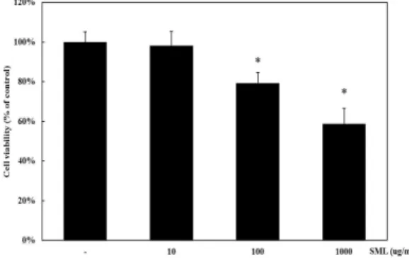
1. 蜈蚣 에테르 추출물의 RAW 264.7 cell에 대한 세포독성 蜈蚣 에테르 추출물(SML)의 세포독성에 관해 알아보기 위 하여 대식세포 RAW 264.7 cell에 蜈蚣 에테르 추출물을 10, 100, 1,000 μg/ml의 농도로 처리하여 24 시간 후에 세포의 생존율을 측정하였다. Fig. 1에 나타난 바와 같이 蜈蚣 에테르 추출물 100 μ g/ml의 농도부터 농도 의존적으로 세포 독성이 관찰되었다.
Fig. 1. The effect of SML ether extracts on cell viability in RAW 264.7.
RAW 264.7 cells were incubated for 24 hrs in the presence or absence of SML ether extracts at indicated dose. Cell viability was measured by MTT assay. Values are mean ± SD of 9 separate experiments performed on 3 different cultures. *p<0.05 vs. control.2. 蜈蚣 에테르 추출물이 RAW 264.7 cell에서 LPS로 유도된 NO 생성에 미치는 영향
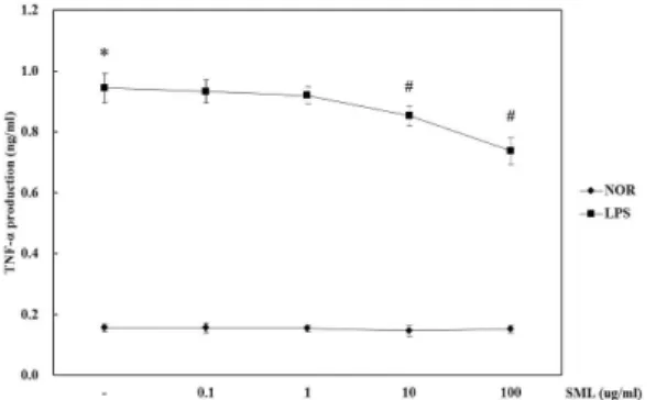
蜈蚣 에테르 추출물이 NO 생성에 미치는 영향을 조사하기 위하여 蜈蚣 에테르 추출물(SML)을 0.1, 1, 10, 100 μg/ml의 농도 로 세포에 처리하여 생성되는 NO양을 측정하였다. 추출물을 전 처리하고 LPS로 자극하여 24 시간 후에 세포 상층액에서 NO의 생성을 측정한 결과 LPS만 처리한 군에서는 LPS 자극 없이 蜈蚣 에테르 추출물 단독 처리군 (NOR군)과 비교하여 NO의 생성량 이 현저하게 증가하였으며, 蜈蚣 에테르 추출물을 10, 100 μg/ml 을 전처리하고 LPS를 처리한 실험군에서는 유의성 있게 NO의 생성을 억제하였다(Fig. 2).
3. 蜈蚣 에테르 추출물이 RAW 264.7 cell에서 LPS로 유도된 iNOS 발현에 미치는 영향
NO 생성 억제기작에 관한 iNOS 단백질의 관련성을 조사하 기 위하여 immunoblot 분석을 이용하여 세포질 내에서의 iNOS 단백질 발현량을 조사하였다. LPS 처리시에는 iNOS 단백질이 발현이 강하게 유도되었으나, LPS에 蜈蚣 에테르 추출물 1 μ g/ml 이상 처리한 실험군에서는 LPS에 의한 iNOS의 발현양이
현저히 감소되는 것을 관찰할 수 있었다(Fig. 3).
Fig. 2. The inhibitory effect of SML ether extracts on LPS-induced NO production.
Cells were pretreated with different dosages of SML ether extracts(0.1 to 100 μg/ml) for 30 min and then incubated with LPS(100 ng/ml) for 24 hrs. NO production from cells was measured by the method of Griess. Values are mean ± SD of 9 separate experiments performed on 3 different cultures. *p<0.05 vs. saline, #p<0.05 vs. the LPS alone. NOR : normal, LPS : LPS-treated group.Fig. 3. The inhibitory effect of SML ether extracts on LPS-induced iNOS expression.
Cells were pretreated with different dosages of SML ether extracts(0.1 to 100 μg/ml) for 30 min and then incubated with LPS(100 ng/ml) for 24 hrs.The level of iNOS protein expression was evaluated using western blotting analysis as described in Material and Method.
4. 蜈蚣 에테르 추출물이 RAW 264.7 cell에서 LPS로 유도된 COX-2 발현에 미치는 영향
Pro-oxidant나 pro-inflammatory stimuli에 의해 MEKK-1, NFκB의 활성화를 경유하여 생성되는 COX-2는 prostaglandin 합성을 증가시켜 염증반응에 있어서 중추적 역할을 한다(23). 본 실험에서 LPS 처리시에는 COX-2 발현량이 강하게 유도 되었으 나, 0.1 ∼ 100 μg/ml 蜈蚣 에테르 추출물 전처리로 LPS에 의한 COX-2 발현량이 현저히 줄어들었다(Fig. 4).
Fig. 4. The inhibitory effect of SML ether extracts on LPS-induced COX-2 expression.
Cells were pretreated with different dosages of SML ether extracts(0.1 to 100 μg/ml) for 30 min and then incubated with LPS(100 ng/ml) for 24 hrs.The level of COX-2 protein expression was evaluated using western blotting analysis as described in Material and Method.
5. 蜈蚣 에테르 추출물이 RAW 264.7 cell에서 LPS로 유도된 p-Iκ B 발현에 미치는 영향
바이러스, 박테리아 감염시 유도되는 염증반응에 의해 활
성화 되는 NFκB는 iNOS나 TNF-α 유전자의 발현에 관련된다
15)
. 정상상태에서 NFκB는 세포질에서 inhibitory molecule인 Iκ Bα, IκBβ, IκBε, p105, p100 등과 결합하여 비활성형으로 존재 하지만, LPS의 자극에 의해 NFκB signaling cascade가 활성화 되면, NFκB가 핵으로 translocation하여 COX-2, iNOS, BclXL, cIAPs 등의 전사를 유도한다
15). 본 실험에서는 IκBα의 phosphorylated form을 측정하여 IκBα 활성에 의한 NFκB 기전 을 확인하였다. LPS 처리시에는 p-IκBα 발현이 유도되었으나, 1, 10, 100 μg/ml SML의 전처리는 LPS에 의한 p—IκBα 발현량 을 감소시켰다(Fig. 5).
Fig. 5. The inhibitory effect of SML ether extracts on LPS-induced phosphorylation of IkBα.
Cells were pretreated with different dosages of SML ether extracts(0.1 to 100 μg/ml) for 30 min and then incubated with LPS(100 ng/ml) for 24 hrs.The level of IkBα phosphorylation was evaluated using western blotting analysis as described in Material and Method.
6. 蜈蚣 에테르 추출물이 RAW 264.7 cell에서 LPS로 유도된 TNF-α 발현에 대한 영향
蜈蚣이 대식세포에 전염증성 사이토카인에 대한 영향을 확 인하기 위하여 TNF-α의 생성을 조사하였다. 蜈蚣 에테르 추출물 을 전처리한 후 LPS로 자극하여 ELISA 방법으로 세포 밖으로 분비되는 TNF-α의 분비량을 관찰하였다. 그 결과 대식세포에서 의 LPS 처리는 TNF-α의 분비를 증가시켰으며 TNF-α의 분비억 제는 蜈蚣 에테르 추출물 10 μg/mL 농도에서 유의성 있는 결과 를 보였다(Fig. 6).
Fig. 6. The inhibitory effect of SML ether extracts on LPS-induced TNF-α production.
Cells were pretreated with different dosages of SML ether extracts(0.1 to 100 μg/ml) for 30 min and then incubated with LPS(100 ng/ml) for 24 hrs.Then TNF-α production was evaluated using ELISA as described in Material and Method.
Values are mean ± SD of 9 separate experiments performed on 3 different cultures.
*p<0.05 vs. saline, #p<0.05 vs. the LPS alone. NOR : normal, LPS : LPS-treated group.
고 찰
蜈蚣은 왕지네과에 속하는 지네류의 전충을 말린 것을 말 하며, 天龍, 浪邸 및 百足 등 많은 이명을 가지고 있는 동물성
한약이다
1). 본 연구에서는 蜈蚣이 RAW 264.7 cell에서 그람음 성 세균 세포외막 성분인 LPS로 유도된 iNOS 발현에 의한 NO 의 생성과 다양한 염증성 세포활성물질의 발현에 미치는 영향 을 연구하였다. NO는 염증성 매개체로서 대식세포의 종양, 박 테리아 파괴능력과 같은 면역반응에 중요한 역할을 한다
20). LPS로 인하여 증가 된 iNOS 발현과 NO의 생산량은, 蜈蚣 에 테르 추출물 1, 10, 100 μg/ml 농도로 처리하였을 때 농도 의존 적으로 iNOS의 발현을 억제했으며 10 μg/ml는 RAW 264.7 cell에 세포독성의 영향 없이 NO의 생성을 유의성 있게 억제하 였다. 이는 蜈蚣 에테르 추출물이 LPS에 의해서 활성화된 RAW 264.7 cell에서 iNOS의 발현을 억제함으로써 NO의 생성 을 억제한 것으로 사료된다.
대표적인 염증매개인자인 COX-2는 prostaglandins, prostacyclin, thromboxanes 등을 포함하는 prostanoids 형성에 관여하는 효소이다. COX-2 억제에 의한 항염증활성이 밝혀졌다
21)
. 본 연구에서 蜈蚣 에테르 추출물의 COX-2에 대한 효과를 확 인하였으며 0.1, 1, 10, 100 μg/ml 농도에서 LPS에 의해 유도되 는 COX-2의 발현을 현저하게 억제했다. 대표적인 전염증성 사 이토카인인 TNF-α는 단핵구세포와 대식세포에서 세포의 분화 와 성장 등의 기능에 관여한다
22). 본 연구에서는 蜈蚣 에테르 추 출물의 LPS에 의한 TNF-α 분비에 대한 활성을 확인하였다. 蜈 蚣 에테르 추출물을 처리한 실험군에서 LPS만을 처리한 대조군 에 비해 TNF-α의 분비를 유의성 있게 억제하는 효과를 보였다.
LPS는 RAW 264.7 세포에서 NF-κB를 활성화 시켜 염증성 사이
토카인의 분비, COX-2, iNOS의 활성화를 유도한다고 알려져 있
다
23). 본 연구에서 蜈蚣 에테르 추출물에 의한 TNF-α 분비억제,
COX-2, iNOS의 활성 억제에 대한 기전으로 NF-κB 경로를 확인
하기 위하여 세포내 NF-κB 억제 단백질인 Iκ-Bα의 활성을 관찰
하였다. 蜈蚣 에테르 추출물 0.1, 1, 10, 100 μg/ml을 전처리 했
을 때 농도 의존적으로 Iκ-Bα 인산화 반응이 억제되었으며 이
결과는 COX-2의 발현억제, NO와 TNF-α의 분비억제 반응 양상
과 유사한 결과를 보였다. 비록 蜈蚣 에테르 추출물 100 μg/ml
농도에서 세포독성이 관찰 되었으나 1 ug/ml 또는 10 μg/ml
蜈蚣 에테르 추출물은 세포독성에 관계없이 여러 염증성 마커를
억제하였다. Jo
7)등에 의한 蜈蚣 물 추출물에 의한 항염증 반응
에서 0.1 ∼ 1 mg/ml의 농도에서 NO 분비억제, iNOS의 발현
억제 및 IL-1β, IL-6 분비억제 및 MAPKs의 활성 억제 효과를
확인하였으나 TNF-α의 분비는 억제하지 않았다. 그러나 본 연
구에서 관찰한 바에 의하면 蜈蚣 에테르 추출물의 유효농도는
10 μg/ml로 유추할 수 있으며 TNF-α 분비억제에 대해서는 물
추출물과 다른 결과를 보였다. 蜈蚣은 quinoline alkaloid로
scolopendrine1, scolopendrine2 가 알려져 있으며, 유독성분으
로 히스타민 유사 물질과 용혈성단백질을 함유한다
12). 또한 지
용성 성분으로 oleic acid, linolenic acid, palmitic acid,
hexadecenoic acid 등 다종의 불포화지방산이 함유되어 있는 것
으로 알려져 있다
2). 본 연구는 蜈蚣의 비수용성 성분에 대한 항
염증 효과에 대한 연구결과로 蜈蚣의 물 추출물에 비해 낮은 농
도에서 항염증활성을 확인하였다.
결 론
蜈蚣 에테르 추출물의 항염증 효과를 확인하기 위하여 LPS 로 유도한 대식세포의 염증 활성 마커를 관찰하였으며 그 결과 蜈蚣 에테르 추출물은 IκBα의 인산화 억제를 통한 NF-κB 경로를 통하여 TNF-α 분비를 억제하였으며, COX-2, iNOS 활성억제 및 NO의 분비를 억제하였다.
감사의 글
본 연구는 2011년도 농림수산식품부 생명산업기술개발 현장 우수기술발굴사업(111102-03)에 의하여 수행되었으며, 이에 감사 드립니다.
참고문헌