Anti-inflammatory Effects of Ethanol Extract of Korean Medicinal Plants at Hwaak Mountain in LPS-induced RAW 264.7 Macrophages
Yun-Mi Kang
1#, Eun-jin Jeon
1, Kyung-Sook Chung
2, Se-Yun Cheon
1, Jong Hyuk Park
3, Yoo-Chang Han
4, and Hyo-Jin An
1*1 : Department of Pharmacology, College of Korean Medicine, Sangji University, 83 Sangjidae-gil, Wonju-si, Gangwon-do 26339, Republic of Korea
2 : Catholic Precision Medicine Research Center, College of Medicine, The Catholic University of Korea, 222, Banpo-daero, Seocho-gu, Seoul, 06591, Republic of Korea
3 : Institute of Natural Cosmetic Industry for Namwon, Jeollabuk-do, 55801, Republic of Korea 4 : Dodan Korean Medicine Clinic, Seoul
ABSTRACT
Objectives : This study was conducted to investigate candidate materials as anti-inflammatory agent from extracts of Korean medicinal plants in Hwaak mountain. Ligustrum obtusifolium (LO) is a Korea medicinal plants that commonly used for robustness and hemostasis. It has been reported that LO has exhibited anti-ischemic, anti-oxidative, anti- hypolipidemic, anti-tumor and hypoglycemic effects. However, LO has not been previously reported to have an anti- inflammatory effect. Therefore, we have evaluated the anti-inflammatory effects of LO and its underlying molecular mechanisms in LPS-induced RAW 264.7 macrophages.
Methods : Cell viability was determined by MTT assay in RAW 264.7 macrophages. Nitric Oxide (NO) was measured with Griess reagent and pro-inflammatory cytokines were detected by ELISA in lipopolysaccharide (LPS)-stimulated RAW 264.7 macrophages. Protein expressions of inducible nitric oxide synthase (iNOS), cyclooxygenase-2 (COX-2) and p65 subunit of nuclear factor-κ B (NF-κ B) were determined by Western blot analysis.
Results : Among 15 extracts of Korean medicinal plants tested, Ligustrum obtusifolium (LO) showed the inhibition of NO production without cytotoxicity. LO reduced the expression levels of iNOS and COX-2 proteins in LPS-simulated RAW 264.7 macrophages in dose-dependent manner. Consistent with these data, LO inhibited the productions of TNF-α , IL-6, and IL-1β in LPS-simulated RAW 264.7 macrophages. Furthermore, LO attenuated the LPS-induced nuclear translocation of p65 NF-κ B in RAW 264.7 macrophages involving suppression of NF-κ B activation.
Conclusions : Taken together, these results suggest that the anti-inflammatory effects of LO is associated with regulation of inflammatory mediators via inhibition of NF-κ B activation in LPS-treated RAW 264.7 macrophages.
1)
Key words : Ligustrum obtusifolium (LO), lipopolysaccharide (LPS), nitric oxide (NO), inducible nitric oxide synthase (iNOS), cyclooxygenase-2 (COX-2), nuclear factor-κ B (NF-κ B)
Ⅰ. Introduction
Inflammation is a response of the organism to injury related to physical or chemical noxious stimuli or microbiological toxins, which is involved in multiple
pathologies such as arthritis, asthma, multiple sclerosis, colitis, inflammatory bowel diseases and atherosclerosis
1). Macrophages play a key role in directing the host immune response to infection related inflammation.
Recruitment and stimulation of macrophages by cytokines
*Corresponding author : Hyo-Jin An, Department of Pharmacology, College of Korean Medicine, Sangji University, 83 Sangjidae-gil, Wonju-si, Gangwon-do, 26339, Republic of Korea.
·Tel:+82-33-738-7503 ·Fax: +82-33-730-0679 ·E-mail : [email protected]
#First author : Yun-Mi Kang, Department of Pharmacology, College of Korean Medicine, Sangji University, 83 Sangjidae-gil, Wonju-si, Gangwon-do, 26339, Republic of Korea.
·Tel:+82-33-738-7503 ·Fax: +82-33-730-0679 ·E-mail : [email protected] ·Received:5 December 2016 ·Revised:24 February 2017 ·Accepted:15 March 2017
and/or microbial products such as lipopolysaccharide (LPS) results in the induction and release of several key immune effector molecules such as nitric oxide (NO), which play crucial roles in the development of immunity against a number of intracellular pathogens
2). LPS, a component of the cell wall of Gram-negative bacteria, stimulates macrophages to produce pro- inflammatory mediators such as tumor necrosis factor alpha (TNF-α), interleukin-6 (IL-6), interleukin-1β (IL-1β ), inducible nitric oxide synthase (iNOS), and cyclooxygenase-2 (COX-2), which trigger a cascade responsible for the inflammatory response
3). These inflammatory cytokines can be regulated by activation of the transcription factors. Among transcription factors, NF-κ B is the most ubiquitous transcription factors that regulate gene expressions involved in cellular proliferation, inflammatory responses, and cell adhesion
4). Activated NF-κB is translocated from the cytoplasm to the nucleus, then binds to the promoter and induces the expression of various inflammatory genes including iNOS, COX-2, inflammatory cytokines, and chemokines
5).
Korean herbal and plants have been widely used by many populations as alternative or complementary medicine in aspects of traditional medicine. For several decades, many studies have been conducted to identify pharmacological effects of Korea medicinal plants
6). Many studies reported that Korean medicinal plants possess various pharmacological effects, including immune-enhancing
7), anti-diabetic
8)and anti-oxidant activities
9). Hwaak mountain is the highest mountain in Gyeonggi-Do in Korea, rises high at the diverging point between Gangwon-do and Gyeonggi-do. It is well known that various Korean herbal plants are distributed in Hwaak mountain. Among Korea medicinal plants, Ligustrum obtusifolium (LO) Siebold & Zucc. is privet, a member of the Oleaceae family, which fruit is called Namjungsil and this is commonly used for robustness and hemostasis in the Republic Korea. It has been reported that the leaf of LO contains a large amount of oleuropein, a phenolic glycoside, which has exhibited anti-ischemic, anti-oxidative, anti-hypolipidemic, anti-tumor and hypoglycemic effects
10). Nonetheless, LO has not been previously reported to have an anti-inflammatory effect. In the present study, as a part of our screening project to evaluate the anti-inflammatory potentials of Korean medicinal plants, we investigated the anti- inflammatory effect of LO in RAW 264.7 macrophage cell line, which can be stimulated with LPS to mimic the condition of inflammation.
Ⅱ. Materials and Methods
1. Chemicals and Reagents
Dulbecco’s modified Eagle’s medium (DMEM), fetal bovine serum (FBS), penicillin, and streptomycin were purchased from Life Technologies Inc. (Grand Island, NY, USA). LPS ( Escherichia coli , serotype 055:B5), 3-(4,5-dimethylthiazol-2-yl)-2,5-diphenyltetrazolium bromide (MTT), N6-(1-Iminoethyl)lysine (NIL), NS-398, and Griess reagent were purchased from Sigma Chemical Co. (St. Louis, MO, USA). Dimethyl sulfoxide (DMSO) was purchased from Junsei Chemical Co., Ltd.
(Tokyo, Japan). iNOS, COX-2, α -tubulin and β -actin monoclonal antibodies were purchased Santa Cruz Biotechnology (Santa Cruz, CA, USA). NF-κ B and PARP antibodies were purchased from Cell Signaling Technology (Danvers, MA, USA). The enzyme immunoassay kits for TNF-α, IL-6, and IL-1β were obtained from R&D Systems (Minneapolis, MN, USA).
2. Preparation of Ethanol Extract of Korean Medicinal Plants
Extracts of Korean medicinal plants in Hwaak mountain were obtained from Institute of Natural Cosmetic Industry for Namwon (Namwon, Jeollabuk- do, Republic of Korea). The dried and powdered 15 plant materials were extracted with 10L 95% EtOH three times by maceration. The extracts were evaporated in vacuo at 40℃ and were filtered, freeze-dried in vaccum. The freeze-dried samples were dissolved in DMSO with the final concentration of 50 ㎎/㎖ for bioassays.
3. Cell Culture
The RAW 264.7 macrophage cell line was obtained from Korea Cell Line Bank (KCLB, Seoul, Republic of Korea). The cells were cultured in DMEM supplemented with 10% FBS, penicillin (100 U/㎖), and streptomycin (100 ㎍/㎖) in a 37℃ and 5% CO2 incubator.
4. MTT Assay for Cell Viability
Cell viability was assessed using the MTT assay.
Briefly, LO-treated cells were incubated for 24 h, and then cells were incubated with MTT solution (5 ㎎/㎖) for 4 h at 37℃. After washing out the supernatant, the insoluble formazan product was dissolved in DMSO.
Cell viability was measured at 570 ㎚ using an Epoch
microplate spectrometer (Biotek, Winooski, VT, USA).
5. NO Assay
NO content was determined indirectly by assaying the culture supernatants for nitrite using the Griess reagent (1% sulfanilamide in 5% phosphoric acid, 1% α - naphthylamide in H
2O). NO production from RAW 264.7 macrophages was a form of NO
2that exists in culture media. A 50 ㎕ amount of cell culture media was mixed with 50 ㎕ of Griess reagent in a 96-well plate, incubated at room temperature for 15 min, and then measured at 540 ㎚ using an Epoch microplate spectrometer (Biotek, Winooski, VT, USA).
6. TNF-α , IL-6, and IL-1β Assays
Culture media were collected about 24 h after treatment with LO and then stored at −70℃. The levels of TNF-α , IL-6, and IL-1β were measured by enzyme immunoassay (EIA) kits according to the manufacturer’s instructions.
7. Preparation of Nuclear Protein Extraction
The cells were treated with LO for 1 h prior to the addition of LPS (1 ㎍/㎖) for 1 h and washed with cold PBS. Nuclear extracts of the cells were prepared as described previously
11).
8. Western Blot Analysis
The cells were resuspended in a commercial lysis buffer (PRO-PREP, Intron Biotechnology, Seoul, Republic of Korea) and incubated for 20 min at 4℃. Cell debris was removed by microcentrifugation, followed by quick freezing of the supernatants. The protein concentration was determined using the Bio-Rad protein assay reagent according to the manufacturer’s instructions (Bio-Rad, Hercules, CA, USA). Aliquots of each protein sample (30 ㎍) were separated on a sodium dodecyl sulfate (SDS) polyacrylamide gel and transferred onto a polyvinylidene fluoride (PVDF) membrane. Membranes were incubated for 1 h with 5% skim milk at room temperature, followed by incubation overnight with a 1:1000 dilution of primary antibody at 4℃. Blots were washed three times with Tween 20/Tris-buffered saline (T/TBS) and incubated with a 1:2500 dilution of horseradish peroxidase-conjugated secondary antibody for 2 h at room temperature. Blots were again washed three times with T/TBS and then developed by enhanced chemiluminescence (GE Healthcare, Waukesha, WI, USA).
9. Statistical Analysis
Data are expressed as the mean ± SD of triplicate experiments. Statistically significant values were
compared using ANOVA and Dunnett’s post hoc test, and p-values less than 0.05 were considered statistically significant.
Ⅲ. Results
1. Anti-inflammatory Effects of Ethanol Extracts of from Korean Medicinal Plants from Hwaak Mountain
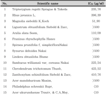
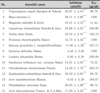
To select candidate of anti-inflammation agents, we investigated the effect of Korean medical plants from Hwaak mountain on cell viability and LPS-induced NO production in RAW 264.7 macrophages. As shown in Table 1, among the ethanol extract of 15plants, 6 plants ( L. obtusifolium Siebold & Zucc.; LO, F. rhynchophylla Hance; FR, S. prunifolia f. simpliciflora Nakai; SP, S.
deltoides Nakai; SD, L. obtusiloba Blume; LOB, A.
mandshuricum Maxim.; AM) had no effect on the cell viability as determined by MTT assay at 500 ㎍/㎖.
These results suggested that ethanol extract of 6 plants indicated nonspecific cytotoxicity in RAW 264.7 macrophages. In addition, our data revealed that 11 plants represent the IC
50value in less than 250 ㎍/㎖
and 4 plants ( T. regelii Sprague & Takeda; TR, R . javanica L.; RJ, M. sieboldii K.Koch ; MS, L. obtusifolium Siebold & Zucc.; LO) show inhibitory ratio on NO production in more than 30% at 125 ㎍/㎖. Considering all of these, LO was selected since it showed nonspecific cytotoxicity and reduction of NO production, and we investigated the anti-inflammatory effect of LO underlying molecular mechanisms in RAW 264.7 macrophages.
No. Scientific name IC50 (㎍/㎖)
1 Tripterygium regelii Sprague & Takeda 235.76
2 Rhus javanica L. 396.39
3 Magnolia sieboldii K.Koch 51.90
4 Ligustrum obtusifolium Siebold & Zucc. >500
5 Aralia elata Seem. 110.09
6 Fraxinus rhynchophylla Hance >500
7 Spiraea prunifolia f. simplicifloraNakai >500
8 Synurus deltoides Nakai >500
9 Lindera obtusiloba Blume >500
10 Sambucus williamsii var. coreana Nakai 225.54
11 Clerodendrum trichotomum Thunb. 425.52
12 Zanthoxylum schinifolium Siebold & Zucc. 410.76
13 Acer mandshuricum Maxim. >500
14 Philadelphus schrenkii Rupr. <10
15 Acer ukurunduense Trautv. & C.A.Mey. <10 Each value represents the mean ± SD (n = 3).
Table 1. Effect of Ethanol Extracts of Korean Medicinal Plants on the Cell Viability in RAW 264.7 Macrophages.
No. Scientific name Inhibition ratio(%)
IC50
(㎍/㎖) 1 Tripterygium regelii Sprague & Takeda 30.67 ± 5.53*** 98.79
2 Rhus javanica L. 39.11 ± 2.96*** >500
3 Magnolia sieboldii K.Koch 52.51 ± 5.12*** 11.44 4 Ligustrum obtusifolium Siebold & Zucc. 34.96 ± 4.55*** 249.66 5 Aralia elata Seem. 28.24 ± 2.81*** 148.18 6 Fraxinus rhynchophylla Hance 15.70 ± 2.24*** >500 7 Spiraea prunifolia f. simplicifloraNakai 11.96 ± 1.29** 167.11 8 Synurus deltoides Nakai 4.48 ± 3.88 >500 9 Lindera obtusiloba Blume 0.00 ± 6.58 239.89 10 Sambucus williamsii var. coreana Nakai 14.91 ± 3.58*** 72.52 11 Clerodendrum trichotomum Thunb. 14.33 ± 1.72*** 243.15 12 Zanthoxylum schinifolium Siebold & Zucc. 20.83 ± 0.95*** 98.79 13 Acer mandshuricum Maxim. 6.03 ± 5.29 188.67 14 Philadelphus schrenkii Rupr. 26.95 ± 1.99*** 99.74 15 Acer ukurunduense Trautv. & C.A.Mey. 17.20 ± 3.58*** >500
Inhibition ratio evaluated at dose of 125㎍/㎖. Each value represents the mean ± SD (n = 3).*p < 0.05, **p < 0.01,***p < 0.001 Table 2. Effect of Ethanol Extracts of Various Korea Compositae Herbs on the LPS-induced NO Production Level in RAW 264.7 Macrophages.
2. Effect of LO on Cell Viability in RAW 264.7 Macrophages
MTT assays were performed to confirm the effect of LO on cell viability in RAW 264.7 macrophages. As shown in Figure 1, treatment with LO (100, 200 and 400 ㎍/㎖) for 24h had no effect on the cell viability in RAW 264.7 macrophages. Accordingly, we investigated the anti-inflammatory effects of LO with concentration 100, 200 and 400 ㎍/㎖ in LPS-stimulated RAW 264.7 macrophages.
Figure 1. Effect of LO on the cell viability in RAW 264.7 macrophages. RAW 264.7 macrophages were treated with different concentrations of LO for 24 h, and their viability was determined using MTT assay. Values represent mean ± S.D. of three independent experiments.
3. Effect of LO on LPS-induced NO Production in RAW 264.7 Macrophages
To investigated the inhibitory effects of LO on LPS- induced NO production in RAW 264.7 macrophages,
cells were treated with or without LO (100, 200 or 400
㎍/㎖) for 1h and then treated with LPS (1 ㎍/㎖) for 24h. As shown in Figure 2, LPS-induced NO production was significantly decreased by LO in a concentration- dependent manner and its effect is similar to NIL used as a positive control inhibitor.
Figure 2. Effect of LO on LPS-induced NO production in RAW 264.7 macrophages. Cells were treated with different concentrations of LO for 1 h prior to the addition of LPS (1 ㎍/㎖), and the cells were further incubated for 24 h. NO levels were determined with Griess reagent. NIL (20 μM) was used as a positive control inhibitor.
The data shown represent the mean ± SD of three independent experiments. ###p < 0.001 vs the control group; ***p < 0.001 vs the LPS-treated group.
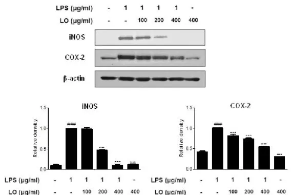
4. Effects of LO on the LPS-induced iNOS and COX-2 Protein Expression in RAW 264.7 Macrophages
Because the inhibition of NO production by LO can result from the suppression of iNOS and COX-2 expression levels
12), we examined the effect of LO on the levels of iNOS and COX-2 protein expressions by Western blot analysis. In unstimulated RAW 264.7 macrophages, the levels of iNOS and COX-2 protein expressions were undetectable or a little. However, the expression levels of iNOS and COX-2 proteins were significantly up-regulated ( P < 0.001) in response to LPS (1 ㎍/㎖), while pretreatment with LO inhibited the expression levels of iNOS and COX-2 proteins ( P < 0.001) in a dose-dependent manner.
5. Effects of LO on the LPS-induced
Production of Pro-inflammatory Cytokines in RAW 264.7 Macrophages
To determine the inhibitory effects of LO on pro-
inflammatory cytokine production induced by LPS, we
investigated its effects on LPS-induced TNF-α , IL-6
and IL-1β productions by using EIA kits. As shown in
Figure 4, LPS significantly increased TNF-α , IL-6 and
IL-1β productions ( P < 0.001), while pretreatment with
LO reduced the LPS-induced these cytokines productions ( P < 0.001) in a dose-dependent manner.
Figure 3. Effects of LO on the LPS-induced iNOS and COX-2 protein expression in RAW 264.7 macrophages. Cells were treated with 100, 200, and 400 ㎍/㎖ LO for 1 h prior to the addition of LPS (1 ㎍/㎖), and the cells were further incubated for 1 h. The protein levels of iNOS and COX-2 were determined by Western blot analysis using specific antibodies. Densitometric analysis was performed using Bio-Rad Quantity One software. The data shown represent the mean ± SD of three independent experiments. ###p < 0.001 vs the control group;
***p < 0.001 vs the LPS-treated group.
Figure 4. Effects of LO on the LPS-induced production of pro- inflammatory cytokines in RAW 264.7 macrophages. Cells were treated with 100, 200, or 400 ㎍/㎖ LO for 1 h prior to the addition of LPS (1 ㎍/㎖), and the cells were further incubated for 24 h.
TNF-α, IL-6, and IL-1β levels were determined by using EIA kits.
The data shown represent mean ± SD of three independent experiments. ###p < 0.05 vs the control group; ***p < 0.001 vs the LPS-treated group.
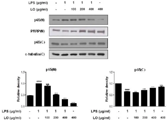
6. Effects of LO on the LPS-induced NF-κ B Activation and the Nuclear Translocation in RAW 264.7 Macrophages
Because NF-κ B is a crucial transcription factor for
the levels of iNOS, and COX-2 protein expression and
pro-inflammatory cytokines productions
13), we studied
the effect of LO on the nuclear translocation of the p65 subunit of NF-κ B in RAW 264.7 macrophages. As shown in Figure 5, LPS induced the translocation of p65 NF-κ B to the nucleus, while pretreatment with LO suppressed this process in a dose-dependent manner.
PARP and α-tubulin were used as internal controls of nuclear and cytosol fraction, respectively. These data indicated that the activation of NF-κB was involved in the inhibition of LPS-induced inflammatory responses in RAW 264.7 macrophages by LO.
Figure 5. Effects of LO on the LPS-induced NF-κB activation and the nuclear translocation in RAW 264.7 macrophages. Cells were pretreated with LO for 1 h prior to the addition of LPS (1 ㎍/㎖) for 1 h. Nuclear (N) and cytosolic (C) extracts were isolated, and the levels of p65 in each fraction were determined by Western blot analysis. PARP and α-tubulin were used as internal controls. Densitometric analysis was performed using Bio-Rad Quantity One software. The data shown represent mean ± SD of three independent experiments.
###p < 0.001 vs the control group; ***p < 0.001 vs LPS-treated group.
Ⅳ. Discussion
The pathology of inflammation is initiated by complex processes triggered by microbial pathogens such as LPS
14). LPS is a strong toll-like receptor 4 (TLR4) signal activator and an endotoxin, an integral outer membrane component of Gram-negative bacteria, and triggers the most potent microbial initiators of inflammatory response, including septic shock, fever, and microbial invasion
15). Macrophages are major cellular targets for LPS action.
LPS stimulates macrophages to produce iNOS, COX-2, HDC, pro-inflammatory cytokines. TNF-α, IL-6, and IL-1β are well-known as pro-inflammatory cytokines in the induction of inflammation in macrophages. These cytokines are associated with the biological functions such as the regulation of cell proliferation, differentiation, and immunity, with the main function being to recruit additional immune cells to inflammatory sites
16, 17). In the present study, it was found that LO is a potent inhibitor on the LPS-induced pro-inflammatory molecules,
including NO, TNF-α , IL-6 and IL-1β in RAW 264.7 macrophages (Figure 2 and 4).
Pro-inflammatory mediators, iNOS and COX-2, play key roles in the pathogenesis of various acute and chronic inflammatory diseases
18). The pharmacological blockade of LPS-inducible inflammatory mediators is an attractive therapeutic strategy for these inflammatory diseases.
iNOS and COX-2 are important enzymes that regulate inflammatory processes. iNOS produces an amount of NO, which is responsible for the toxicity of activated macrophages during inflammatory conditions. COX-2 is also an inducible enzyme that produces PGs during inflammation, and accumulating evidence indicates that PGE
2, which is one of the most abundant PGs, is involved in the pain, edema, and vessel permeability associated with inflammatory diseases
19). In this study, our data indicated that LO suppressed the expression levels of iNOS and COX-2 proteins in LPS-stimulated RAW 264.7 macrophages (Figure 3).
In general, LPS activates TLR4, which activates
MyD88- and TRIF-dependent pathways, in addition to activating NF-κ B, phosphorylation of mitogen activated protein kinase (MAPK) and activator protein 1(AP-1)
20,21)