Characterization of Bacillus mojavensis KJS-3 for the Promotion of Plant Growth
Kang Min Kim, Jie Liu, Youn Suk Go and Jae Seon Kang*
Department of Pharmacy, Kyungsung University, Busan 608-736, Korea Received June 2, 2015 /Revised July 7, 2015 /Accepted July 8, 2015
Scientists have recently shown an interest in the characteristics of Bacillus mojavensis strains because of their increasing use in plants as a defense against diseases and mycotoxins. We have shown here that B. mojavensis KJS-3 possesses the typical characteristics of B. mojavensis strains including a strong resistance to high temperatures (≤50°C), tolerance to high salt concentrations (7% NaCl), ethanol tol- erance (40% ethanol), and pH range for growth (pH 5-9). B. mojavensis KJS-3 has been used for the production of cyclic lipopeptides including important antifungal substances such as surfactin, iturin, and fengycin. Polymerase chain reaction analysis in this study showed that B. mojavensis KJS-3 can be used for the production of fengycin and the findings of LC-MS/MS analyses suggest that B. moja- vensis KJS-3 can be used to produce iturin and surfactin. Antifungal activity analys is confirmed that B. mojavensis KJS-3 has antifungal effects on Botrytis cinerea, Rhizoctonia solani AG-4, Sclerotinia scle- rotiorum, and Colletotricum goeosporioides. A microscopy assessment of the roots of wild ginseng plants planted together with B. mojavensis KJS-3 revealed that the roots contained B. mojavensis KJS-3, con- firming the bacteria to be a plant growth promoting endophyte (PGPE) which acts against plant dis- eases and mycotoxins. Our findings lead us to conclude that B. mojavensis KJS-3 can be produced at an industrial level as a microbial pesticide or microbial fertilizer.
Key words :
Antifungal activity, Bacillus mojavensis KJS-3, plant pathogenic fungi, plant growth promoting endophyte
*Corresponding author
*Tel : +82-51-663-4882, Fax : +82-51-663-4809
*E-mail : [email protected]
This is an Open-Access article distributed under the terms of the Creative Commons Attribution Non-Commercial License (http://creativecommons.org/licenses/by-nc/3.0) which permits unrestricted non-commercial use, distribution, and reproduction in any medium, provided the original work is properly cited.
Journal of Life Science 2015 Vol. 25. No. 8. 910~916 DOI : http://dx.doi.org/10.5352/JLS.2015.25.8.910
Introduction
B. mojavensis strains discovered in the Mojave Desert and found to have antibacterial and antifungal activities have been broadly utilized to protect plants against diseases and mycotoxins [1, 2]. B. mojavensis strains were confirmed to be a novel species distinct from Bacillus subtilis, Bacillus amy- loliquefaciens, Bacillus licheniformis, and Bacillus atrophaeus [1, 18]. However, based on Bergey’s Manual of Systematic Bacteriology which serves as an aid in the identification of those bacteria that have been described and cultured, these Bacillus species are more closely related to each other, and exhibit similar biochemical characteristics.
B. mojavensis KJS-3, a new B. mojavensis strain, was initially found in food waste and has been registered in the Korean Culture Center of Microorganisms (KCCM) under the ac-
cession number KCCM10961P [5] with specific patterns of isoquinones. Previous studies have shown that B. mojavensis KJS-3 exhibits almost all typical characteristics of B. moja- vensis strains: the KJS-3 strain bacteria are rod-shaped, Gram-positive, and endospore-forming aerobic bacteria.
Bacillus species are used in many medical, pharmaceutical, agricultural, and industrial processes that take advantage of the bacteria’s wide range of physiological characteristics and their ability to produce a host of enzymes, antibiotics, and other metabolites [5, 12, 13, 16]. The rhizosphere, a narrow region of soil that is directly influenced by root secretions, is associated with microbial activity [3]. Based on the differ- ent positions in plant micro-ecosystems, plant growth pro- moting microorganisms can categorized as either plant growth promoting rhizobacteria (PGPR) or PGPE, where PGPRs grow on or around the roots and PGPEs grow inside the roots, specifically in the intercellular space of roots.
These microorganisms affect plant growth in three different
ways: (1) by synthesizing and providing particular com-
pounds to the plants [7], (2) by facilitating the uptake of
certain nutrients from the environment [4], and (3) by pro-
tecting plants from certain diseases [11]. The potential of
PGPEs to improve plant health has led to a lot of research
into the applied use of these bacteria as microbial pesticides, primarily in agricultural crops [8]. The potential for micro- bial pesticides to reduce the need for chemicals such as chemical pesticides makes them important in the develop- ment of sustainable agricultural practices.
The overall objectives of this study were (1) to examine the biochemical characteristics of B. mojavensis KJS-3, pre- dominantly resistance characteristics including survival at high temperatures, pH tolerance, salt tolerance, and ethanol tolerance; (2) to identify antifungal substances extracted from B. mojavensis KJS-3 and assess the potential anti- microbial activity of these substances; (3) to demonstrate that B. mojavensis KJS-3 functions as a PGPE to protect plants against diseases.
Materials and Methods
Bacterial strains and media
The bacterial strain B. mojavensis KJS-3 (KCCM 10961P) was obtained from Dr. Jae Seon Kang (Department of Pharmacy, Kyungsung University, Korea). Bacteria were grown in tryptic soy broth (TSB) for routine use. For colony selection for bacterial culture, bacterial strains were grown on tryptic soy agar (TSA), from which single colonies were transferred to tubes containing 5 ml TSB and grown aerobi- cally in a shaking incubator (160 rpm) overnight at 37°C.
All reagents were purchased from Sigma-Aldrich (St. Louis, MO, USA) and all microorganisms were obtained from the KCCM and the Korean Agricultural Culture Collection (KACC).
High temperature tolerance
B. mojavensis KJS-3 was cultured in 5 ml TSB medium overnight, after which 100 μl preculture was diluted with 900 μl sterile distilled water. The preculture was continually diluted in sterile distilled water in this way to a final bacte- rial concentration of 10
3CFU/ml.
The final diluted solution (500 μl) was plated onto TSA plates which were incubated at various temperatures (20-50°C) and bacterial growth on the plates was assessed the following day.
Acid and alkali tolerance
To determine the optimum pH range for B. mojavensis KJS-3 growth, the bacteria was cultured in 8 ml TSB medium overnight, and six tubes each containing 100 ml TSB medium
were adjusted to different pHs (pH 2, 4, 6, 7, 8, and 9) using 6 M HCl and 3 M NaOH. An aliquot (1 ml) of the overnight TSB culture of B. mojavensis KJS-3 was added to each of the six tubes which were then incubated (160 rpm) for 8 hr. After 8 hr the optical densities (600 nm) of the six cultures were measured using a UV-Vis spectrophotometer (UV-Mini1240;
Shimadzu, Japan), allowing for the growth status of B. moja- vensis KJS-3 at each pH to be determined.
Salt tolerance
B. mojavensis KJS-3 was cultured in 10 ml TSB medium overnight. A different amount of NaCl (1, 3, 5, 7, and 9 g) was added to each of five tubes containing 100 ml TSB me- dium each and another tube of 100 ml TSB medium lacking NaCl was included as a negative control. After high pressure sterilization of the six tubes of medium, 1 ml of the overnight TSB culture of B. mojavensis KJS-3 was added into each of the six tubes which were then incubated (160 rpm) for 8 hr. After 8 hr, the optical densities (600 nm) of the six cul- tures were measured using a UV-Vis spectrophotometer.
Ethanol tolerance
B. mojavensis KJS-3 endospores were produced using the method described by Choi et al. [5]. Endospore powder (0.01 g) was added to 1 ml sterile distilled water to produce a bacterial suspension. Preparations of 20, 40, 80, and 95%
ethanol were sterilized by autoclaving, after which 100 μl bacterial suspensions were added into each ethanol solution (20, 40, 80, and 95%). The bacteria-containing ethanol sol- utions were continually diluted in sterile distilled water to a final concentration of 10
6CFU/ml, after which 500 μl of each final dilution was plated on to a TSA plate and cultured at 37°C overnight. The following day the bacterial growth on the plates was assessed.
Extraction and identification of antifungal substances
Most Bacillus strains produce antibiotics such as iturin,
fengycin, and surfactin. For the assessment of potential anti-
biotic production by B. mojavensis KJS-3, several colonies of
the bacteria were transferred into tubes containing 5 ml TSB
medium and grown aerobically in a shaking incubator (180
rpm) overnight at 37°C. The 5 ml of preculture was trans-
ferred to a 1 l triangular flask containing TSB medium and
the B. mojavensis KJS-3 culture was then incubated, shaking,
for 24 hr at 25°C. After 24 hr the culture was subjected to
centrifugation at 8,000 rpm for 15 min and 500 ml of the
resulting supernatant was adjusted to pH 2 using 6 M HCl.
Methanol was used to dissolve 150 mg of the resulting pre- cipitate and the methanol was subsequently concentrated us- ing a rotary evaporator. Evaporator bottoms were extracted using methanol and then reconcetrated by using a vacuum evaporator. 10 mg of residue was antimicrobial substances of B. mojavensis KJS-3.
After extraction and purification from the B. mojavensis KJS-3 culture, 10 mg of residue for the potential detection of iturin and surfactin was dissolved in methanol and the sample was further diluted to a final concentration of 100 μg/ml for analysis by LC-MS/MS (Agilent 6410 Triple Quadrupole LC/MS; Agilent Technologies, Santa Clara, CA, USA). For LC, the chromatographic conditions were as fol- lows: Gemini NX-C185 μm stationary phase (150×4.6 mm i.d.; Phenomenex, Torrance, CA, USA); mobile phase: A=wa- ter with 0.1 % TFA and B =acetonitrile with 0.1 % TFA; elu- tion mode: gradient 0-3 min (B=50%), 3-25 min (B =100%).
For MS, the interface conditions used were as follows: the MS was set to electrospray ionization (ESI) mode with a mass range of 100-1,500 ms; SIM polarity in positive mode, gas temperature 350°C, gas flow 10 l/min, nebulizer pres- sure 40 psi, capillary voltage 4.8 kV, and iturin A (molecular weight 1044.6 [M+H]
+) and surfactin (molecular weight 1022.3 and 1036.3 [M+H]
+) as internal standards.
Polymerase chain reaction (PCR) amplifications for the identification of fengycin were performed using a PCR Thermal Cycler (M-2325; Takara Bio, Otsu, Japan). PCR am- plifications were performed in 20 μl ready mix PCR reaction mixtures containing 1 tube TOP simple DryMIX-Tenuto (Enzynomics, Daejeon, Korea), 1 μl genomic DNA template, 1 μl forward primer (FENTD1F, 5′-tttggcagcaggagaagttt- 3′), 1 μl reverse primer (FEND1R, 5′-gctgtccgttctgctttttc- 3′), and distilled water to make up the 20 μl reaction vol- ume [17]. The mixture was gently mixed by vortexing and then subjected to centrifugation for the entire mixture to be collected at the bottom of the tube. The PCR program used was with denaturation, annealing and extention cycle. The amplified DNA was then evaluated by agarose gel electro- phoresis and subsequent ethidium bromide staining.
Anti-fungalactivity
Four phytopathogenic fungi (Botrytis cinerea KACC 40573, Rhizoctonia solani AG-4 KACC 40142, Sclerotinia sclerotiorum KACC 41065, and Colletotrichum gloeosporioides KCCM 11220) were included in the antifungal activity analysis carried out
in this study. The phytopathogenic fungi were cultured on potato dextrose agar (PDA) medium at 37°C, while B.
mojavensisKJS-3 was cultured aerobically in 5 ml TSB me- dium in a shaking incubator (180 rpm) overnight at 37°C.
For each of the phytopathogenic fungi, a small piece of agar was cut out after 48 hr of culture and transferred onto a new PDA plate. On each of these new plates, a line of B.
mojavensis KJS-3 culture was drawn and all plates were cul- tured at 37°C for 48 hr.
PGPE of B. mojavensis KJS-3
Wild white ginseng was harvested from the jiri mountains in Hamyang-gun, Korea and cultivated wild ginseng seeds were then combined with B. mojavensis KJS-3 as follows: 10 kg deshelled ginseng seeds were mixed with 1 kg corn starch, 10 g B. mojavensis KJS-3 (1×10
9CFU/g), and 1 l dis- tilled water. The ginseng seeds combined with the bacteria were then planted on land surface of farmland, cultivated for 3 years, and harvested. The roots of the plants were washed with sterile water, after which they were divided with a sterile razor on a clean bench and observed under a microscope (100×, MTV-33K9HN; Mintron, New Taipei City, Taiwan).
Results and Discussion
Stability of B. mojavensis KJS-3
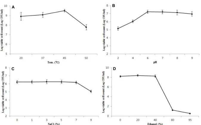
The tolerance of B. mojavensis KJS-3 to different pH, tem-
perature, and salt conditions is shown in Fig. 1. The results
of the temperature tolerance analysis revealed the bacterial
strain to have a growth temperature range of 20-50°C and
an optimum growth temperature of 45°C (Fig. 1A). B. moja-
vensis KJS-3 was found to survive in neutral or slightly acidic
pHs, with the optimal pH range for growth being pH 6-8
(Fig. 1B). No significant decreases in cell viability were ob-
served at neutral (pH 6.0, 7.0, and 8.0) pHs. As shown in
the results (Fig.1C), B. mojavensis KJS-3 cell viability is not
affected by NaCl concentrations ≤7%, indicative of the bac-
teria being highly tolerant to salt. Microbial growth is greatly
affected by external environmental conditions and salt is a
naturally occurring element in soils and water [10]. Salt tol-
erance is therefore essential for the survival of micro-
organisms in soil and water [10]. With increasing human
development, soil salinization is one of the most important
problems resulting from land degradation and basic envi-
ronmental problems in arid and semi-arid regions [10]. The
A B
C D
Fig. 1. Viability of B. mojavensis KJS-3 cultivated with various (A) Temperature (°C), (B) pHs, (C) NaCl (%), (D) ethanol (%) on TSB medium.
Fig. 2. Identification of Fengycin produced from B. mojavensis KJS-3 on TSB medium by PCR analysis. (A) Marker, (B) B. mojavensis KJS-3, (C) Bacillus subtilis KCCM 11316 (positive control).
results presented here are consistent with those previously reported regarding the viability of B. mojavensis KJS-3 endo- spores under conditions of varied pH, temperature, and NaCl concentration [5]. Ethanol is commonly used to kill bacteria including B. mojavensis KJS-3. The endospores of B.
mojavensis KJS-3, however, were found to have a strong toler- ance to ethanol, surviving in solutions containing ≤40%
ethanol (Fig. 1D). These findings have implications for in- dustrial fermentation which is widely applied in terms of fermentation by microorganisms such as probiotics to pro- duce microbial products useful to humans.
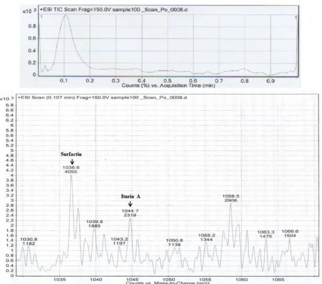
Identification of fengycin, iturin, and surfactin B. subtilis KCCM 11316 was used as a positive control for fengycin detection by PCR analysis in this study. In the agar- ose gel visualizing the resulting PCR products, aprominent band representing the fengycin synthetase gene of B. subtilis was observed at 1.2 kbp and a corresponding band, also at 1.2 kbp, was observed for B. mojavensis KJS-3 (Fig. 2). In the LC-MS/MS analyses carried out, the fractions containing standards (surfactin and iturin A) were found to contain qua- si-molecular ions at m/z = 1022.3 and 1036.3 ([M+H]
+) for surfactin and at m/z = 1044.6 ([M+H]
+) for iturin A. Based
on the peaks of the standard samples and the molecular
weights detected for the test substances (surfactin: 1036.3 m/z
[M+H]
+; iturin A: 1044.7m/z [M+H]
+; Fig. 3) it can be con-
cluded that the surfactin and iturin A produced by B. moja-
Fig. 3. Identification of Surfactin and Iturin A produced from B. mojavensis KJS-3 on TSB medium by LC-MS/MS. LC (upper) and MS/MS (below) data.
vensis KJS-3 are similar to commercially available surfactin and iturin A standards, respectively. These findings there- fore indicate that fengycin, surfactin, and iturin A can be produced by B. mojavensis KJS-3. Cyclic lipopeptides includ- ing surfactin, iturin, and fengycin are the major classes of biosurfactants which are known to be produced by Bacillus species [15]. In the LC-MS/MS spectrum, well-resolved groups with peaks between 1,000 and 1060 m/z were ob- served, and these peaks can be attributed to the isoform en- sembles of surfactins, iturins, and fengycins, which are in- cluded in the well-known surfactin products produced by Bacillus strains [21]. The cyclic lipopeptides have fur- thermore been shown to have higher antimicrobial and anti- fungal activity against Gram-positive cocci than against Gram-negative bacilli [6, 20].
Antifungal activity of B. mojavensis KJS-3 Cultured B. mojavensis KJS-3 were found to exert anti- fungal activity against Botrytis cinerea KACC 40573, Rhizocto- nia solani AG-4 KACC 40142, Sclerotinia sclerotiorum KACC 41065, and Colletotrichum gloeosporioides KCCM 11220. B. mo- javensis KJS-3 was found to inhibit the growth of all four fungal species (Fig. 4): inhibitive belts were clearly formed and B. mojavensis KJS-3 was shown to inhibit the in vitro growth of the mycelia of these phytopathogenic fungi.
Several strains of B. mojavensis have been shown to inhibit
the in vitro growth of Fusarium moniliforme; however, all
strains of B. mojavensis produce different antifungal sub-
stances [1]. The antifungal compounds produced by Bacillus
spp., such as fengycin, iturin, and surfactins, have been ex-
tensively studied for potential biocontrol activity [14].
Fig. 4. Antifungal activity of the cocultivated B. mojavensis KJS-3 (Vertical line) on mycelia growth of (A) Botrytis cinerea, (B) Rhizoctonia solani AG-4, (C) Sclerotinia sclerotiorum, (D) Colletotrichum gloeosporioides. All strains are culti- vated on PDA medium.
Fig. 5. Identification of B. mojavensis KJS-3 cocultivated with wild white ginseng seeds on land surface of farmland for 3 years throughout microscopy. Bar = 10 μm.
B. mojavensis KJS-3 as a PGPE
An unknown bacterial strain suspected to be B. mojavensis KJS-3 was observed inside the roots of cultivated wild white ginseng plants by microscopy (Fig. 5). To confirm the iden- tity of the selected bacterial strains, the enzymes produced by the unknown strains were shown to be the same as those produced by B. mojavensis KJS-3. B. mojavensis KJS-3 as a plant endophyte is able to enter plant roots and survive in the intercellular space, and furthermore, as a PGPE, exerts a protective effect on plants. Many studies have demon- strated PGPEs to be effective biocontrol agents for plant pro- tection in vitro and in vivo and field application of suspen- sions of Bacillus spp. in the growth season of plants more over resulted in significantly reduced disease incidence [1,
9, 14, 19]. PGPEs are rhizosphere bacteria that can enhance plant growth in a wide variety of applications such as micro- bial pesticides, microbial fertilizers, and animal feed additives. This study confirms that B. mojavensis KJS-3 is a promising probiotic that can be produced at an industrial level as a microbial product. The potential use of B. moja- vensis KJS-3 as a biological control agent for field applica- tions is supported by the presently reported results of labo- ratory bioassay analyses, which serve as relative predictors of resistance to pathogenic fungi. The findings of this study indicate that the industrial production of B. mojavensis KJS-3 may have wide applications in various areas, especially in agriculture.
References
1. Bacon, C. W. and Hinton, D. M. 2002. Endophytic and bio- logical control potential of Bacillus mojavensis and related species. Biol. Control 23, 274-284.
2. Bacon, C. W. and Hinton, D. M. 2007. Potential for control of seedling blight of wheat caused by Fusarium graminearum and related species using the bacterial endophyte Bacillus mojavensis. Biocontrol. Sci. Technol. 17, 81-94.
3. Bloemberg, G. V. and Lugtenberg, B. 2001. Molecular basis of plant growth promotion and biocontrol by rhizobacteria.
Curr. Opin. Plant Biol. 4, 343-350.
4. Cakmakci, R., Dönmez, F. Aydın, A. and Sahin, F. 2006.
Growth promotion of plants by plant growth-promoting rhizobacteria under greenhause and two different field soil conditions. Soil. Biol. Biochem. 38, 1482-1487.
5. Choi, S. M., Park, M. H., Jung, T. S., Moon, K. H., Kim, K. M. and Kang, J. S. 2011. Characterization of Bacillus moja- vensis KJS-3 for industrial applications. Arch. Pharm. Res. 34, 289-298.
6. Fernandes, P. A. V., Arruda, I. R. D., Santo, A. F. A. B. D., Araújo, A. A. D., Maior, A. M. S. and Ximenes, E. A. 2007.
Antimicrobial activity of surfactants produced by Bacillus subtilis R14 against multidrug-resistant bacteria. Braz. J.
Microbiol. 38, 704-709.
7. Glick, B. R. 1995. The enhancement of plant growth by free-living bacteria. Can. J. Microbiol. 41, 109-117.
8. Hallmann, J., Qualt-Hallmann, A., Mahaffee, W. F. and Kloepper, J. W. 1997. Bacterial endophytes in agricultural crops. Can. J. Microbiol. 43, 895-914.
9. Jang, Y., Kim, S. G. and Kim, Y. H. 2011. Biocontrol efficacies of Bacillus species against cylindrocarpon destructans caus- ing ginseng root rot. Plant Pathol. J. 27, 333-341.
10. Kassas, M. 1977. Arid and semi-arid lands: problems and prospects. Agro-ecosyst. 3, 185-204.
11. Khan, M. A., Gul, B. and Weber, D. J. 2002. Improving seed germination of Salicorniarubra (Chenopodiaceae) under sal- ine conditions using germination regulating chemicals.
West. N. Am. Naturalist 62, 101-105.
초록:식물 성장 촉진에 사용에 있어 Bacillus mojavensis KJS-3의 특징
김강민․유걸․고윤석․강재선*
(경성대학교 약학과)
최근 식물 성장에 있어 곰팡이 독소에 관련된 질병에 효과가 있는 Bacillus mojavensis 균주 사용의 보고가 있다.
우리는 B. mojavensis KJS-3균주의 다양한 온도, 염도, 에탄올, pH에서 성장하는 특징을 확인 하였다. B. mojavensis KJS-3균주는 Polymerase chain reaction 분석에 의해 fengycin을 LC-MS/MS 분석을 통해서는 iturin 및 surfactin 와 같은 cyclic lipopeptides를 생산함을 확인 하였다. B. mojavensis KJS-3균주는 식물 유해 곰팡이 균주인 Botrytis cinerea, Rhizoctonia solani AG-4, Sclerotinia sclerotiorum, Colletotricum goeosporioides에 항곰팡이 효과가 있음을 확인 하였고 이 결과를 바탕으로 인삼재배에 있어 B. mojavensis KJS-3를 사용하여 성장을 관찰한 결과 뿌리 내에서 성 장하여 plant growth promoting endophyte가 있음을 알 수 있었다. 이러한 특징들에 의해 미생물 농약 및 비료로 사용할 수 있을 것이다.
12. Kim, K. M., Jung, T. S., Ok, S., Ko, C. Y. and Kang, J. S.
2011. In vitro Characterization study of Bacillus mojavensis KJS-3 for a potential probiotic. Food Sci. Biotechnol. 20, 1155- 1159.
13. Kim, K. M., Jung, T. S., Ok, S., Ko, C. Y. and Kang, J. S.
2012. Evaluation of genotoxicity of Bacillus mojavensis KJS-3 on culture supernatant for use as a probiotic. Mol. Cell.
Toxicol. 8, 77-81.
14. Li, Y., Han, L., Zhang, Y., Fu, X., Chen, X., Zhang, L., Mei, R. and Wang, Q. 2013. Biological control of apple ring rot on fruit by Bacillus amyloliquefaciens 9001. Plant Pathol. J. 29, 168-173.
15. Mukherjee, A. K. and Das, K. 2005. Correlation between di- verse cyclic lipopeptides production and regulation of growth and substrate utilization by Bacillus subtilis strains in aparticular habitat. FEMS Microbiol. Ecol. 54, 479-489.
16. Pyo, J. S., Shrestha, S. (Amatya), Park, S. H. and Kang, J.
S. 2014. Biological control of plant growth using the plant growth-promoting rhizobacterium Bacillus mojavensis KJS-3.
J. Life Sci. 24, 1308-1315.
17. Ramarathnam, R., Bo, S., Chen, Y., Fernando, W. G., Xuewen, G. and de Kievit, T. 2007. Molecular and biochemical de-
tection of fengycin and bacillomycin D-producing Bacillus SPP., antagonistic to fungal pathogens of canola and wheat.
Can. J. Microbiol. 53, 901-911.
18. Roberts, M. S., Nakumora, L. K. and Cohan, F. M. 1994.
Bacillus mojavensis sp.Nov., distinguishable from Bacillus subtilis by sexual isolation, divergence in DNA sequence, and differences in fatty acid composition. Int. J. Syst.
Bacteriol. 44, 256-264.
19. Ryu, H., Park, H., Suh, D. S., Jung, G. H. and Park, K. 2014.
Biological control of Colletotrichum panacicola on Panax gin- seng by Bacillus subtilis HK-CSM-1. J. Ginseng Res. 38, 215- 219.
20. Vanittanakom, N., Loeffler, W., Koch, U. and Jung, G. 1986.
Fengycin--a novel antifungal lipopeptide antibiotic pro- duced by Bacillus subtilis F-29-3. J. Antibiot. 39, 888-901.
21. Vater, J., Kablitz, B., Wilde, C., Franke, P., Mehta, N. and Cameotra, S. S. 2002. Matrix-assisted laser desorption ion- ization-time of flight mass spectrometry of lipopeptide bio- surfactant in whole cells and culture filtrates of Bacillus sub- tilis C-1 isolated from petroleum sludge. Appl. Environ.
Microb. 68, 6210-6219.