Growth Promotion of Pavlova viridis by Bacteria Isolated from the Microalga
Sarker Anowarul Kabir Ahamed
1, Jin-Joo Kim
2, Tae-O Choi
3and Tae-Jin Choi
1*
1Department of Microbiology, Pukyong National University, 45, Yongso-ro, Nam-Gu. Busan 608-737, Korea
2Department of Fisheries Biology, Pukyong National University, Busan 608-737, Korea
3Chloland Co. Limited., 395
Hambakgeum-gil,
Dongbu-myeon,
Geoje-si,
Gyeongsangnam-do 656-851, Korea Received March 16, 2015 /Revised April 6, 2015 /Accepted April 6, 2015
The marine microalga Pavlova viridis can grow fast and has the ability to accumulate essential nu- trients for culturing marine animals, such as EPA and DHA, and it has been used as food for raring larval fish and prawn. The symbiotic relationship between the flagellate microalga Pavlova viridis and its associated bacteria was investigated. An axenic culture of P. viridis was obtained by repeated treat- ment of the microalga with an antibiotic cocktail. The axenic status was confirmed after sub-culturing three times in a sterile f/2 medium without an antibiotic. The axenic alga was then co-inoculated with five bacteria, arbitrarily designated as I1–I5, isolated from the alga to test the growth promotion of the algae. All bacterial strains promoted the growth of P. viridis, and bacterial isolate I3 was the most effective among the five bacteria tested. The cell number of P. viridis in the co-culture with I3 was significantly higher than that of the control culture. A sequence analysis of the 16S rRNA gene iso- lated from I3 revealed a 97% nucleotide sequence similarity to that of Citrobacter sp. The growth of strain I3 was also significantly enhanced by co-culturing with P. viridis, indicating a symbiotic relation- ship between the microalga and its associated bacterium. The association between the microalga and bacterium was confirmed by scanning electron microscopy.
Key words :
Aaxenic, Citrobacter sp., coculture, Pavlova viridis, symbiosis
*Corresponding author
*Tel : +82-51-629-5617, Fax : +82-51-629-5619
*E-mail : [email protected]
This is an Open-Access article distributed under the terms of the Creative Commons Attribution Non-Commercial License (http://creativecommons.org/licenses/by-nc/3.0) which permits unrestricted non-commercial use, distribution, and reproduction in any medium, provided the original work is properly cited.
Journal of Life Science 2015 Vol. 25. No. 5. 568~576 DOI : http://dx.doi.org/10.5352/JLS.2015.25.5.568
Introduction
Microalgae are microscopic unicellular organisms found typically in marine and fresh waters, and they usually coex- ist with bacteria [18]. They are primary producers that affect the nutrient cycles in aquaculture ecosystems [8]. Microalgae are indispensable natural food sources in aquaculture for all growth stages of bivalve, crustacean, and fish species, and they also serve as food for zooplankton to continuously sup- port the food web. Microalgae provide energy and nutrients to marine organisms that need those for growth and development. Because they possess well-balanced nutrient contents, they have been used for the enrichment of nu- trients in zooplanktons before these are fed to juvenile fish, and are very important in the culture of diverse aquatic ani- mals [3, 4, 26].
Microalgae are used for various other purposes as well.
For example, they are good sources of highly valuable bio- active compounds, polyunsaturated fatty acid, antioxidants, and pharmaceuticals [13]. Algae are also known for their conspicuous color pigments. They produce not only chlor- ophyll, the photosynthetic pigment, but also phycobilipro- teins and carotenoids that are used to protect skin from dam- age due to sunlight, as well natural food colorants with ap- plications in, e.g., cosmetics [16, 22].
The marine microalga Pavlova viridis used in this study can grow quickly, and owing to its high contents of poly- unsaturated fatty acids, EPA, and DHA, it is used as food for rearing larval fish and prawn [19]. It has the ability to accumulate essential nutrients for culturing marine animals [5].
The isolation and culture of microalgae is often accom-
panied by bacterial contamination. Axenic algal cultures are
essential for physiological, chemical, molecular, or taxo-
nomic studies and the determination of zooplankton food
preference and algal histories [23]. As some aquatic bacteria
can inhibit algal growth or cause lysis of algal cells, estab-
lishing axenic algal cultures is important to protect the algae
from such algicidal action [8]. A few methods are used for
the establishment of axenic microalgae cultures, although
obtaining an axenic culture from a highly contaminated algal
culture is an arduous job [6, 7]. Treatment with antibiotics is the most common approach to attain axenicity of micro- algae from bacteria [7].
Many bacteria live on the cell surface of microalgae and they have diverse effects on the growth of the host algae.
The relationships between microalgae and bacteria vary de- pending on the species and environmental conditions [18].
Bacteria associated with microalgae may have positive or negative impacts on the growth of microalgae. Many reports have demonstrated that aquatic bacteria can cause algal cells to lyse, or exert inhibitory effects on the growth of micro- algae via detrimental chemicals produced by the bacteria [2, 12, 18]. In addition to these suppressive actions, influential or stimulatory effects between bacteria and microalgae have also been reported [10, 21, 24]. Watanabe et al. [27] Watanabe et al. [27] reported a strong positive effect on the growth of the microalga Chlorella sorokiniana when cocultured with the bacterium Microbacterium trichotecenolyticum. Moreover, Riquelme et al. [21] Riquelme et al. [21] reported the fastest growth of the alga Asterionella glacialis with Pseudomonas sp.;
they assumed that the bacteria produce chemicals like vita- mins and lipoprotein, which promote the growth of microalgae. Also, eight bacterial strains separated from Chlorella ellipsoidea had growth-promoting effects in co- culture with the microalga [20].
In the current study, we isolated and characterized bac- teria associated with P. viridis and studied their interactions by coculturing the axenic alga with and without the isolated bacteria.
Materials and Methods
Microalga culture and identification
The marine microalga P. viridis was obtained from the Korea Marine Microalgae Culture Center, Pukyong National University, Busan, Korea. The alga was cultured in f/2 me- dium [14, 15] at 20°C under a 16:8 hr light:dark cycle with a light intensity of 30 μmol m
–2s
–1[1]. For the genetic iden- tification of host alga, genomic DNA was isolated according to a previously described method (http://www.gbiogene.
com). The 18S rRNA gene of the alga was amplified by PCR (HS Prime Taq premix 2×; Genet Bio, Cheonan-si, Korea) using primers 512F (5’-ATTCCAGCTCCAATA GCG-3’) and 978R (5’-GACTACGATGGTATCTAATC-3’). The PCR con- ditions were as follows: 94°C for 2 min, followed by 35 cycles at 94°C for 2 min, 52°C for 45 s, and 72°C for 1 min, followed
by a final extension at 72°C for 10 min. The PCR products were visualized in a 1% agarose gel. The amplified DNA fragments were purified from agarose gels using a Gel SV kit (Gene All, Seoul, Korea) and were sequenced using 18S rRNA gene primers (512F/978R; G&C Bio, Daejon, Korea).
Homology analysis was carried out using BLAST (http://
blast.ncbi.nlm.nih.gov/Blast.cgi).
Isolation of bacteria
To obtain bacterial isolates from the original algal culture, 100-μl algal cultures of P. viridis were spread on Luria–
Bertani (LB) agar plates and incubated at 37°C for 72 hr.
Colonies showing different morphology and color were streaked again on LB plates and incubated at 37°C for 72 hr. After three passages on LB plates, glycerol stocks of sin- gle colonies were prepared in LB broth containing 20% glyc- erol and then stored at -80°C for further study.
Establishing an axenic culture of P. viridis The isolated bacteria were tested against the five anti- biotics listed in Table 1. LB agar plates with different concen- trations of antibiotic were prepared and single colonies were inoculated and incubated at 37°C for 72 hr. Antibiotic sus- ceptibility was identified by the growth of bacterial colonies.
An antibiotic cocktail was prepared based upon the anti- biotic resistance for the cleaning of P. viridis. An axenic cul- ture of P. viridis was established by following the method described by Hong et al. [17] Hong et al. [17]. Well-grown alga in f/2 medium was transferred to f/2 medium with antibiotic cocktail. After 8-10 days, the alga was transferred again to fresh f/2 medium containing the same antibiotic cocktail. Axenic status was verified through spreading the culture on LB agar plates followed by incubation at 37°C for 7 days. These procedures were repeated until no bacterial growth occurred.
Identification of bacterial isolates
The taxonomic identification of bacterial isolates was per-
formed by sequence analysis of the 16S rRNA gene. An over-
night culture of 3-5 ml was centrifuged at 13,000 ×g for 1
min, after which the pellet was suspended in 500 μl of ex-
traction buffer [400 mM NaCl, 20 mM Trizma Base, 5 mM
EDTA (pH 8.0), 1% SDS]. Next, 50 μl of 20% SDS and 500
μl of phenol:chloroform:isoamylalcohol (25:24:1) was added
to the suspension. The mixture was vortexed for 1 min and
then centrifuged at 13,000 rpm for 5 min. The supernatant
was transferred to a new tube and 0.1 volumes of 3 M so- dium acetate and 0.6 volumes of isopropanol were added to the tube and mixed gently by inverting, followed by cen- trifugation at 13,000 rpm for 20 min. Finally, the dried DNA was dissolved in 50 μl of dH
2O. The 16S rRNA gene from bacteria was amplified by PCR (HS Prime Taq premix 2 ; Genet Bio) using 27F (5’-AGAGTT TGATCCTGGCTCAG-3’) and 1492R (5’-GGTTACCTTGTTA CGACTT-3’) [24] primers under the same PCR conditions as described above. The PCR product was sequenced using 16S rRNA gene primers (27F and 1492R). Homology analysis was carried out using EzTaxon (http://www.ezbiocloud.net/).
Culture of bacterial isolate I3
Suitable growth conditions for bacterial strain I3 were de- termined by optimizing the temperature, NaCl concen- tration, and pH. For each comparison, a 40-μl aliquot of bac- terial isolate I3 culture freshly grown at 37°C overnight in LB broth was used to inoculate 4 ml of LB broth. The opti- mum temperature was determined by inoculating a 40-µl ali- quot of bacterial isolate I3 culture to 4 ml of LB broth and incubating the culture at 4°C, 20°C, 25°C, 30°C, 37°C, or 45°C with shaking at 150 rpm for 24 hr. The optimum NaCl con- centration for bacterial growth was examined by preparing LB broth containing 0%, 1%, 2%, 3%, 4%, 5%, 7%, and 10%
NaCl and inoculating 4 ml of broth with a 40-μl aliquot of bacterial isolate I3 culture, followed by incubation at 37°C with shaking at 150 rpm for 24 hr. The optimum pH was examined by preparing LB broth containing 1% NaCl with the pH adjusted to 3, 4, 5, 6, 7, 8, 9, or 10; then 4 ml of broth was inoculated with a 40-μl aliquot of bacterial isolate I3 culture and incubated at 37°C with shaking at 150 rpm for 24 hr. Three replications were made for each temperature.
Bacterial growth at each temperature was monitored after 24 hr by measuring the absorbance at 600 nm using a spec- trophotometer (MBA 2000; Perkin-Elmer, Wellesley, MA, USA). Means were determined from three replicates.
Algal growth assays with five bacterial strains To determine the growth promoting effect of the bacteria, 100 ml of f/2 medium was inoculated with axenic P. viridis and cultured for 7 days at 20°C as described above. Five bacterial isolates were inoculated separately, with each in- oculum consisting of 4 ml cultured in LB broth overnight at 37°C. The f/2 medium (100 ml) was inoculated with 1 ml of algal culture containing 7.08×10
4cells/ml plus 1 ml
each of the five bacterial suspensions that had been adjusted to an optical density of 0.5 or 1 ml of LB broth as the control.
Cultures were prepared in triplicate and incubated at 20°C for 10 days, and the growth of P. viridis was monitored daily by counting the cells using a hemocytometer.
Algal growth with bacteria-free filtrate
The bacterial filtrate was prepared by growing the I3 iso- late in 25 ml of LB broth for 3 days at 37°C and 150 rpm.
The culture was centrifuged at 4,000 × g for 10 min, and filtered twice through 0.2-μm polycarbonate filters (Minisart, Goettingen, Germany). Then, a 1-ml aliquot of this bac- teria-free filtrate or LB broth as a negative control was added to 100 ml of f/2 medium containing 7.02×10
4algal cells/ml prepared as described above; the mixtures were incubated at 20°C for 8 days. Each culture was prepared in triplicate and the algal cells were counted daily for up to 8 days using a hemocytometer.
For comparative assays of algal growth with different amount of filtrates, 1, 2.5, 5.0, and 10.0 ml of bacteria-free filtrates of I3 were added to 98, 96.5, 94, and 89 ml of f/2 medium, respectively, containing 7.02×10
4algal cells/ml.
The same amounts of LB broth were used as controls with- out bacteria-free filtrate. The cells were cultured at 20°C and the cells were counted after 5 days using a hemocytometer.
Growth promotion of the alga and bacteria in coculture
The inoculums of P. viridis and bacterial isolate I3 were prepared as described above. Bacterial suspension (1 ml; op- tical density 0.5) and 1 ml of axenic P. viridis bearing initial cells of 7.06×10
4were inoculated to 100 ml of fresh f/2 me- dium as a coculture treatment. Controls were prepared sim- ilarly using the bacteria alone or alga alone. All cultures were prepared in triplicate. Three replicates of 100-μl sam- ples were taken each day from the P. viridis single culture and coculture. Samples were serially diluted and algal cells were counted using a hemocytometer. For the bacterial sin- gle culture and bacteria–algal cocultures, 100-μl samples were taken daily and serially diluted by 10
4–10
5using f/2 medium; then 100 μl of each dilution was spread on LB agar plates and incubated at 37°C for 3 days to count the colony forming units (CFU) of bacteria.
Scanning electron microscopy (SEM) method
The association of the bacteria and algae in coculture was
verified by SEM observation. One milliliter of 2% osmium tetroxide was added to 14 ml of overnight coculture for fix- ation and then stored at 4°C overnight. The cells were col- lected by low-speed centrifugation at 800 g for 20 min, and the supernatant was discarded. The cells were re- suspended with 15 ml of distilled water and poured onto a 0.2-μm cellulose acetate membrane filter of 47 mm diame- ter (Toyo Roshi Kaisha, Tokyo, Japan) connected to a vac- uum through a bottle-top filter (Thermo Scientific, Billerica, MA, USA). After rinsing with 1 ml of distilled water, the membrane was removed, folded sample-side-in, and placed in a 1.8-ml microcentrifuge tube. The cells were sequentially dehydrated with 1.5 ml of 20%, 30%, 50%, 70%, 80%, 85%, 90%, 95%, and 100% ethanol for 30 min each. After two more dehydration steps at 100% ethanol for 30 min, the cells were stored overnight in 100% ethanol. After replacing the ethanol with isoamyl alcohol twice for 1 hr, the samples were dried at critical point, coated with gold–palladium, and observed under SEM (JSM-6490LV; JEOL, Tokyo, Japan).
Results
Identification of the host
To identify the host alga, genomic DNA was prepared from the pure culture of the microalga and used for PCR amplification. A DNA fragment of 490 bp was obtained and its sequence was determined without cloning. Homology analysis was carried out from the GenBank database using BLAST. The results indicated 99% nucleotide sequence iden- tity to that of the 18S rRNA sequence of P. viridis (GenBank accession number HQ877913).
Isolation and identification of bacteria
Five bacterial isolates were obtained from the microalga P. viridis and arbitrarily designated I1–I5. Genomic DNA was prepared from each bacterial isolate for amplification of the partial 16S rRNA (~1,450 bp). The sequences were determined and compared with sequences in the database using EzTaxon. The 16S rRNA sequence of bacterial strains I1–I5 showed 99.950%, 99.034%, 99.69%, 99.217%, and 98.825% sequence identity to that of Kocuria marina (AY 211385), Staphylococcus cohnii (D83361), Citrobacter freundii (ANAV01000046), Micrococcus yunnanensis (Fj214355), and Staphylococcus haemolyticus (L37600), respectively.
Antibiotic resistance test and cleaning of microalga To prepare an antibiotic cocktail to remove bacteria from the alga, five different antibiotics (60 μg/ml ampicillin, 75 μg/ml chloramphenicol, 30 μg/ml kanamycin, 30 μg/ml streptomycin, and 30 μg/ml tetracycline) were tested sepa- rately against the isolated bacteria. Bacterial strains I2, I3, and I4 were susceptible to all antibiotics at the above concentrations. Bacterial isolate I1 demonstrated suscepti- bility to all antibiotics except ampicillin. Bacterial isolate I5 was susceptible to all antibiotics except streptomycin.
Therefore, a working stock of antibiotics containing 2.5 mg/ml of chloramphenicol, 2.0 mg/ml ampicillin, 1 mg/ml of kanamycin, streptomycin, and tetracycline was prepared to achieve the appropriate final concentrations, and this was used to remove these bacteria from the host, P. viridis. The bacterial flora in the microalga prior to antibiotic treatment was 8.1×10
5cells/ml. However, no bacterial colony was ob- served when the algal culture was spread on LB plates after 10 days of culture in f/2 medium containing the antibiotic cocktail. The antibiotic treatment was repeated two more times to confirm that the culture of the microalga was axenic at least in current bacterial culture conditions
Test for growth promotion of P. viridis by isolated bacteria
Each of the five bacterial isolates was inoculated with P.
viridis containing 7.08×10
4cells/ml in f/2 medium. All bacte- rial isolates significantly increased the growth of P. viridis over 10 days, although the stimulatory effect varied among isolates. Bacterial isolate I3 showed the highest growth pro- motion activity among the five isolates tested (Fig. 1). The growth of P. viridis increased by approximately twofold compared to that of the control culture by the most effective bacterial isolate (I3), whereas the other bacterial isolates in- creased the growth of the microalga by about 0.5–1.5-fold.
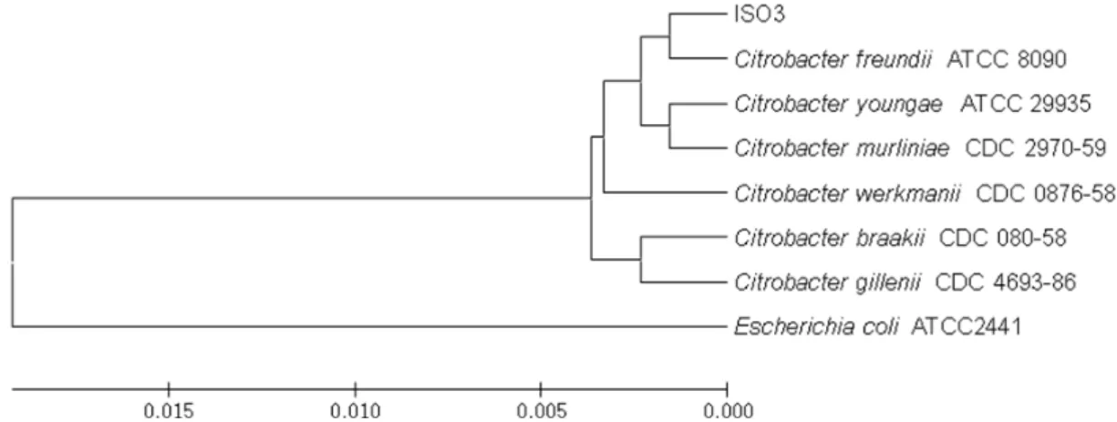
Growth and molecular characteristics of bacterium I3, Citrobacter sp.
Isolate I3 whose 16S rRNA gene showed 99.69% sequence
similarity to that of Citrobacter freundii also formed a close
cluster with this species among other Citrobacter species in
an UPGMA phylogenetic tree (Fig. 2). Therefore, this isolate
was designated as Citrobactor sp. without further bio-
chemical test for species identification. The isolate 3 ap-
peared on LB medium as a translucent colony with a smooth
edge. The bacterium showed slow growth at 4°C and the
Fig. 1. Growth promotion of P. viridis by five bacterial isolates.
Axenic P. viridis was cocultured with five different bacte- rial isolates, I1 (―□―), I2 (―○―), I3 (―▼―), I4 (―
△―), and I5 (― ■―). Data shown represent the means
± SD from three replicates.
Fig. 2. An UPGMA phylogenetic tree constructed based on the 16S rRNA gene of the isolate 3. Citrobacter species showing the high sequence similarity in the analysis using Eztaxon were included and Escherichia coli was included as an outgroup.
The result is from 1,000 bootstraps.
Fig. 3. Growth promotion of the bacterial isolate I3 and microalga P. viridis in coculture. The growth of bacteria in single culture (―●―) and in coculture (―○―), and the growth of P. viridis in single culture (―△―) and in co- culture (―▼―) are represented as the means ± SD from three replicates.
growth rate increased with rising temperature from 25°C to 30°C. The optimum temperature for growth was 37°C and the growth declined drastically with further increases of temperature. Considerable growth was observed at 0% NaCl and the fastest growth was achieved at 1% NaCl; it de- creased gradually with further increases of NaCl concen- tration and very little growth was recorded at 10% NaCl.
Slow growth was detected at pH 3, but this increased pro- gressively with increasing pH; the fastest growth rate was reached at pH 7.0, followed by a gradual decline with fur- ther increases in pH, and only slight growth was observed at pH 10.
Growth promotion of bacteria and the alga in coculture
The growth of bacteria I3 (Citrobacter sp.) and the host P. viridis in coculture was measured to investigate their sym- biotic relationship. Initially, the lag phase of bacterial growth was almost the same in coculture and in single culture.
However, the growth of bacteria in coculture was much fast-
er than that in single culture at the end of the culture period
(Fig. 3). The exponential growth phase was also longer than
that in single culture. A significant difference was observed
between the two conditions. Similarly, bacterial isolate I3
markedly promoted the growth of P. viridis in coculture, and
the exponential growth of P. viridis lasted longer in coculture
than in single culture (Fig. 3).
Fig. 4. Growth promotion of P. viridis with different amounts of bacteria-free filtrate from bacterial isolate I3. Data rep- resent the means ± SD from three replicates. Black box:
LB + P. viridis; gray box: filtrate + P. viridis; dark gray:
f/2 + P. viridis.
Fig. 5. Comparison of the growth of microalga P. viridis in pure culture (―○―) and in the presence of 10% bacteria-free filtrate from bacterial isolate I3 (―●―). Data represent the means ± SD from three replicates.
Growth promotion of the alga with bacteria-free Filtrate
The bacteria-free filtrate of bacteria I3 was mixed with a pure culture of P. viridis and the effect on algal growth was investigated for 8 days. There was no bacterial growth when the filtrate was added to fresh LB and incubated at 37°C indicating the sterility of the filtrate. Significant growth enhancement was observed in the algal culture containing bacteria-free filtrate compared to alga in f/2 medium con- taining same amount of LB broth. Increased algal growth was observed with increasing the filtrate concentration, but no significant growth difference was observed between 5%
and 10% filtrate (Fig. 4). The growth curves for P. viridis in f/2 medium containing LB or 10% bacterial filtrate are depicted in Fig. 5. At the beginning, no difference in growth was observed between the two cultures. However, after 2 days, the microalga cultured with filtrate displayed faster growth with a longer period of exponential growth. After 8 days, the cell number in the f/2 medium containing 10%
of bacterial filtrate was twice that observed in the same me- dium containing LB broth (Fig. 5). There was no bacteria growth in the filtrate after incubation of 5ml aliquots of the filtrate for 8 days
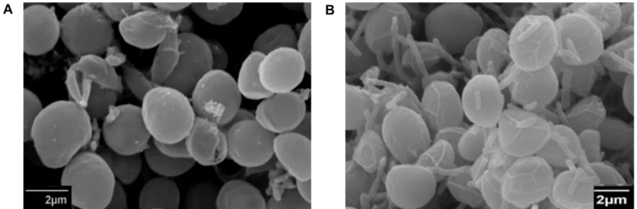
Observation by SEM
The intimate relationship between bacterial isolate I3 and P. viridis was observed under SEM. The average size of the alga cultured under two different conditions was similar
with an average diameter of 2.8 μm. Bacterial isolate I3 ex- hibited direct adhesion to the surface of alga cells (Fig. 6).
The size of the bacteria on the algal surface was about 3.5×0.7 μm. Other than the presence of bacteria on the sur- face of the alga, no morphological difference existed between the alga in coculture and that in single culture.
Discussion
Microalgae and bacteria are the most influential organ- isms with respect to numbers in all aquatic environments and together control nutrient cycling [8]. Many bacteria are associated with microalgae and they may have progressive or suppressive effects on the growth of host algae. The inter- actions between microalgae and bacteria are variable, de- pending on the species and environmental conditions [18, 21].
Riquelme et al. [21] Riquelme et al. [21] reported that bac-
terial strains of Flavobacterium sp. inoculated in axenic cul-
ture of the marine diatom A. glacialis successfully promoted
the growth of microalga. They also reported that the natu-
rally occurring bacterial strain Pseudomonas sp. promoted the
growth of microalga, whereas Vibrio sp. did not affect algal
growth. In another report, several bacterial strains isolated
from the benthic diatom Nitzschia sp. were cocultured with
the diatom, and among many bacterial strains, only
Alcaligenes sp. showed significant growth-promoting effects
on Nitzschia sp. [12]. Furthermore, the marine bacterium
Flavobacterium sp. increased the growth of the diatom
Chaetoceros gracilis but was algicidal toward the microalga
Gymnodinium mikimotoi. Unexpectedly, this bacterium has no
A B
Fig. 6. Scanning electron microscope pictures of P. viridis in pure culture (A) and in coculture with bacterial isolate I3. Scale bar:
2 μm.
obvious effects on the growth of Isochrysis galbana and Pavlova lutheri [24]. All of these results implied that the inter- actions between bacteria and microalgae are species-specific [12, 18].
In the current study, five bacterial isolates were obtained from a culture of P. viridis and these isolates were sub- sequently cocultured with the cleaned microalga. Each of the bacterial isolates promoted the growth of P. viridis, which exhibited positive effects on the growth of the associated bacteria.
The most effective bacterial isolate, I3, was identified as Citrobacter sp. by sequence analysis of its 16S rRNA gene.
Coculture with a bacteria-free filtrate from this isolate also revealed a stimulating effect on the growth of the microalga, and this effect was dose-dependent. These results are in- dicative of a bacteria–algae association. Bacteria are known to produce metabolites such as vitamins that play an im- portant role in the growth of microalgae [21]. Many micro- algae require extra nutrients such as biotin, thiamine, and cobalamin as growth factors [9]. One survey reported that 171 of the 326 algal species examined required exogenous vitamin B12 for their growth. Microalgae collect vitamin B12 from associated bacteria, implying symbiotic interactions [9].
Nutrients are regenerated by heterotrophic activity through microorganisms in the environment. Heterotrophic demineralization supplies continuous nutrients and algae al- so produce some organic compounds to contribute to the smooth running of the nutrient cycle. This phenomenon is critical in a symbiotic relationship [8, 28]. Croft et al. [9] Croft et al. [9] separated Halomonas sp., a vitamin B
12-producing bacterium, from a non-axenic culture of Amphidinium oper- culatum and cocultured it with two auxotrophic vitamin B
12-requiring algae, Porphyridium purpureum and A. oper-
culatum, in a mineral medium without organic carbon sources. They found that vitamin B
12was supplied by Halomonas sp. to the algae in exchange for the products of photosynthesis and that this interaction was symbiotic.
The results of our coculture experiment between P. viridis and Citrobacter sp. indicated the promotion of growth of both the microalga and bacterial strain. The growth of the bacteria in coculture was much faster than that of the single culture at the end of the culture period, and the growth of P. viridis was also faster in coculture than in single culture. The bac- teria-free filtrate of bacterial strain I3 also promoted the growth of the microalga. These results suggest that the alga produces some organic compounds that increase the growth of the bacterium, and that the bacterial strain produces some metabolites that promote the growth of the microalga, in- dicating that these two organisms have developed a sym- biotic relationship and future study for the identification of the substances involved in the interaction will clarify the relationships. Because the interactions between algae and bacteria occur in an aquatic environment where growth-pro- moting metabolites can diffuse quickly, close contact is re- quired between the two organisms. Such close association was confirmed by SEM observation, which demonstrated the growth of bacteria on the algal surface. Direct attachment of symbiotic bacteria on the surface of microalga and indirect attachment on the sheath produced by the microalga has been observed in other microalga–bacteria combinations [28].
The findings of this study indicated a close symbiotic rela-
tionship between the microalga P. viridis, which is important
in aquaculture, and the bacterium Citrobacter sp., which was
isolated from the microalga. This information can be useful
in the mass culture of P. viridis for increasing the microalgal
population for potential use in aquaculture.
Acknowledgment
This work was supported by a Research Grant of Pukyong National University (Year 2014, Grant Number CD20140449).
References
1. Agrawal, S. C. and Sarma, Y. S. 1982. Effects of nutrients present in bold's basal medium on the green alga Stigeoclonium pascheri. Folia Microbiol. 27, 131-137.
2. Barker, K. H. and Herson, D. S. 1978. Interactions between diatom Thallasiosira pseudonanna and an associated pseudo- monad in a mariculture system. Appl. Environ. Microbiol. 35, 791-796.
3. Brown, M. R. and Farmer, C. L. 1994. Riboflavin content of six species of microalgae used in mariculture. J. Appl.
Phycol. 6, 61-65.
4. Brown, M. R., Jeffrey, S. W., Volkman, J. K. and Dunstan, G. A. 1997. Nutritional properties of microalgae for mariculture. Aquaculture 151, 315-331.
5. Chen, B. L., Huang, Q., Lin, X. J., Shi, Q. and Wu, S. 1998.
Accumulation of Ag, Cd, Co, Cu, Hg, Ni, and Pb in Pavlova viridis Tseng (Haptophyceae). J. Appl. Phycol. 10, 371-376.
6. Cho, J. Y., Choi, J. S., Kong, I. S., Park, S. I., Kerr, R. G.
and Hong, Y. K. 2002. A procedure for axenic isolation of the marine microalga Isochrysis galbana from heavily con- taminated mass cultures. J. Appl. Phycol. 14, 385-390.
7. Connell, L. and Cattolico, R. A. 1996. Fragile algae: axenic culture of field-collected samples of Heterosigma carterae.
Mar. Biol. 125, 421-426.
8. Cole, J. J. 1982. Interactions between bacteria and algae in aquatic ecosystems. Ann. Rev. Ecol. Syst. 13, 291-314.
9. Croft, M. T., Lawrence, A. D., Raux-Deery, E., Warren, M.
J. and Smith, A. G. 2005. Algae acquire vitamin B12 through a symbiotic relationship with bacteria. Nature 483, 90-93.
10. Ferrier, M., Martin, J. L. and Rooney-Varga, J. N. 2002.
Stimulation of Alexandrium fundyense growth by bacterial as- semblages from the Bay of Fundy. J. Appl. Microbiol. 92, 706-716.
11. Frank, J. A., Reich, C. I., Sharma, S., Weisbaum, J. S., Wilson, B. A. and Olsen. G. J. 2008. Critical evaluation of two pri- mers commonly used for amplification of bacterial 16S rRNA genes. Appl. Environ. Microbiol. 74, 2461-2470.
12. Fukami, K., Nishijima, T. and Ishida, Y. 1997. Stimulative and inhibitory effects of bacteria on the growth of microalgae. Hydrobiologia 358, 185-191.
13. Gouveia, L., Batista, A. P., Suousa, I., Raymuado, A. and dan Bandarra, N. M. 2008. Microalgae in novel food prod- ucts, pp. 75-111. In: K. N. Papadopoulos (ed.), Food chem- istry research developments: Nova Science Publishers: NY,
USA.
14. Guillard, R. L. and Ryther, J. H. 1962. Studies of marine planktonic dioatoms: I. Cyclotella nana Hustedt and Dettonula confervacea (Cleve) Gran. Can. J. Microbiol. 8, 229-239.
15. Guillard, R. R. L. 1975. Culture of phytoplankton for feeding marine invertebrates, pp. 29-60. In: Smith, W. L. and Chanley, M. H. (eds.), Culture of marine invertebrate ani- mals: Plenum Press: NY. USA.
16. Hallmann, A. 2007. Algal transgenics and biotechnology.
Transgenic Plant J. 1, 81-98.
17. Hong, J. W., Choi, H. G., Kang, S. H. and Yoon, H. S. 2010.
Axenic purification and cultivation of an Arctic cyanobacte- rium, Nodularia spumigena KNUA005, with cold tolerance potential for sustainable production of algae-based biofuel.
Algae 25, 99-104.
18. Jones, A. K. 1982. The interaction of algae and bacteria, pp.189-247. In: Bull, A. T. and Slater, J. H. (eds.), Microbial Interactions and Communities: Academic Press: London.
UK.
19. Lu, K. H. and Lin, X. 2000. Screening of fatty acid composi- tion of the 13 microalgae and their application in artificial feeding of mitten crab. J. Ningbo Univ. (NSEE) 14, 27-32.
20. Park, Y., Je, K. W., Lee, K., Jung, S. E. and Choi, T. J. 2008.
Growth promotion of Chlorella ellipsoidea by co-inoculation with Brevundimonas sp. isolated from the microalga. Hydrobi- ologia 598, 219-228.
21. Riquelme, C. E., Fukami, K. and Ishida, Y. 1988. Effects of bacteria on the growth of a marine diatom, Asterionella glacialis. Bull. Japan Soc. Microbiol. Ecol. 3, 29-34.
22. Spolaore, P., Joannis-Cassan, C., Duran, E. and Isambert, A.
2006. Commercial applications of microalgae. J. Biosci.
Bioeng. 101, 87-96.
23. Stanier, R. Y., Kunisawa, R., Mandel, M. and Cohen-Bazire, G. 1971. Purification and properties of unicellular blue- green algae (order Chroococcales). Bacteriol. Rev. 35, 171-205.
24. Suminto, I. and Hirayama, K. 1996. Effects of bacterial coex- istence on the growth of a marine diatom Chaetoceros gracilis.
Fisheries Sci. 62, 40-43.
25. Suminto, I. and Hirayama, K. 1997. Application of a growth- promoting bacteria for stable mass culture of three marine microalgae. Hydrobiologia 358, 223-230.
26. Volkman, J. K., Jeffrey, S. W., Nichols, P. D., Rogers, G. I.
and Garland. C. D. 1989. Fatty acids and lipid composition of 10 species of microalgae used in mariculture. J. Exp. Mar.
Biol. Ecol. 128, 219-240.
27. Watanabe, K., Takihana, N., Aoyagi, H., Hanada, S., Wata- nabe, Y., Ohmura, N., Saiki, H. and Tanaka, H. 2005.
Symbiotic association in Chlorella culture. FEMS Microbiol.
Ecol. 51, 187-196.
28. Watanabe, K., Imase, M., Sasaki, K., Ohmura, N., Saiki, H.
and Tanaka, H. 2006. Composition of the sheath produced by the green alga Chlorella sorokiniana. Lett. Appl. Microbiol.
42, 538-543.
초록:파블로바 비리디스로부터 분리한 세균에 의한 미세조류의 생장 촉진
사커 아노와룰 아하메드
1․김진주
2․최태오
3․최태진
1*
(1부경대학교 미생물학과, 2부경대학교 수산생물학과, 3㈜클로랜드)