- 44 -
KISEP Original Article J Rhinol 11(1,2), 2004
Correlation between Presumed Chronic Sinusitis-Induced Pain and Paranasal Sinus Computed
Tomographic Findings in Korea
Myoung Gu Hwang, M.D., Sung Wan Kim, M.D., Seung Keun Yeo, M.D., Kyung Sup Rho, M.D., Chang Il Cha, M.D. and Joong Saeng Cho, M.D.
ABSTRACT
The correlation between facial pain and/or headache in patients with chronic sinusitis and localized findings on paranasal sinus computed tomography (CT) are poorly understood. So we prospectively evaluated the relationship of paranasal sinus pain symp- toms with CT imaging. Fifty eight patients with headache and/or facial pain rated their pain in 9 areas at the time of CT scan- ning and 2 months after endoscopic sinus surgery (ESS). We scored the degree of air-fluid level, mucosal thickening, and mucus retention cysts using a grade scale of severity. The ostiomeatal unit, middle meatus and nasofrontal duct were also evaluated for patency. Bivariate analysis was performed to evaluate the relationship between patients’ pain, that was improved after ESS and CT findings. Among 58 patients who had facial pain and/or headache at the time of CT scan, the pain improved in 51 patients after ESS. Bivariate analysis failed to show any relationship between pain symptoms and CT findings in 51 patients. This study suggests that findings on CT do not routinely correlate with the patients’ symptoms of facial pain or headache. CT should therefore be reserved for delineating the anatomy and degree of sinus disease before surgical intervention.
KEY WORDS
:Chronic sinusitis・Computed tomography.
INTRODUCTION
Headache and facial pain are symptoms commonly noticed in chronic sinusitis patients. Headache and fa- cial pain are the main symptoms in acute sinusitis and are well known to have a close relationship with the affected site and site of pain. However, in the case of chronic sinusitis it is difficult to finalize the diagnosis with only a simple physical examination and therefore is very dependent on PNS CT findings. Many doubtful questions have been presented about the relation of the pain site and affected area, however, definite studies
based on this area have not yet been executed. Therefore in this study we wish to inquire the correlation of the degree of headache and facial pain with PNS CT fin- dings in chronic sinusitis patients.
MATERIAL AND METHOD
This study was carried out on 151 patients diagnosed as chronic sinusitis and underwent endoscopic sinus surgery at the Department of Otorhinolaryngology Head and Neck Surgery at Kyung Hee University from Ja- nuary 2002 to June 2002. Among theses patients, pa- tients who underwent endoscopic sinus surgery and showed improvement of preoperative symptoms such as headache and facial pain two months after surgery were selected for this study. Patients with medical histories of neruologic headaches, psychiatric and general medi- cal diseases were excluded. The areas of headache and facial pain were divided into 9 areas and the pain score was obtained for each area before PNS CT was carried out in chronic sinusitis patients diagnosed based on na- Department of Otolaryngology, College of Medicine, Kyunghee
University, Seoul, Korea
Address correspondences and reprint requests to Sung Wan Kim, M.D., Department of Otorhinolaryngology Head and Neck Sur- gery, College of Medicine, Kyunghee University, #1 Hoeki-dong Dongdaemoon-Gu, 130-702, Seoul, Korea
Tel:82-2-958-8474, Fax:82-2-958-8470 E-mail:[email protected]
Accepted for publication on June 29, 2004
Hwang et al:Pain and Computed Tomography in Chronic Sinusitis / 45
sal symptoms lasting for more than 3 months who are scheduled to undergo endoscopic sinus surgery. The pain areas were divided into the right forehead, left forehead, right temple, left temple, right cheek, left ch- eek, top of head, between the eyes and back of the head.
The degree of pain was scored between 0 and 5 points depending on the degree of the symptoms. The same scoring method was carried out on patients who showed improvement of headache and facial pain two months after surgery. The affected areas on PNS CT were also scored before surgery. The degree of air-fluid level, mucosal thickening and existence of mucus retention cysts of the right frontal sinus, left frontal sinus, right anterior ethmoid, left anterior ethmoid, right posterior ethmoid, left posterior ethmoid, right maxillary sinus, left maxillary sinus, right sphenoid sinus and left sphe- noid sinus were scored 0 points if none existed, 1 point when below 1/3, 2 points when 2/3 and 3 points when above 2/3. The patency of the nasofrontal duct, middle meatus and ostiomeatal unit (OMU) was also exami- ned. Thereafter, the correlation of the site of pain and PNS CT findings were compared as explained in the following. To begin with, pain of the right forehead and temple were related and compared to the right frontal sinus, pain of the left forehead and temple to the left frontal sinus, pain between the eyes to the bilateral an- terior and posterior ethmoid sinuses, pain of the right cheek to the right maxillary sinus, pain of the left cheek to the left maxillary sinus, pain of the top of the head to the bilateral posterior ethmoid and sphenoid sinuses and lastly, pain of the back of the head was related and compared to all sinuses. Also, in the case of the patency of the opening sites revealed on PNS CT, the Pearson corrleation coefficient was obtained by correlating pain of the right temple and cheek with the patency of the right OMU, nasofrontal duct and middle meatus, pain of the left temple and cheek with the left OMU, naso- frontal duct and middle meatus, pain of bilateral temples
and between the eyes with bilateral nasofrontal ducts, pain of bilateral temples, between the eyes and both cheeks with bilateral middle meatuses, pain of the right cheek with the right OMU and lastly correlating the pain of the left cheek with the left OMU. Statistical significance of differences was tested by SPSS software version 11.0 (SPSS Inc., Chicago, IL, USA). A p value of <0.05 was considered to be significant.
RESULTS
Among the 151 patients diagnosed with chronic sinu- sitis who underwent endoscopic sinus surgery at our hospital from January to June of 2002, 58 patients complained of headache and facial pain. Among them, 27 men (average age:45.68 years old) and 24 women (average age:32.25 years old), a total of 51, showed improvement of headache and facial pain two months after surgery which revealed the possibility that the symptoms orginated from sinusitis. The Pearson corre- lation coefficient was 0.131 for pain of the right fore- head and temple in correlation with the right frontal sinus, 0.233 for pain of the left forehead and temple in correlation with the left frontal sinus, 0.049 for pain between the eyes in correlation with the bilateral ante- rior and posterior ethmoid sinuses, -0.007 for right cheek pain in correlation of the right maxillary sinus, 0.148 for left cheek pain in correlation of the left maxil- lary sinus, 0.238 for pain of the top of the head in correlation of the bilateral posterior ethmoid and sphe- noid sinuses, and 0.093 for pain of the back of the head in correlation of all sinuses which showed no statistical significant corrleation (Table 1). Also the Pearson corrleation coefficient the site of pain and patency of openings were each 0.093 for right temple and cheek pain in correlation to the patency of the right OMU, nasofrontal duct and middle meatus, -0.068 for left temple and cheek pain in correlation to the patency
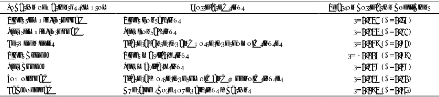
Table 1. Correlation between patient's location of pain symptoms and findings on sinus CT
Location of patient’s symptom Correlated sinus Pearson correlation coefficient
Right temple/forehead Right frontal sinus -r=0.131 (p=0.57)
Left temple/forehead Left frontal sinus -r=0.233 (p=0.11)
Between eyes Bilateral anterior and posterior ethomoid sinuses -r=0.049 (p=0.82)
Right cheek Right maxillary sinus r=-0.007 (p=0.79)
Left cheek Left maxillary sinus -r=0.148 (p=0.48)
Top of head Bilateral posterior ethmoid and sphenoid sinuses -r=0.238 (p=0.10)
Back of head Average scores over all sinus locations -r=0.093 (p=0.65)
46 / J Rhinol 11(1,2), 2004
of the left OMU, nasofrontal duct and midle meatus, 0.225 for pain of bilateral temples and between the eyes in correlation to the patency of bilateral nasofrontal ducts, -0.276 for pain of bilateral temples, between the eyes, bilateral cheeks in correlation to the patency of bilateral middle meatuses, 0.001 for right cheek pain in correlation to the patency of the right OMU and 0.136 for left cheek pain in correlation to the patency of the left OMU with this also revealing no statistical signifi- cant correlation (Table 2).
DISCUSSION
Pain and tenderness of the invaded area are the main symptoms in acute sinusitis patients. For example, cheek pain and toothache of a set of teeth are common in acute maxillary sinusitis and pain and tenderness of the forehead that is severe in the morning and subsides in the afternoon is commonly noticed in acute frontal sinusitis. In acute ethmoid sinusitis, deep pain of the orbit and root of the nose are commonly noticed and numerous symptoms in the form of pansinusitis are noticed in acute sphenoid sinusitis.
1)On the other hand, the medical history and affection time are mostly uncer- tain in many cases of chronic sinusitis. There are also many asymptomatic cases and there are reports that state that the degree of pain and affected area have no correlation with what is revealed on PNS CT.
2)Howe- ver, such a research has not been carried out within the country and therefore our team carried forward this study and apart from existing studies, this study was based on patients with a definite diagnosis of chronic sinusitis who showed improvement after endoscopic sinus sur- gery so that patients with headache and/or facial pain followed by different reasons other than chronic sinusi- tis were completely excluded from the experiment.
For its low cost, simple x-ray is primarily used for the diagnosis of chronic sinusitis, however, there are
many cases where it does concur with PNS CT fin- dings.
3)Some authors, on the other hand, have stated that simple x-ray shows a concurrence rate of 78.4% in the maxillary sinus, 71.1% in the frontal sinus and 52.6% in the ethmoid sinus.
4)There are especially many cases of false positive diagnoses in the ethmoid sinus.
5)For these reasons, PNS CT has been presented as the most accurate test for the diagnosis of chronic sinusitis and is still being used as the most trusted diagnosis method.
6)Also, the nasal endoscopy, which has been recently developed, allows detalied observation of the nasal cavity and surroundings of the middle turbinate and its accuracy has been proven to be very high.
7)It has shown an 87% concurrence rate with PNS CT findings when carried out by an experienced otorhino- laryngologist for the diagnosis of chronic sinusitis
8)and it is being used as a useful diagnostic tool along with PNS CT.
There have been concerns arising about the limited information PNS CT has been presenting dealing with the disease status of the sinus in chronic sinusitis patients, however, it has been stated that in the case of an upper respiratory infection, abnormal findings may be found in PNS CT.
9)Also, abnormal findings were found in PNS CT in 27% of adults with no rhiological symp- toms.
10)There have also been reports that state that only 47% of patients with chronic sinusitis symptoms revealed abnormal findings in PNS CT.
11)The limita- tions of this study were the restrictions of PNS CT along with revealing only a certain temporary point of the process course of chronic sinusitis. Therefore, the diagnosis of chronic sinusitis must be carried out not only by PNS CT but also through a complete and tho- rough examination of the patient’s medical history, phy- sical findings, endoscopic tests as so forth.
This study suggests that findings on CT do not rou- tinely correlate with the patients’ symptoms of facial pain or headache. CT should therefore be reserved for
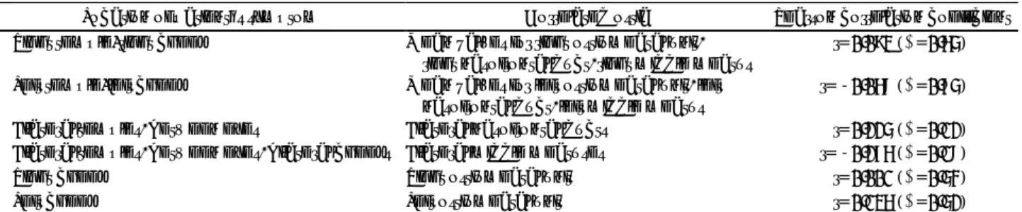
Table 2. Correlation between patient’s location of pain symptoms and occlusion of sinus ostia on sinus CT
Location of patient’s symptom Correlated ostia Pearson correlation coefficient Right temple/right cheek Mean values for right ostiomeatal unit,
right nasofrontal duct, right middle meatus
-r=0.093 (p=0.65) Left temple/left cheek Mean values for left ostiomeatal unit, left
nasofrontal duct, left middle meatus r=-0.068 (p=0.75)
Bilateral temples, between eyes Bilateral nasofrontal ducts -r=0.225 (p=0.12)
Bilateral temples, between eyes, bilateral cheeks Bilateral middle meatuses r=-0.276 (p=0.18)
Right cheek Right ostiomeatal unit -r=0.001 (p=0.54)
Left cheek Left ostiomeatal unit -r=0.136 (p=0.52)
Hwang et al:Pain and Computed Tomography in Chronic Sinusitis / 47
delineating the anatomy and degree of sinus disease before surgical intervention.
CONCLUSION
This study did not show any kind of corrleation bet- ween preoperative symptom sites and degree with PNS CT findings revealed before surgery in chronic sinusitis patients who have shown improvement after endosco- pic sinus surgery. Therefore, in the case of treatment, especially surgical treatment for chronic sinusitis, we find it difficult to presume the affected sinus by only the state of pain appealed by the patients. Hence, we believe that a generalized thorough examination of the affected area along with the degree of pain through a preoperative PNS CT must be preceded before surgery.
REFERENCES
1) Ferguson BJ. Acute and chronic sinusitis: How to ease symptom and
locate the cause. Sinusitis 1998;97:45-8.
2) Timothy B, Jay P, Franz JW. Relationship between patient-based description of sinusitis and paranasal sinus computed tomographic findings. Arch Otolaryngol Head Neck Surg 1997;123:1189-92.
3) Brocian RC. Sinusitis. Western H Med 1993;158:615-6.
4) Iinuma T, Hirota Y, Kase Y. Radio-opacity of the paranasal sinuses.
Conventional views and CT. Rhinology 1994;32:134-6.
5) Lloyd GAS, Lund VJ, Scadding GK. CT of paranasal sinuses and functional endoscopic surgery: A critical analysis of 100 sympto- matic patients. J Laryngol Otol 1991;105:503-10.
6) Davidson TM, Brahme FJ, Gallagher ME. Radiographic evaluation for nasal dysfuction: computed tomography versus plain films. Head Neck 1989;11:405-9.
7) Kamal RH. Nasal endoscopy in chronic maxillary sinusitis. J Laryn- gol Otol 1989;103:275-8.
8) Roberts DN, Hampal S, East CA, Lloyd GAS. The diagnosis of inflammatory sinonasal disease. J Laryngol Otol 1995;109:27-30.
9) Gwaltney JM Jr, Phillips CD, Miller RD, Riker DK. Computed to- mographic study of the common cold. N Engl J Med 1994;330:25-30.
10) Flinn J, Chapman ME, Wightman AJA, Maran AGD. A prospective analysis of incidental paranasal sinus abnormalities on CT head scans. Clin Otolaryngol 1994;19:287-9.
11) Stankiewicz JA, Chow JM. A diagnostic dillema for chronic rhino - sinusitis: Definition accuracy and validity. Am J Rhinol 2002;16:
199-202.