ꠏꠏꠏꠏꠏꠏꠏꠏꠏꠏꠏꠏꠏꠏꠏꠏꠏꠏꠏꠏꠏꠏꠏꠏꠏꠏꠏꠏꠏꠏꠏꠏꠏꠏꠏꠏꠏꠏꠏꠏꠏꠏꠏꠏꠏꠏꠏꠏ
<접수일:2002년 5월 14일, 심사통과일:2002년 6월 18일>
※통신저자:이 신 석
광주광역시 동구 학1동 8번지 전남대학교 의과대학 내과학교실
Tel:062) 220-6591, Fax:062) 225-8578, E-mail:[email protected]
루푸스에서 유리형 Fas는 질병의 활성도를 반영하는가?
서남대학교 의과대학 내과학교실, 전남대학교 의과대학 내과학교실*
김 윤 아․이 신 석*․박 미 정*
= Abstract =
Do Increased Levels of Soluble Fas Correlate with Disease Activity in Systemic Lupus Erythematosus?
Yun-A Kim, M.D., Shin-Seok Lee, M.D.*, Mi-Jeong Park, M.D.*
Department of Internal Medicine, Seonam University College of Medicine, Gwangju Department of Internal Medicine*, Chonnam National University
Medical School, Gwangju, Korea
Objective: To determine whether levels of soluble Fas (sFas) are elevated in patients with systemic lupus erythematosus (SLE) and correlated with clinical disease activity.
Methods: Serum samples were obtained from 62 SLE patients and 39 normal controls. We measured sFas levels using an enzyme-linked immunosorbent assay. Disease activity variables including Systemic Lupus Erythematosus Disease Activity Index (SLEDAI) were examined.
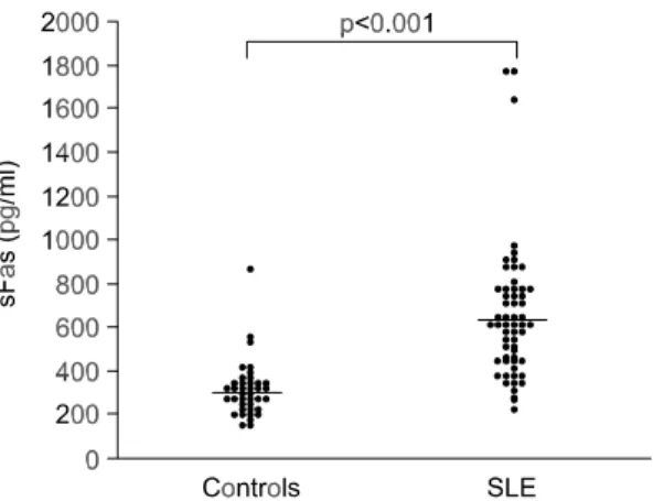
Results: The mean age of the patients was 32.1 years (range 13∼60 years) and the mean disease duration was 3.0 years (range 0.2∼10.0 years); 1 patient was male (1.6%). The median serum sFas concentration was 610.0 pg/ml for SLE patients and 292.9 pg/ml in controls. The serum sFas concentration was significantly higher in SLE patients than in controls (p<0.001).
The Receiver Operating Characteristics (ROC) curve showed that the area under the curve for sFas was 0.911. Using the cut-off value of 370.5 pg/ml, the sensitivity was 0.871 and the specificity was 0.846. However, we could not find any correlation between sFas levels and disease activity variables such as SLEDAI, complement, and anti-dsDNA antibodies. There was no significant difference in sFas levels between active (SLEDAI ≥8) and inactive (SLEDAI <8) SLE patients.
서 론
전신성홍반성루푸스(이하 루푸스)는 다클론성 B세 포의 활성으로 자가항체 및 면역복합체 생성이 증가 하여 여러 장기에 손상을 입히고, 만성적인 경과를 보이는 자가면역질환이다1). 자가반응성(autoreactive) B세포는 정상인에서도 존재하나, 그 수는 증식을 시 키는 자극이 없으면 며칠 내에 세포가 죽음으로써 조절된다. 이러한 조절과정을 세포자멸(apoptosis or programmed cell death)이라고 하며 자가관용(self-toler- ance)확립에 중요하다2). 최근에 세포자멸의 이상이 자가면역질환 특히 루푸스의 발생과 밀접한 관계가 있는 것으로 보고되고 있다.
Fas (APO-1/CD95)는 림프구, 섬유아세포, 내피세 포, 상피세포 등 여러 세포의 표면에 발현하여 세포 자멸을 매개하는 세포 표면 단백 항원이다3). 루푸스 의 병인으로 Fas가 관련이 있을 것이라는 가설은 루 푸스의 동물 모델인 MRL lpr/lpr 마우스와 gld/gld 마우스에서 비롯되었다4,5). Fas 유전자의 돌연변이가 있는 lpr/lpr 마우스와 Fas ligand (이하 FasL)에 돌연 변이가 있는 gld/gld 마우스에서 항DNA 항체의 증가 와 림프절 종대, 루푸스 신장염, 활막염 등 인간의 루푸스와 유사한 증상이 나타나는 것으로 보아 Fas 와 FasL의 이상이 자가면역 세포의 자연적인 세포자 멸에 장애를 초래하여 루푸스가 발생하는 것으로 가 정해 볼 수 있다. 인간에서는 Fas의 변이에 의한 림 프구의 세포자멸 이상으로 autoimmune lymphoprolifera- tive syndrome (ALPS)이 보고된 바 있지만6) 실제 루 푸스로 진단된 경우는 극히 드문 것으로 알려져 있 다7). 또한 루푸스 환자를 대상으로 한 많은 연구에 서 Fas의 발현이 오히려 증가되어 있고8), 아직까지 Fas 나 FasL의 기능에 있어 고정된 결함은 밝혀진 것이 없기 때문에 인간의 루푸스에서 Fas와 FasL의 역할 에 대해서는 이론이 많은 실정이다.
Cheng 등9)은 Fas mRNA의 transcript variant 중 exon 6가 splicing으로 탈락되는 형태의 Fas를 발견하였고,
이러한 형태의 Fas는 세포막 내 부분이 없어서 세포 외로 분비되는 유리형 Fas (soluble Fas, 이하 sFas)로 Fas/FasL의 결합을 저해하여 세포자멸을 억제한다고 하였다. sFas는 특히 루푸스 환자의 혈청에서 증가되 어 있다고 알려져 있으나 아직까지 이견이 있고 루 푸스에 있어 특이적이지 않다는 주장도 있어10,11) sFas 의 역할에 대해서는 좀 더 연구되어야 할 필요가 있 다고 본다.
이에 저자들은 루푸스 환자를 대상으로 혈청 sFas 의 발현 양상을 살펴보고, 이를 질병의 활성도와 관 련지어봄으로써 sFas의 임상적 의의를 알아보고자 하였다.
대상 및 방법 1. 대상환자
전남대학교병원 류마티스내과에 내원하였던 62명 의 루푸스 환자들을 대상으로 혈청 sFas의 농도를 측정하였으며 정상 성인 39명을 대조군으로 하였다.
루푸스의 진단은 미국 류마티스학회가 정한 진단기 준12)을 따랐다.
2. sFas의 측정
환자와 대조군의 혈액은 채혈하고 실온에서 30분 동안 방치한 후 원심분리하였으며 분리된 혈청은 sFas를 측정할 때까지 -70oC 냉동고에 보관하였다.
sFas의 농도는 sFas ELISA kit (QuantikineⓇ, R&D Sys- tem, Inc., USA)를 이용하여 측정하였다. 요약하면, murine 항Fas 단일클론항체로 도포된 96개 well에 검체 희석액으로 10배 희석한 환자의 혈장이나 표준액을 100μl씩 넣고 2시간 동안 실온에서 배양한 후 세척 완충액으로 씻어내었다. 이후 horseradish peroxidase가 conjugation되어 있는 다클론성 항Fas 항체 200μl를 각각의 well에 넣고 실온에서 2시간 동안 배양시킨 후 세척하였다. 이후 각각의 well에 기질액 200μl를 넣 고 실온에서 30분 동안 반응시킨 후 반응정지액(2N Conclusion: These results suggest that sFas levels are elevated in SLE patients, but sFas does not reflect disease activity.
ꠏꠏꠏꠏꠏꠏꠏꠏꠏꠏꠏꠏꠏꠏꠏꠏꠏꠏꠏꠏꠏꠏꠏꠏꠏꠏꠏꠏꠏꠏꠏꠏꠏꠏꠏꠏꠏꠏꠏꠏꠏꠏꠏꠏꠏꠏꠏꠏꠏꠏꠏꠏꠏꠏꠏꠏꠏꠏꠏꠏꠏꠏꠏꠏꠏꠏꠏꠏꠏꠏꠏꠏꠏꠏꠏꠏꠏꠏꠏꠏꠏꠏꠏꠏꠏꠏ Key Words: Systemic lupus erythematosus, Soluble Fas, Disease activity
sulfuric acid) 50μl를 넣고 450 nm에서 흡광도를 측 정하였다. 표준액을 이용하여 표준 곡선을 구하였으 며, 이를 이용하여 환자 혈청의 sFas 농도를 계산하 였고 sFas의 최저 측정치는 20 pg/ml이었다.
3. 임상 지표
대상 환자들은 혈액을 채취할 때 질병의 활성도를 평가하기 위해 신체검사와 혈액 및 소변 검사를 같 이 시행하였다. SLE Disease Activity Index (이하 SLEDAI)13)를 측정하였고 SLEDAI가 8 이상인 경우 를 활성형 루푸스, 8 미만인 경우를 비활성형 루푸 스로 정의하였으며14) 루푸스로 인한 주요장기의 손 상 정도를 알아보기 위해 Systemic Lupus Interna- tional Collaborating Clinics/American College of Rheu- matology Damage Index (이하 SLICC/ACR)15)을 함께 측정하였다. 혈액검사는 전혈구 및 감별혈구 계산 치, 화학검사, C3, C4, CH50, 항dsDNA 항체를 측정 하였다.
4. 통계분석
결과는 중앙값과 범위로 표시하였고, 통계처리는 Window용 SPSS 프로그램(SPSS, Chicago, IL)을 사용 하여 분석하였다. 루푸스 환자와 정상 대조군의 혈 청 sFas 농도는 Mann-Whitney U test를 사용하여 비 교하였고, receiver operating characteristics (ROC)곡선 을 이용하여 루푸스 환자에서 혈청 sFas의 임계치 (cut-off value)를 구하여 민감도와 특이도를 계산하였 다. sFas 농도와 질병의 활성도를 나타내는 임상지표
들과의 관련성은 Spearman 상관계수를 구하여 알아
보았고, 활성형 루푸스와 비활성형 루푸스에서 sFas 농도는 Mann-Whitney U test를 사용하여 비교하였다.
p<0.05일 때 통계적으로 유의한 것으로 간주하였다.
결 과 1. 대상 환자의 임상적 특성
혈청 sFas의 농도를 측정하였던 루푸스 환자는 62 명으로 남자 1명, 여자 61명이었으며 환자의 평균 연령은 32.1세(범위 13∼60세)였고 평균 유병 기간은
3.0년(범위 0.2∼10.0년)이었다(표 1). 활성형 루푸스 Table 1. Clinical and laboratory characteristics in patients
with SLE
ꠧꠧꠧꠧꠧꠧꠧꠧꠧꠧꠧꠧꠧꠧꠧꠧꠧꠧꠧꠧꠧꠧꠧꠧꠧꠧꠧꠧꠧꠧꠧꠧꠧꠧꠧꠧꠧꠧꠧꠧꠧꠧꠧꠧꠧꠧꠧꠧ
Variables SLE (n=62)
ꠏꠏꠏꠏꠏꠏꠏꠏꠏꠏꠏꠏꠏꠏꠏꠏꠏꠏꠏꠏꠏꠏꠏꠏꠏꠏꠏꠏꠏꠏꠏꠏꠏꠏꠏꠏꠏꠏꠏꠏꠏꠏꠏꠏꠏꠏꠏꠏ
Male/Female (n) 1/61
Age (years, mean (range)) 32.1 (13∼60) Disease duration (years, mean(range)) 3.0 (0.2∼10.0) Anti-dsDNA (IU/mL, mean±SEM) 40.1±7.7 SLEDAI (mean±SEM) 6.2±0.7 SLICC/ACR (mean±SEM) 0.1±0.1 SLEDAI≥8 (n, %) 20 (32.3) ꠏꠏꠏꠏꠏꠏꠏꠏꠏꠏꠏꠏꠏꠏꠏꠏꠏꠏꠏꠏꠏꠏꠏꠏꠏꠏꠏꠏꠏꠏꠏꠏꠏꠏꠏꠏꠏꠏꠏꠏꠏꠏꠏꠏꠏꠏꠏꠏ SEM: standard error mean
Fig. 2. Receiver operating characteristics (ROC) curve for sFas.
Fig. 1. Serum concentration of sFas in patients with SLE and normal controls. Each point represents an individual patient, and bars denote the median.
환자는 20명(32.3%)이었고 비활성형 루푸스 환자는 42명(67.7%)이었다.
2. 루푸스 환자의 혈청 sFas 발현 양상
루푸스 환자의 혈청 sFas 농도는 610.0 pg/mL (중 앙값, 범위 221.4∼1776.7 pg/mL)로 정상 대조군의 292.9 pg/mL (중앙값, 범위 150.0∼864.3 pg/mL)에 비 해 통계적으로 유의하게 증가되어 있었다(그림 1).
혈청 sFas의 ROC 곡선하 면적은 0.911이었고 ROC 곡 선을 이용하여 구한 혈청 sFas의 임계치는 370.5 pg/
mL이었으며, 민감도와 특이도는 각각 0.871, 0.846이 었다(그림 2).
3. 혈청 sFas와 질병 활성도
증가된 혈청 sFas 농도가 루푸스의 활성도를 반영 하는지 알아보기 위해 질병 활성도를 반영하는 여러 임상지표들과의 상관관계를 살펴보았는데 혈청 sFas
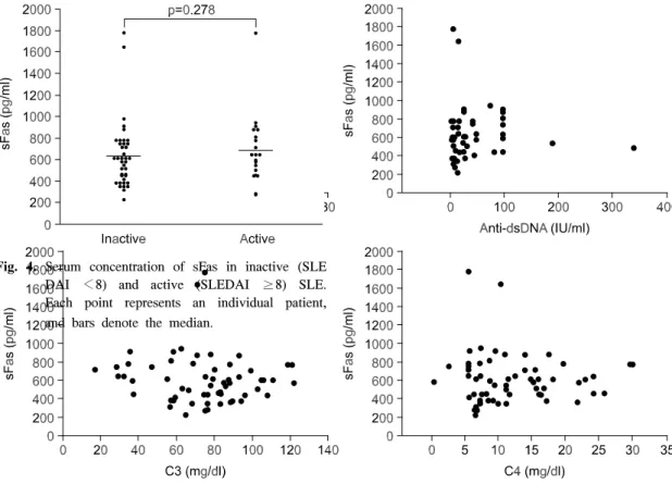
는 C3, C4, CH50, 항dsDNA 항체 그리고 SLEDAI와 유 의한 상관관계를 보이지 않았다(그림 3). 또한 루푸 스의 손상지수인 SLICC/ACR과도 유의한 상관관계 를 보이지 않았다. 활성형 루푸스와 비활성형 루푸 스 두 군 간의 혈청 sFas 농도는 각각 684.8 pg/ml (중 앙값, 범위 269.1∼1776.7 pg/ml), 593.3 pg/ml (중앙 값, 범위 221.4∼1776.7 pg/ml)로 양 군 간에 유의한 차이는 없었다(그림 4).
고 찰
본 연구에서는 루푸스 환자들을 대상으로 혈청 sFas의 농도를 측정하였는데 정상 대조군에 비해 유 의하게 증가되어 있었다. 이는 Cheng 등9)이 처음으로 10명의 루푸스 환자를 대상으로 한 연구에서 sFas가 증가되어 있다는 결과와 일치한다. 한편, Goel 등10) 은 루푸스와 류마티스관절염 등 자가면역질환 환자 Fig. 3. Relationship between serum concentration of sFas and SLE Disease Activity Index (SLEDAI), anti-dsDNA,
C3, C4 levels in patients with SLE.
Fig. 4. Serum concentration of sFas in inactive (SLE DAI <8) and active (SLEDAI ≥8) SLE.
Each point represents an individual patient, and bars denote the median.
들에서 측정한 sFas가 정상 대조군과 차이가 없다고 하였고, Knipping 등11)도 루푸스와 연소성 류마티스 관절염 환자에서 측정한 sFas가 정상 대조군과 차이 가 없어 sFas는 루푸스의 발병과 관련이 없다고 주 장하였다. 하지만 이후로 보고된 많은 연구들16-26)에 서 sFas는 루푸스 환자에서 정상 대조군과 다른 자가 면역질환에 비해 유의하게 증가되어 있음이 밝혀져 있다. 이러한 차이는 검사시기나 대상환자의 수, 그 리고 인종에 따른 차이라고 하기보다는 ELISA 측정 법에 따른 차이일 가능성이 높다. Goel 등10)과 Knipping 등11)이 사용한 방법으로 측정이 가능한 sFas의 최저 치는 2 ng/ml인 반면 이후로 보고된 논문들에서 사 용된 방법은 20 pg/ml까지 측정이 가능한 것으로 되 어있다. 국내에서는 송 등27)이 30명의 루푸스 환자 들을 대상으로 sFas를 측정하여 대조군과 유의한 차 이가 없다고 하였는데 이러한 결과도 외국의 연구에 서처럼 ELISA의 민감도에 따른 차이일 것으로 판단 된다.
루푸스 환자에서 증가되어 있는 sFas는 FasL와 결 합하여 Fas-FasL 결합에 의한 세포자멸을 방해하고 이로 인해 자가반응성의 B 세포가 사멸되지 않고 살아남아 루푸스가 발병할 가능성이 있다. 실제로 Fas 유전자에 결함이 있는 lpr/lpr 마우스와 FasL 유 전자에 결함이 있는 gld/gld 마우스에서는 인간의 루 푸스와 유사한 증상이 나타난다. 하지만 인간에서 Fas 유전자의 결함에 의해 발병하는 Canale-Smith 증 후군은 일반적인 루푸스 환자들의 증상과 차이가 있 고6) 인간에서 루푸스는 Fas와 같은 하나의 유전자 이상에 의해 발병하기보다는 최소한 4개 이상의 유 전자 이상이 복합적으로 관여하여 발병하는 것으로 알려져 있다28). 또한 B 세포의 세포자멸 이상, apo- ptotic body의 제거능력 감소, IL-8, IL-10과 같은 B 세포를 자극하는 사이토카인의 증가 등이 루푸스의 발병과 더 관련이 있는 것으로 보고되고 있다29). 따 라서 루푸스 환자들에서 증가되어 있는 sFas는 세포 자멸에 의한 조각들이 제대로 제거되지 않고 남아있 기 때문에 증가하는 것으로 가정해볼 수 있다. 림프 구가 활성화되면 Fas의 발현이 상향 조절되고 Fas 양성인 세포들은 활성화에 따른 사멸(activation-in- duced death)로 세포사하게 된다. 이때 Fas의 유리형 인 sFas가 나오게 되고 이것이 제대로 제거되지 않
기 때문에 루푸스 환자에서 sFas가 증가한다는 것이 다. 종합해 보면, 루푸스 환자에서 증가되어 있는 sFas 는 루푸스의 직접적인 원인이라고 하기보다는 이차 적인 현상일 가능성이 높고 루푸스의 활성도에 따라 sFas의 농도가 변화할 가능성이 있다.
본 연구에서는 62명의 루푸스 환자들을 대상으로 sFas의 농도를 측정하여 sFas가 루푸스의 활성도를 반영하는지 살펴보았는데 항dsDNA 항체, 보체, SLEDAI 등 루푸스의 활성도를 반영하는 것으로 알 려져 있는 지표들과 sFas가 서로 유의한 상관관계를 보이지 않았고 활성형과 비활성형 루푸스군 간에도 sFas의 농도에는 차이가 없었다. Jodo 등18)은 루푸스 환자에서 증가되어 있는 sFas가 항dsDNA 항체, SLEDAI와는 순상관관계를, 보체와는 역상관관계를 나타내어 질병의 활성도를 반영한다고 하였고 Bijl 등22)도 sFas가 SLEDAI와 순상관관계를 나타내어 질 병의 활성도와 관련이 있음을 보고한 바 있다. 반면, Goel 등10)과 Rose 등19)은 sFas가 루푸스의 활성도와 는 서로 관련이 없다고 하였고 Al-Maini 등25)은 sFas 가 루푸스의 활성도를 반영하는 것이 아니라 루푸스 로 인한 주요 장기의 손상 정도를 반영한다고 하였 다. 한편, van Lopik 등23)은 루푸스 환자를 대상으로 전향적인 연구를 시행하였는데 경과 관찰 도중 질환 이 악화된 환자들과 악화되지 않았던 환자들을 각각 두 군으로 나누어 sFas의 농도를 비교한 결과 재발 한 환자군에서 sFas가 의의 있게 증가된 것을 관찰 하였고, 개개인의 루푸스 환자에서 연속적으로 측정 한 sFas의 결과와 SLEDAI와는 서로 관계가 없다고 하였다. 이는 sFas가 질환의 활성도를 반영하기보다 는 처음부터 sFas가 높은 환자에서 재발의 위험이 높다는 것을 나타낸다고 주장하였다. sFas가 루푸스 의 활성도에 미치는 영향에 관한 여러 연구 결과들 이 이처럼 서로 일치하지 않는 것은 대상환자의 수, 유병기간의 차이, 질병 활성도를 평가하는 지표의 차이, sFas 측정 방법 등 여러 가지 다양한 원인에 기인된다고 볼 수 있고 실제로 sFas가 루푸스의 발 생과 진행에 있어 그다지 중요한 역할을 하지 않을 가능성도 높다.
한편, van der Linden 등26)과 Al-Maini 등25)은 sFas 가 루푸스의 활성도보다는 손상 정도를 반영한다고 하였는데 본 연구에서는 루푸스의 손상지수인 SLICC/
ACR과 sFas는 서로 유의한 상관관계를 보이지 않았 다. 이는 대상환자들의 평균 유병 기간이 3년으로 비교적 초기의 환자들이 많아 손상지수가 높지 않았 던 것도 하나의 원인으로 생각되며 좀 더 다양한 유 병 기간을 가진 환자들을 대상으로 연구가 진행되었 더라면 다른 결과가 나올 수도 있었을 것으로 예상 된다.
본 연구의 결과를 정리하면, 루푸스 환자에서 증 가되어 있는 sFas는 루푸스의 활성도나 손상 정도를 반영하지 않았다. 루푸스에서 증가되어 있는 sFas의 역할에 대해서는 앞으로 좀 더 연구되어야 할 필요 가 있다고 보며 Fas/FasL30), Bcl-2/bax31), caspases32), poly (ADP-ribose) polymerase (PARP)에 대한 항체 존 재33) 등 아포토시스에 영향을 주는 다른 여러 인자 들과도 관련지어서 그 역할을 살펴 볼 필요가 있다 고 생각된다.
결 론
저자들은 62명의 루푸스 환자들과 39명의 정상 대 조군을 대상으로 혈청 내 sFas를 ELISA법으로 측정 한 결과 루푸스 환자의 혈청 sFas 농도는 610.0 pg/ml (중앙값, 범위 221.4∼1776.7 pg/ml)로 정상 대조군의 292.9 pg/ml (중앙값, 범위 150.0∼864.3 pg/ml)에 비 해 통계적으로 유의하게 증가되어 있었으나 루푸스 의 활성도를 반영하는 여러 임상지표들과는 유의한 상관관계를 보이지 않았다. 이와 같은 연구 결과는 sFas가 루푸스의 일차적인 원인이라기보다는 이차적 인 현상일 가능성을 시사한다고 본다.
REFERENCES
1) Kotzin BL. Systemic lupus erythematosus. Cell 1996;
85:303-6.
2) Cohen JJ, Duke RC, Fadok VA, Sellins KS. Apoptosis and programmed cell death in immunity. Annu Rev Immunol 1992;10:267-93.
3) Trauth BC, Klas C, Peters AM, Matzku S, Moller P, Falk W, et al. Monoclonal antibody-mediated tumor regression by induction of apoptosis. Science 1989;
245:301-5.
4) Watanabe-Fukunaga R, Brannan CI, Copeland NG, Jenkins NA, Nagata S. Lymphoproliferation disorder in mice explained by defects in Fas antigen that
mediates apoptosis. Nature 1992;356:314-7.
5) Takahashi T, Tanaka M, Brannan CI, Jenkins NA, Copeland NG, Suda T, et al. Generalized lymphoproli- ferative disease in mice, caused by a point mutation in the Fas ligand. Cell 1994;76:969-76.
6) Drappa J, Vaishnaw AK, Sullivan KE, Chu JL, Elkon KB. Fas gene mutations in the Canale-Smith syn- drome, an inherited lymphoproliferative disorder asso- ciated with autoimmunity. N Engl J Med 1996;335:
1643-9.
7) Vaishnaw AK, Toubi E, Ohsako S, Drappa J, Buys S, Estrada J, et al. The spectrum of apoptotic defects and clinical manifestations, including systemic lupus erythematosus, in humans with CD95 (Fas/ APO-1) mutations. Arthritis Rheum 1999;42:1833- 42.
8) Lorenz HM, Grunke M, Hieronymus T, Herrmann M, Kuhnel A, Manger B, et al. In vitro apoptosis and expression of apoptosis-related molecules in lympho- cytes from patients with systemic lupus ery- thematosus and other autoimmune diseases. Arthritis Rheum 1997;40:306-17.
9) Cheng J, Zhou T, Liu C, Shapiro JP, Brauer MJ, Kiefer MC, et al. Protection from Fas-mediated apoptosis by a soluble form of the Fas molecule.
Science 1994;263:1759-62.
10) Goel N, Ulrich DT, St Clair EW, Fleming JA, Lynch DH, Seldin MF. Lack of correlation between serum soluble Fas/APO-1 levels and autoimmune disease.
Arthritis Rheum 1995;38:1738-43.
11) Knipping E, Krammer PH, Onel KB, Lehman TJ, Mysler E, Elkon KB. Levels of soluble Fas/APO- 1/CD95 in systemic lupus erythematosus and juvenile rheumatoid arthritis. Arthritis Rheum 1995;38:1735-7.
12) Hochberg MC. Updating the American College of Rheumatology revised criteria for the classification of systemic lupus erythematosus. Arthritis Rheum 1997;40:1725.
13) Bombardier C, Gladman DD, Urowitz MB, Caron D, Chang CH. Derivation of the SLEDAI. A disease activity index for lupus patients. The Committee on Prognosis Studies in SLE. Arthritis Rheum 1992;35:
630-40.
14) Miret C, Font J, Molina R, Garcia-Carrasco M, Filella X, Ramos M, et al. Bcl-2 oncogene (B cell lym- phoma/leukemia-2) levels correlate with systemic lupus erythematosus disease activity. Anticancer Res 1999;19:3073-6.
15) Gladman DD, Urowitz MB, Goldsmith CH, Fortin P, Ginzler E, Gordon C, et al. The reliability of the
Systemic Lupus International Collaborating Clinics/
American College of Rheumatology Damage Index in patients with systemic lupus erythematosus. Arthritis Rheum 1997;40:809-13.
16) Okubo M, Ishida H, Kasukawa R. Elevated levels of soluble Fas in systemic lupus erythematosus: comment on the article by Knipping et al. Arthritis Rheum 1996;39:1612-4.
17) Tokano Y, Miyake S, Kayagaki N, Nozawa K, Mori- moto S, Azuma M, et al. Soluble Fas molecule in the serum of patients with systemic lupus erythematosus.
J Clin Immunol 1996;16:261-5.
18) Jodo S, Kobayashi S, Kayagaki N, Ogura N, Feng Y, Amasaki Y, et al. Serum levels of soluble Fas/APO-1 (CD95) and its molecular structure in patients with systemic lupus erythematosus (SLE) and other auto- immune diseases. Clin Exp Immunol 1997;107:89-95.
19) Rose LM, Latchman DS, Isenberg DA. Elevated soluble fas production in SLE correlates with HLA status not with disease activity. Lupus 1997;6:717-22.
20) Nozawa K, Kayagaki N, Tokano Y, Yagita H, Okumura K, Hasimoto H. Soluble Fas (APO-1, CD95) and soluble Fas ligand in rheumatic diseases. Arthritis Rheum 1997;40:1126-9.
21) Papo T, Parizot C, Ortova M, Piette JC, Frances C, Debre P, et al. Apoptosis and expression of soluble Fas mRNA in systemic lupus erythematosus. Lupus 1998;7:455-61.
22) Bijl M, van Lopik T, Limburg PC, Spronk PE, Jaegers SM, Aarden LA, et al. Do elevated levels of serum- soluble fas contribute to the persistence of activated lymphocytes in systemic lupus erythematosus? J Auto- immun 1998;11:457-63.
23) van Lopik T, Bijl M, Hart M, Boeije L, Gesner T, Creasy AA, et al. Patients with systemic lupus erythematosus with high plasma levels of sFas risk relapse. J Rheumatol 1999;26:60-7.
24) Courtney PA, Crockard AD, Williamson K, Mc- Connell J, Kennedy RJ, Bell AL. Lymphocyte apoptosis in systemic lupus erythematosus: relation- ships with Fas expression, serum soluble Fas and disease activity. Lupus 1999;8:508-13.
25) Al-Maini MH, Mountz JD, Al-Mohri HA, El-Ageb EM, Al-Riyami BM, Svenson KL, et al. Serum levels of soluble Fas correlate with indices of organ damage in systemic lupus erythematosus. Lupus 2000;9:132-9.
26) van der Linden MW, van Lopik T, Aarden LA, Westendorp RG, Huizinga TW. Soluble CD95 con- centrations are increased in patients with severe
systemic lupus erythematosus, but not in their first degree relatives. Ann Rheum Dis 2001;60:237-41.
27) 송관규, 이영호. 전신성 홍반성 낭창 환자에서 유리형 Fas 단백질에 대한 연구. 대한류마티스학회지 1996;3:
50-6.
28) Sullivan KE. Genetics of systemic lupus erythem- atosus. Clinical implications. Rheum Dis Clin North Am 2000;26:229-56.
29) Hahn BH. An overview of the pathogenesis of systemic lupus erythematosus. In: Wallace DJ, Hahn BH, eds. Dubois' Lupus Erythematosus. 6th ed. p. 87-96, Philadelphia, Lippincott Williams & Wilkins, 2002.
30) Suzuki N, Ichino M, Mihara S, Kaneko S, Sakane T.
Inhibition of Fas/Fas ligand-mediated apoptotic cell death of lymphocytes in vitro by circulating anti-Fas ligand autoantibodies in patients with systemic lupus erythematosus. Arthritis Rheum 1998;41:344-53.
31) Gatenby PA, Irvine M. The bcl-2 proto-oncogene is overexpressed in systemic lupus erythematosus. J Auto- immun 1994;7:623-31.
32) Rosen A, Casciola-Rosen L. Macromolecular sub- strates for the ICE-like proteases during apoptosis. J Cell Biochem 1997;64:50-4.
33) Decker P, Isenberg D, Muller S. Inhibition of caspase- 3-mediated poly (ADP-ribose) polymerase (PA RP) apoptotic cleavage by human PARP autoantibodies and effect on cells undergoing apoptosis. J Biol Chem 2000;275:9043-6.