INTRODUCTION
Systemic lupus erythematosus (SLE) is a multisystem autoim-
mune disease with diverse clinical manifestations involving variable tissues and organs, such as the skin, hematologic sys- tem, cardiovascular system, lungs, musculoskeletal system, and kidneys.1 Recent studies have provided evidence that physical activity should be considered as a crucial factor in understand- ing disease progression, manifested as disability, fatigue, and quality of life in patients with SLE.2-5 Considering the effect of physical activity on disease activity or damage, physical activity was found to be related with an increased risk of organ dam- age, but not disease activity.6,7 In contrast, low aerobic activity was not found to be correlated with overall disease activity and organ damage.8 Multiple studies have shown a prevalence of less physical activity in 23–59% of patients with SLE.2,9-11 Given the evidence linking physical activity and unfavorable SLE-re- lated clinical progression, defining the effect of physical activity
Self-Reported Physical Activity Is Associated with Lupus Nephritis in Systemic Lupus Erythematosus:
Data from KORean Lupus Network (KORNET) Registry
Seong-Kyu Kim
1, Jung-Yoon Choe
1, and Shin-Seok Lee
21Division of Rheumatology, Department of Internal Medicine, Arthritis & Autoimmunity Research Center, Daegu Catholic University School of Medicine, Daegu;
2Department of Rheumatology, Chonnam National University Medical School, Gwangju, Korea.
Purpose: The aim of this study was to identify the associations among physical activity, disease activity, and organ damage in pa- tients with systemic lupus erythematosus (SLE).
Materials and Methods: A total of 415 patients with SLE were consecutively enrolled from the KORean lupus Network (KORNET) registry. This registry assessed clinical features, disease activity [Systemic Lupus Erythematosus Disease Activity Index 2000 (SLE- DAI-2K)], and organ damage [Systemic Lupus International Collaborating Clinics/American College of Rheumatology (SLICC/
ACR) Damage Index (SDI)] upon enrollment in the study. Self-reported physical activity was measured by the International Physi- cal Activity Questionnaire. Statistical analyses were conducted using the Mann-Whitney U test and multivariate logistic regression analysis.
Results: A significant difference in vigorous activity was noted between patients with lupus nephritis (LN) (n=93) and those with- out LN (n=322) (p=0.012), but not in moderate and walking activities. In contrast, no differences in physical activity, walking, moderate, and vigorous intensity, according to SLEDAI-2K and SDI were found. In addition to younger age (p=0.032), high physi- cal component summary of SF-36 (p=0.004) and SLEDAI-2K (p=0.038), and less vigorous physical activity were associated with LN (p=0.024). However, cardiovascular disease was not associated with physical activity in SLE patients.
Conclusion: This study showed that patients with LN had less vigorous physical activity than patients without LN. The results sug- gest that lupus nephritis might be associated with physical activity.
Key Words: Systemic lupus erythematosus, lupus nephritis, physical activity, SLEDAI-2K, SLICC/ACR Damage Index
pISSN: 0513-5796 · eISSN: 1976-2437
Received: May 8, 2018 Revised: July 3, 2018 Accepted: July 4, 2018
Corresponding author: Seong-Kyu Kim, MD, PhD, Division of Rheumatology, De- partment of Internal Medicine, Arthritis and Autoimmunity Research Center, Daegu Catholic University School of Medicine, 33 Duryugongwon-ro 17-gil, Nam-gu, Dae- gu 42472, Korea.
Tel: 82-53-650-3465, Fax: 82-53-629-8248, E-mail: [email protected]
•The authors have no financial conflicts of interest.
© Copyright: Yonsei University College of Medicine 2018
This is an Open Access article distributed under the terms of the Creative Com- mons Attribution Non-Commercial License (https://creativecommons.org/licenses/
by-nc/4.0) which permits unrestricted non-commercial use, distribution, and repro- duction in any medium, provided the original work is properly cited.
Yonsei Med J 2018 Sep;59(7):857-864 https://doi.org/10.3349/ymj.2018.59.7.857
on disease status in SLE patients should be of great importance.
Various methods have been developed to measure physical activity. Early studies in patients with SLE used self-reported estimates. The International Physical Activity Questionnaire (IPAQ)2,12 or accelerometer measures12,13 were used according to the purposes of each study. Comparing the two methods to assess their association in SLE patients, Ahn, et al.12 concluded that the IPAQ was useful to provide descriptive data and was modestly correlated with accelerometer measurements. This study also suggested that an accelerometer may be helpful in detecting changes in physical activity in therapeutic interven- tion trials. However, these two estimates have limitations: sub- jective responses, abdominal adiposity, underestimated report- ing in activities, such as swimming and bicycling, and cost- effectiveness in large population studies.
There is insufficient data about the relationship between physical activity and disease activity/organ damage in SLE pa- tients, although debate over this issue remains.6-8 In our cross- sectional study, we used self-reported IPAQ estimates to inves- tigate the association between physical activity and SLE-specific disease activity/organ damage measurements in patients with SLE.
MATERIALS AND METHODS
Subjects
Patients enrolled in the KORean lupus Network (KORNET) registry were consecutively recruited from rheumatology out- patient clinics at four university-based medical centers be- tween January 2014 and December 2015. A total of 505 patients with SLE who met the 1982 revised and 1997 updated Ameri- can College of Rheumatology classification criteria for SLE14,15 were initially identified from this registry. However, only 415 SLE patients were enrolled in this study: 90 patients were ex- cluded for failure to complete the questionnaire on self-report- ed measured physical activity. All patients provided written in- formed consent for enrollment in the registry. The Institutional Review Board of all four medical centers approved the registry protocol (IRB No. CR-14-123-L).
Clinical data collection
The KORNET registry collected clinical information through medical record review and interviews with each patient. Upon enrollment in the KORNET registry, each patient’s demograph- ic data and health behaviors, such as smoking and alcohol consumption, were recorded. Body mass index, marital sta- tus, and education duration were identified. Questionnaires related to health status were also assessed using Beck’s De- pression Inventory16 and the 36-item Short Form Health Survey (SF-36).17 Serologic markers included in this study were eryth- rocyte sedimentation rate (mm/hr), C-reactive protein (mg/dL), complement 3 (mg/dL), complement 4 (mg/dL), and CH50 (U/
mL). Positivity for anti-double stranded DNA (anti-dsDNA) an- tibody was also identified. In addition, anti-rheumatic medica- tions being taken at the time of enrollment, such as hydroxy- chloroquine, corticosteroids, methotrexate, azathioprine, mycophenolate mofetil, and tacrolimus, were recorded. Enrolled patients with hypertension, ischemic heart disease, myocardial infarction, congestive heart failure, and/or a cardiac arrhyth- mia were defined as having cardiovascular disease (CVD). The definitions for each CVD were as follows: Hypertension was di- agnosed if diastolic blood pressure was over 90 mm Hg or sys- tolic blood pressure was 140 mm Hg or more when not taking or if taking anti-hypertensive medicine. Ischemic heart disease included stable or unstable angina pectoris based on clinical features or laboratory findings. Myocardial infarction was de- fined as a non-ST segment elevation or ST segment elevation myocardial ischemia diagnosed by abnormal changes in elec- trocardiogram, chest pain, and elevated cardiac enzymes. Con- gestive heart failure was diagnosed by echocardiography and clinical findings related with impaired heart function. Cardiac arrhythmia included premature heartbeat, tachyarrhythmia, or bradycardia identified by electrocardiogram.
SLE-related disease activity using the SLE Disease Activity Index 2000 (SLEDAI-2K)18 and organ damage using the Sys- temic Lupus International Collaborating Clinics/American College of Rheumatology (SLICC/ACR) Damage Index (SDI)19 were evaluated by a rheumatologist (SK Kim). According to dis- ease activity and organ damage scores, this study was classified into two groups: inactive (SLEDAI<2) versus active (SLEDAI≥
2) and non-damaged (SDI<1) versus damaged (SDI≥1). The Charlson comorbidity index (CCI) score is defined as the sum of the comorbidity scores.20 CCIa is age-adjusted value of CCI.
Assessment of physical activity
The IPAQ, developed by the World Health Organization in 1998 (https://sites.google.com/site/theipaq/scoring-protocol), was utilized as a self-reported questionnaire for physical activity surveillance.21 For recording physical activity for seven days, pa- tients used the 7- item short-form IPAQ instead of the 27-item long form. A Korean version of the short-form IPAQ has been confirmed through validation against both the long-form IPAQ and accelerometer measurements.22
Metabolic equivalent of task (MET) values and formula for computation of MET-minutes/week were measured from the IPAQ assessment. The short-form IPAQ was categorized as fol- lows: walking MET-minutes/week (3.3×walking-intensity ac- tivity minutes×walking-intensity days), moderate MET-minutes/
week (4.0×moderate-intensity activity minutes×moderate- intensity days), and vigorous MET-minutes/week (8.0×vigorous- intensity activity minutes×vigorous-intensity days).23 Total physical activity MET-minutes/week was the sum of walking, moderate, and vigorous MET-minutes/week scores. In addi- tion, we assigned three categorical scores: category 1 low, cate- gory 2 moderate, and category 3 high, according to levels of
physical activity.
Statistical analysis
Statistical analysis for continuous variables was assessed by two normality tests: the Kolmogorov-Smirnov and Shapiro-Wilk tests. The data were not normally distributed and were de- scribed as median±interquartile range (IQR) for continuous variables. For categorical variables, data are expressed as a number (%). The statistical differences between two groups [lu- pus nephritis (LN) (+) vs. LN (-), CVD (+) vs. CVD (-), SLEDAI- 2K<2 vs. SLEDAI-2K≥2, and SDI=0 vs. SDI≥1] were calculated using the chi-square test or Fisher’s exact test for categorical variables and the Mann-Whitney U test for continuous vari- ables. Binary logistic regression analysis was used to deter- mine risk factors related to LN and CVD. All statistical analy- ses were conducted using IBM SPSS Statistics 19.0 (IBM Corp., Armonk, NY, USA). Statistical significance was considered at a p value less than 0.05.
RESULTS
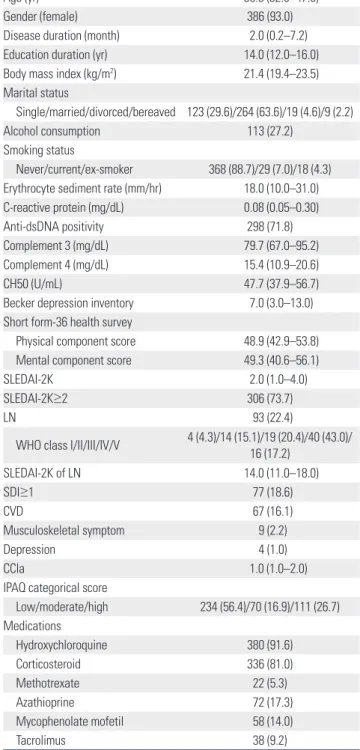
Table 1 shows the baseline demographic data, clinical features, and laboratory findings in the 415 SLE patients. Median age at enrollment was 39.8 years (IQR 32.0–47.0) and 93.0% were fe- male (n=386). Notably, 93 patients with LN (22.4%) were iden- tified, all of whom were pathologically diagnosed in the course of the disease. Median SLEDAI-2K of all patients was 2.0 (IQR 1.0–4.0). The percentage of patients with SLEDAI-2K
≥2 and with at least one organ damaged (≥1 of SDI) was 73.7%
(n=306) and 18.6% (n=77), respectively. In total, 16% of pa- tients (n=67) had at least one manifestation of CVD.
Comparison of characteristics between patients with and without LN is illustrated in Table 2. There were significant dif- ferences in age, marital status, anti-dsDNA positivity, physical component summary of SF-36, frequency of CVD, and use of corticosteroid, methotrexate, mycophenolate mofetil, and ta- crolimus between the two groups. However, the frequency for some disease activity and damage-related variables including SLEDAI-2K ≥2 and SDI ≥1 were similar between the two groups.
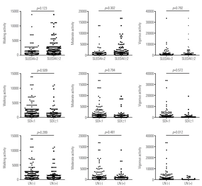
The differences in physical activity based on IPAQ scores according to SLEDAI-2K (<2 vs. ≥2), SDI (<1 vs. ≥1), and LN (presence vs. absence) were assessed (Fig. 1). There were no significant differences in walking, moderate, and vigorous physical activity between active (SLEDAI-2K≥2) and inactive (SLEDAI-2K<2) (p>0.05) patients. Similarly, there were no dif- ferences in each physical activity score between organ-dam- aged (SDI≥1) and non-organ-damaged (SDI<1) (p>0.05) pa- tients. Walking and moderate activity showed similar levels despite the presence or absence of LN. However, patients with LN showed less vigorous activity, compared to those without LN (p=0.012). In addition, there were no differences in num-
bers of patients in low, moderate, and high categories between patients with and without LN (p>0.05). Patients with CVD also showed similar levels of each physical activity score, compared to those without CVD (p>0.05 of all, data not shown).
Table 1. Baseline Characteristics of Enrolled Subjects (n=415)
Variables Results
Age (yr) 39.8 (32.0–47.0)
Gender (female) 386 (93.0)
Disease duration (month) 2.0 (0.2–7.2)
Education duration (yr) 14.0 (12.0–16.0)
Body mass index (kg/m2) 21.4 (19.4–23.5)
Marital status
Single/married/divorced/bereaved 123 (29.6)/264 (63.6)/19 (4.6)/9 (2.2)
Alcohol consumption 113 (27.2)
Smoking status
Never/current/ex-smoker 368 (88.7)/29 (7.0)/18 (4.3) Erythrocyte sediment rate (mm/hr) 18.0 (10.0–31.0) C-reactive protein (mg/dL) 0.08 (0.05–0.30)
Anti-dsDNA positivity 298 (71.8)
Complement 3 (mg/dL) 79.7 (67.0–95.2)
Complement 4 (mg/dL) 15.4 (10.9–20.6)
CH50 (U/mL) 47.7 (37.9–56.7)
Becker depression inventory 7.0 (3.0–13.0) Short form-36 health survey
Physical component score 48.9 (42.9–53.8)
Mental component score 49.3 (40.6–56.1)
SLEDAI-2K 2.0 (1.0–4.0)
SLEDAI-2K≥2 306 (73.7)
LN 93 (22.4)
WHO class I/II/III/IV/V 4 (4.3)/14 (15.1)/19 (20.4)/40 (43.0)/
16 (17.2)
SLEDAI-2K of LN 14.0 (11.0–18.0)
SDI≥1 77 (18.6)
CVD 67 (16.1)
Musculoskeletal symptom 9 (2.2)
Depression 4 (1.0)
CCIa 1.0 (1.0–2.0)
IPAQ categorical score
Low/moderate/high 234 (56.4)/70 (16.9)/111 (26.7) Medications
Hydroxychloroquine 380 (91.6)
Corticosteroid 336 (81.0)
Methotrexate 22 (5.3)
Azathioprine 72 (17.3)
Mycophenolate mofetil 58 (14.0)
Tacrolimus 38 (9.2)
SLEDAI-2K, Systemic Lupus Erythematosus Disease Activity Index 2000; LN, lupus nephritis; SDI, Systemic Lupus International Collaborating Clinics/Amer- ican College of Rheumatology (SLICC/ACR) Damage Index; CVD, cardiovascu- lar disease; IPAQ, International Physical Activity Questionnaire; CCIa, age-ad- justed Charlson comorbidity index.
Data are described as medians with interquartile range or number (%).
Logistic regression analysis was performed to determine the risk factors associated with LN (Table 3). Younger age, shorter disease duration, higher physical component score (PCS) of SF-36, higher SLEDAI-2K, and less vigorous physical activity were significantly associated with the presence of LN in uni- variate logistic regression analysis. However, multivariate lo- gistic regression analysis showed that younger age, higher PCS of SF-36, higher SLEDAI-2K, and less vigorous physical activity were associated with LN. Age, gender, and BMI were
noted to be related to CVD in multivariate logistic regression analysis (data not shown). Remarkably, physical activity mea- sured by the IPAQ estimate was not related to CVD.
DISCUSSION
Studies have established that physical activity could be limit- ed by a high level of disease activity, poor functional disability, Table 2. Comparison of Characteristics according to LN
Variables LN
p value
Absence (n=322) Presence (n=93)
Age (yr) 40.5 (33.0–48.3) 35.0 (28.0–44.0) 0.003
Gender (female) 301 (93.5) 85 (91.4) 0.488
Disease duration (month) 2.0 (0.2–8.1) 1.9 (0.2–6.1) 0.209
Education duration (yr) 14.0 (12.0–16.0) 14.0 (12.0–16.0) 0.149
Body mass index (kg/m2) 21.4 (19.3–23.4) 21.1 (18.9–24.2) 0.458
Marital status 0.019
Single/married/divorced/bereaved 84 (26.1)/216 (67.1)/16 (5.0)/6 (1.9) 39 (41.9)/48 (51.6)/3 (3.2)/3 (3.2)
Alcohol consumption 90 (28.0) 23 (24.7) 0.539
Smoking status 0.455
Never/current/ex-smoker 284(88.2)/25(7.8)/13(4.0) 84(90.3)/4(4.3)/5(5.4)
Erythrocyte sediment rate (mm/hr) 19.0 (11.0–31.0) 16.0 (9.0–28.8) 0.161
C-reactive protein (mg/dL) 0.09 (0.06–0.30) 0.07 (0.05–0.30) 0.646
Anti-dsDNA positivity 156 (48.4) 64 (68.8) 0.001
Complement 3 (mg/dL) 81.3 (67.2–95.4) 77.4 (63.9–95.1) 0.254
Complement 4 (mg/dL) 15.6 (11.3–20.7) 15.0 (9.9–20.6) 0.390
CH50 (U/mL) 47.3 (38.5–56.4) 50.1 (36.4–58.7) 0.353
Becker depression inventory 8.0 (4.0–14.0) 5.0 (3.0–11.0) 0.060
Short form-36 health survey
Physical component score 8.3 (42.1–53.4) 51.1 (44.7–55.1) 0.010
Mental component score 48.8 (42.1–53.5) 50.7 (44.1–56.8) 0.090
SLEDAI-2K 2.0 (1.0–4.0) 4.0 (0.0–6.0) 0.145
SLEDAI-2K≥2 237 (73.6) 69 (74.2) 0.909
SDI≥1 56 (17.4) 21 (22.6) 0.257
CVD 42 (13.0) 25 (26.9) 0.001
Musculoskeletal symptom 7 (2.2) 2 (2.2) 0.674
Depression 3 (0.9) 1 (1.1) 0.639
CCIa 1.0 (1.0–2.0) 1.0 (1.0–2.0) 0.194
IPAQ categorical score 0.295
Low/moderate/high 177 (55.0)/53 (16.5)/92 (28.6) 57 (61.3)/1 7(18.3)/19 (26.7)
Medications
Hydroxychloroquine 295 (91.6) 85 (91.4) 0.947
Corticosteroid 253 (78.6) 83 (89.2) 0.021
Methotrexate 21 (6.5) 1 (1.1) 0.025
Azathioprine 58 (18.0) 14 (15.1) 0.507
Mycophenolate mofetil 21 (6.5) 37 (39.8) < 0.001
Tacrolimus 11 (3.4) 27 (29.0) < 0.001
LN, lupus nephritis; SLEDAI-2K, Systemic Lupus Erythematosus Disease Activity Index 2000; SDI, Systemic Lupus International Collaborating Clinics/American College of Rheumatology (SLICC/ACR) Damage Index; CVD, cardiovascular disease; IPAQ, International Physical Activity Questionnaire; CCIa, age-adjusted Charl- son comorbidity index.
Data are described as medians with interquartile range or number (%).
and organ damage in patients with SLE.2,5-7 Nevertheless, there has been insufficient data about the relationship be- tween physical activity and clinical features in SLE patients.
The main objective of this study was to clarify an association between physical activity assessed by the IPAQ scores and dis- ease activity/organ damage in SLE patients. We observed that physical activity was not associated with SLE-specific mea- sures like SLEDAI and SDI. However, patients with LN were shown to perform less vigorous physical activity, compared to those without LN.
There is evidence of a close relationship between physical inactivity and high disease activity or specific organ involve- ment in patients with SLE. Several clinical studies have re- vealed that exercise or regular physical activity, without the in- terference of disease activity, improves quality of life, fatigue, and other SLE-related symptoms.5,24 Eriksson, et al.7 demon- strated less low to moderate intensity physical activity in SLE patients with SDI ≥2, compared to matched controls. In con-
trast, they did not find a significant difference in physical activ- ity between patients with high disease activity (SLEDAI>5) or low disease activity (SLEDAI≤5) and controls. Two different 12-week randomized trials in small populations with SLE dem- onstrated no significant changes in disease activity after an ex- ercise training program.25,26 Although there was a marked dif- ference in cutoffs for SLEDAI and SDI scores compared to a former study,7 this study failed to confirm the difference of phys- ical activity based on IPAQ scores. The beneficial role of exercise and physical activity in SLE-related disease activity or organ damage should be confirmed in a larger study population.
Physical activity is an independent risk factor for CVD in the general population. Regular physical activity has beneficial health effects and also contributes to decreased mortality risk and development of morbidity.27 The data from the Framing- ham Heart Study indicate that moderate to high physical activ- ity leads to increased total and CVD-free life expectancy, com- pared to low physical activity.28 Premature CVD is attributed SLEDAI<2
p=0.123
p=0.509
p=0.289
p=0.302
p=0.794
p=0.481
p=0.792
p=0.572
p=0.012 SDI<1
LN (-) LN (+) LN (-) LN (+) LN (-) LN (+)
SDI<1 SDI≥1 SDI<1
SDI≥1 SDI≥1
SLEDAI<2 SLEDAI≥2
SLEDAI≥2 SLEDAI<2 SLEDAI≥2
15000
10000
5000 0
15000
10000
5000 0
15000
10000
5000 0
20000 15000 10000 5000 0 20000 15000 10000 5000 0 20000 15000 10000 5000 0
40000 30000 20000 10000 0
40000 30000 20000 10000 0
40000 30000 20000 10000 0
Walking activityWalking activityWalking activity Moderate activityModerate activityModerate activity Vigorous activityVigorous activityVigorous activity
Fig. 1. Comparison of individual physical activity levels according to disease activity, organ damage, and LN. Each unit was described as MET minutes/
week. p values were calculated by Mann-Whiney U test. SLEDAI-2K, Systemic Lupus Erythematosus Disease Activity Index 2000; SDI, Systemic Lupus International Collaborating Clinics/American College of Rheumatology (SLICC/ACR) Damage Index; LN, lupus nephritis; MET, metabolic equivalent of task.
as the most common cause of mortality in SLE patients affect- ed by the disease for over 5 years.29 Earlier studies on physical activity have primarily focused on the risk of CVD in SLE pa- tients.6,30 A multicenter inception cohort study found low phys- ical activity level (score<28) to be a potent risk factor for coro- nary artery disease in 278 SLE patients who were followed for 3 years (71.6% increased risk from enrollment).6 Volkmann, et al.30 demonstrated that physical activity, assessed from self-re- ports by calculating METS per week, was negatively correlated with carotid intima-media thickness and number of carotid plaques, thus implicating the harmful effect of low physical activity on subclinical atherosclerosis in SLE. Subsequently, they recommended increased exercise to reduce the risk of ath- erosclerosis in SLE patients. These findings might be compati- ble with a sedentary lifestyle as a traditional risk factor for CVD in general.31 SLE-induced cognitive impairment has been known to be caused by multiple complex factors, such as in- flammation and lower energy expenditure. Physical inactivity coupled with obesity was associated with an increased risk of cognitive dysfunction in female patients with SLE.2 We found no association between physical activity and the risk of CVD dis-
ease in this study. Based on these results, future studies identify- ing the role of physical activity and other lifestyles in the risk of CVD are warranted.
LN is the most debilitating manifestation of SLE and is as- sociated with increased morbidity and mortality.1 The patho- genesis of LN is well understood: formation of glomerular im- mune complexes, B cell activation with autoantibodies, and inflammatory mediators.32 Although the precise mechanism for the favorable effect of physical activity has not been clearly determined, there is evidence that physical inactivity could induce an increased inflammatory response. Prolonged mu- rine restraint stress induced changes in inflammatory cyto- kine profiles, increasing IL-6 and reducing IL-10, indicating an enhanced inflammatory cascade.33 In contrast, daily mod- erate exercise attenuated systemic inflammation in response to lipopolysaccharide injection in mice.34 Recently, Aqel, et al.35 demonstrated that daily moderate exercise improves the in- flammatory response of LN in NZM2410/J mice through block- ing the expression of inflammatory mediators IL-6, tumor ne- crosis factor-α, CXCL1, and anti-dsDNA antibodies. Their findings also implicated the possibility of adjunct therapeutic Table 3. Regression Analysis for Risk Factors Associated with Lupus Nephritis
Variables Univariate analysis Multivariate analysis*
Coefficient (B) (95% CI for B) p value Exp(B) [95% CI for Exp(B)] p value
Age -0.139 (-0.009– -0.002) 0.005 0.975 (0.953–0.998) 0.032
Gender (ref.=male) 0.034 (-0.102–0.214) 0.489 0.759 (0.300–1.924) 0.562
Disease duration -0.118 (-0.004–0.000) 0.016 0.982 (0.962–1.003) 0.085
Education duration 0.055 (-0.006–0.020) 0.263
Body mass index -0.028 (-0.016–0.009) 0.564
Erythrocyte sediment rate -0.052 (-0.004–0.001) 0.305
C-reactive protein -0.017 (-0.080–0.057) 0.743
Complement 3 -0.054 (-0.003–0.001) 0.269
Complement 4 -0.040 (-0.007–0.003) 0.420
CH50 0.051 (-0.002–0.005) 0.361
Becker depression inventory -0.054 (-0.008–0.002) 0.274
Short form-36 health survey
Physical component score 0.134 (0.002–0.013) 0.006 1.055 (1.018–1.094) 0.004
Mental component score 0.078 (-0.001–0.007) 0.111
SLEDAI-2K 0.110 (0.002–0.026) 0.026 1.080 (1.004–1.161) 0.038
SDI 0.051 (-0.029–0.095) 0.296 1.217 (0.843–1.757) 0.294
Musculoskeletal symptom 1.0111 (0.206–4.952) 0.989 1.566 (0.304–8.077) 0.592
Depression 0.865 (0.089–8.417) 0.901 0.646 (0.060–6.917) 0.646
CCIa 1.191 (0.851–1.668) 0.308 1.313 (0.903–1.909) 0.154
Physical activity by IPAQ
Walking 0.038 (0.000–0.000) 0.444
Moderate -0.111 (0.000–0.000) 0.912
Vigorous -0.121 (-0.222– -0.025) 0.014 1.000 (0.999–1.000) 0.024
Total -0.044 (0.000–0.000) 0.374
SLEDAI-2K, Systemic Lupus Erythematosus Disease Activity Index 2000; SDI, Systemic Lupus International Collaborating Clinics/American College of Rheumatology (SLICC/ACR) Damage Index; IPAQ, International Physical Activity Questionnaire; CCIa, age-adjusted Charlson comorbidity index; CI, confidence interval.
*Multivariate regression analysis was performed using covariates, including age, gender, disease duration, physical component score of SF-36, SLEDAI-2K, SDI, musculoskeletal symptom, depression, and CCIa.
strategy for LN. Compatible with these observation, we found that patients with LN performed less vigorous physical activity.
There are several limitations that should be considered to understand this result. First, this study used only self-reported IPAQ estimates to assess levels of physical activity. Because our study contained a large number of patients, we could not perform objective measurements with an accelerometer. The IPAQ estimate has some advantages in studies that enroll many patients and also for acquisition of discriminable data related with the types of each physical activity, whereas estimation using an accelerometer is not cost-effective. Overall, moderate correlation between self-reported and objectively measured physical activity was confirmed through a cross-sectional study that included 129 patients with SLE.12 Second, healthy and dis- ease control groups were not included in this analysis. Ap- proximately 56% of patients were classified into the low cate- gory in the assessment of physical activity in our registry, which is consistent with earlier evidence showing less physical activi- ty in patients with SLE.9,10 Control groups with inactive, healthy subjects are needed to compare the levels of physical activity in patients with SLE. Third, our results were acquired through cross-sectional analysis. Therefore, we could not identify wheth- er enforcement of physical activity hinders disease activity and lessens organ damage measured through SLEDAI and SDI.
Some debate over the effect of therapeutic intervention through a regular exercise program or physical activity on SLE-related symptoms and disease activity remains.5,24-26 The possibility for exercise and physical activity as therapeutic modalities should be assessed in a prospective study. Considering 90 patients were excluded with missing data for physical activity, the study population may lack representativeness. Patients with a high disease activity or severe disease impairment may be less will- ing to participate in the questionnaire for physical activity. Ex- cluding such patients who do not complete the questionnaire introduces a high likelihood of selective bias. In addition, physi- cal activity according to disease activity or severity may be un- derestimated as a result of this study.
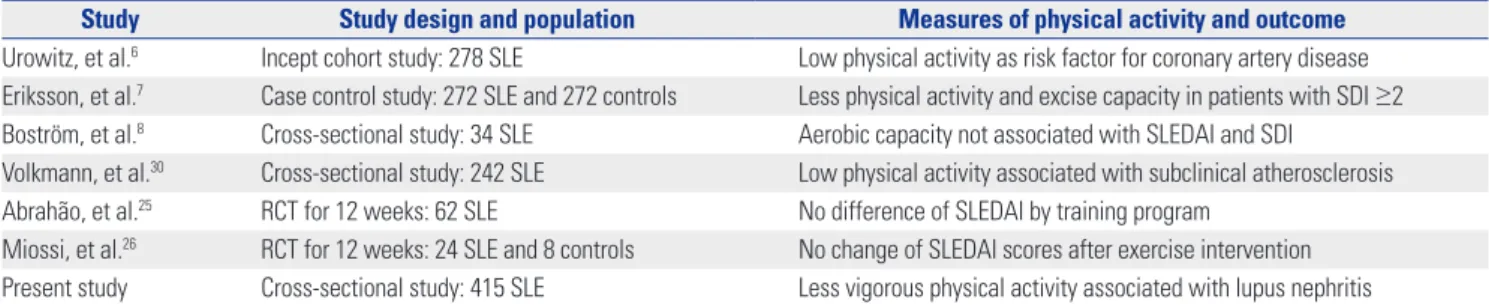
There is still debate over the effect of physical activity in the clinical status of patients with SLE (Table 4). Using the self-re- ported questionnaire IPAQ, this study was performed to iden- tify the associations between physical activity and disease ac-
tivity and organ damage in a population of patients with SLE.
We found that patients with LN showed no difference in walk- ing and moderate physical activity, compared to patients with- out LN. Patients with LN also tended to be reluctant to conduct vigorous physical activity. Efforts to strengthen the intensity of physical activity should be encouraged to avoid incremental exposure to SLE-related morbidity and mortality.
ACKNOWLEDGEMENTS
This work was supported by the Research Program funded by the Korea Centers for Disease Control and Prevention (grant number 2014-E63002-00).
ORCID
Seong-Kyu Kim https://orcid.org/0000-0002-7780-0167
REFERENCES
1. Tsokos GC. Systemic lupus erythematosus. N Engl J Med 2011;365:
2110-21.
2. Katz P, Julian L, Tonner MC, Yazdany J, Trupin L, Yelin E, et al.
Physical activity, obesity, and cognitive impairment among wom- en with systemic lupus erythematosus. Arthritis Care Res (Hobo- ken) 2012;64:502-10.
3. Rodríguez Huerta MD, Trujillo-Martín MM, Rúa-Figueroa Í, Cuel- lar-Pompa L, Quirós-López R, Serrano-Aguilar P; Spanish SLE CPG Development Group. Healthy lifestyle habits for patients with sys- temic lupus erythematosus: a systemic review. Semin Arthritis Rheum 2016;45:463-70.
4. Pettersson S, Boström C, Eriksson K, Svenungsson E, Gunnarsson I, Henriksson EW. Lifestyle habits and fatigue among people with systemic lupus erythematosus and matched population controls.
Lupus 2015;24:955-65.
5. O’Dwyer T, Durcan L, Wilson F. Exercise and physical activity in systemic lupus erythematosus: a systematic review with meta- analyses. Semin Arthritis Rheum 2017;47:204-15.
6. Urowitz MB, Gladman D, Ibañez D, Fortin P, Sanchez-Guerrero J, Bae S, et al. Accumulation of coronary artery disease risk factors over three years: data from an international inception cohort. Ar- thritis Rheum 2008;59:176-80.
7. Eriksson K, Svenungsson E, Karreskog H, Gunnarsson I, Gustafs- son J, Möller S, et al. Physical activity in patients with systemic lupus erythematosus and matched controls. Scand J Rheumatol 2012;
Table 4. Summary for the Effect of Physical Activity on Disease Activity and Organ Damage
Study Study design and population Measures of physical activity and outcome Urowitz, et al.6 Incept cohort study: 278 SLE Low physical activity as risk factor for coronary artery disease Eriksson, et al.7 Case control study: 272 SLE and 272 controls Less physical activity and excise capacity in patients with SDI ≥2 Boström, et al.8 Cross-sectional study: 34 SLE Aerobic capacity not associated with SLEDAI and SDI
Volkmann, et al.30 Cross-sectional study: 242 SLE Low physical activity associated with subclinical atherosclerosis Abrahão, et al.25 RCT for 12 weeks: 62 SLE No difference of SLEDAI by training program
Miossi, et al.26 RCT for 12 weeks: 24 SLE and 8 controls No change of SLEDAI scores after exercise intervention Present study Cross-sectional study: 415 SLE Less vigorous physical activity associated with lupus nephritis
SLE, systemic lupus erythematosus; SLEDAI, Systemic Lupus Erythematosus Disease Activity Index; SDI, Systemic Lupus International Collaborating Clinics/Ameri- can College of Rheumatology (SLICC/ACR) Damage Index; RCT, randomized controlled study.
41:290-7.
8. Boström C, Dupré B, Tengvar P, Jansson E, Opava CH, Lundberg IE.
Aerobic capacity correlates to self-assessed physical function but not to overall disease activity or organ damage in women with sys- temic lupus erythematosus with low-to-moderate disease activity and organ damage. Lupus 2008;17:100-4.
9. Petri M, Spence D, Bone LR, Hochberg MC. Coronary artery dis- ease risk factors in the Johns Hopkins Lupus Cohort: prevalence, recognition by patients, and preventive practices. Medicine (Balti- more) 1992;71:291-302.
10. Costenbader KH, Wright E, Liang MH, Karlson EW. Cardiac risk factor awareness and management in patients with systemic lupus erythematosus. Arthritis Rheum 2004;51:983-8.
11. dos Santos Fde M, Borges MC, Correia MI, Telles RW, Lanna CC.
Assessment of nutritional status and physical activity in systemic lupus erythematosus patients. Rev Bras Reumatol 2010;50:631-8.
12. Ahn GE, Chmiel JS, Dunlop DD, Helenowski IB, Semanik PA, Song J, et al. Self-reported and objectively measured physical ac- tivity in adults with systemic lupus erythematosus. Arthritis Care Res (Hoboken) 2015;67:701-7.
13. Legge A, Blanchard C, Hanly JG. Physical activity and sedentary behavior in patients with systemic lupus erythematosus and rheu- matoid arthritis. Open Access Rheumatol 2017;9:191-200.
14. Tan EM, Cohen AS, Fries JF, Masi AT, McShane DJ, Rothfield NF, et al. The 1982 revised criteria for the classification of systemic lu- pus erythematosus. Arthritis Rheum 1982;25:1271-7.
15. Hochberg MC. Updating the American College of Rheumatology revised criteria for the classification of systemic lupus erythema- tosus. Arthritis Rheum 1997;40:1725.
16. Richter P, Werner J, Heerlein A, Kraus A, Sauer H. On the validity of the Beck Depression Inventory. A review. Psychopathology 1998;
31:160-8.
17. Ware JE Jr, Sherbourne CD. The MOS 36-item short-form health survey (SF-36). I. Conceptual framework and item selection. Med Care 1992;30:473-83.
18. Alarcón GS, McGwin G Jr, Bartolucci AA, Roseman J, Lisse J, Fes- sler BJ, et al. Systemic lupus erythematosus in three ethnic groups.
IX. Differences in damage accrual. Arthritis Rheum 2001;44:2797- 806.
19. Gladman D, Ginzler E, Goldsmith C, Fortin P, Liang M, Urowitz M, et al. Systemic lupus international collaborative clinics: devel- opment of a damage index in systemic lupus erythematosus. J Rheumatol 1992;19:1820-1.
20. Charlson ME, Pompei P, Ales KL, MacKenzie CR. A new method of classifying prognostic comorbidity in longitudinal studies: de- velopment and validation. J Chronic Dis 1987;40:373-83.
21. Craig CL, Marshall AL, Sjöström M, Bauman AE, Booth ML, Ain- sworth BE, et al. International physical activity questionnaire:
12-country reliability and validity. Med Sci Sports Exerc 2003;35:
1381-95.
22. Chun MY. Validity and reliability of Korean version of international
physical activity questionnaire short form in the elderly. Korean J Fam Med 2012;33:144-51.
23. Hernández-Hernández V, Ferraz-Amaro I, Díaz-González F. In- fluence of disease activity on the physical activity of rheumatoid arthritis patients. Rheumatology (Oxford) 2014;53:722-31.
24. Mancuso CA, Perna M, Sargent AB, Salmon JE. Perceptions and measurements of physical activity in patients with systemic lupus erythematosus. Lupus 2011;20:231-42.
25. Abrahão MI, Gomiero AB, Peccin MS, Grande AJ, Trevisani VF.
Cardiovascular training vs. resistance training for improving qual- ity of life and physical function in patients with systemic lupus er- ythematosus: a randomized controlled trial. Scand J Rheumatol 2016;45:197-201.
26. Miossi R, Benatti FB, Lúciade de Sá Pinto A, Lima FR, Borba EF, Prado DM, et al. Using exercise training to counterbalance chro- notropic incompetence and delayed heart rate recovery in sys- temic lupus erythematosus: a randomized trial. Arthritis Care Res (Hoboken) 2012;64:1159-66.
27. Pate RR, Pratt M, Blair SN, Haskell WL, Macera CA, Bouchard C, et al. Physical activity and public health. A recommendation from the Centers for Disease Control and Prevention and the American College of Sports Medicine. JAMA 1995;273:402-7.
28. Franco OH, de Laet C, Peeters A, Jonker J, Mackenbach J, Nus- selder W. Effects of physical activity on life expectancy with car- diovascular disease. Arch Intern Med 2005;165:2355-60.
29. Svenungsson E, Jensen-Urstad K, Heimbürger M, Silveira A, Hamsten A, de Faire U, et al. Risk factors for cardiovascular dis- ease in systemic lupus erythematosus. Circulation 2001;104:1887- 93.
30. Volkmann ER, Grossman JM, Sahakian LJ, Skaggs BJ, FitzGerald J, Ragavendra N, et al. Low physical activity is associated with proin- flammatory high-density lipoprotein and increased subclinical atherosclerosis in women with systemic lupus erythematosus. Ar- thritis Care Res (Hoboken) 201;62:258-65.
31. Lewandowski LB, Kaplan MJ. Update on cardiovascular disease in lupus. Curr Opin Rheumatol 2016;28:468-76.
32. Peutz-Kootstra CJ, de Heer E, Hoedemaeker PJ, Abrass CK, Bruijn JA. Lupus nephritis: lessons from experimental animal models. J Lab Clin Med 2001;137:244-60.
33. Voorhees JL, Tarr AJ, Wohleb ES, Godbout JP, Mo X, Sheridan JF, et al. Prolonged restraint stress increases IL-6, reduces IL-10, and causes persistent depressive-like behavior that is reversed by re- combinant IL-10. PLoS One 2013;8:e58488.
34. Kasawara KT, Cotechini T, Macdonald-Goodfellow SK, Surita FG, Pinto E Silva JL, Tayade C, et al. Moderate exercise attenuates lipo- polysaccharide-induced inflammation and associated maternal and fetal morbidities in pregnant rats. PLoS One 2016;11:e0154405.
35. Aqel SI, Hampton JM, Bruss M, Jones KT, Valiente GR, Wu LC, et al.
Daily moderate exercise is beneficial and social stress is detrimen- tal to disease pathology in murine lupus nephritis. Front Physiol 2017;8:236.