© 2012 The Korean Academy of Medical Sciences.
This is an Open Access article distributed under the terms of the Creative Commons Attribution Non-Commercial License (http://creativecommons.org/licenses/by-nc/3.0) which permits unrestricted non-commercial use, distribution, and reproduction in any medium, provided the original work is properly cited.
pISSN 1011-8934 eISSN 1598-6357
Methimazole-Induced Bullous Systemic Lupus Erythematosus:
A Case Report
Bullous systemic lupus erythematosus (SLE) is a kind of LE-non-specific bullous skin disease that is rarely induced by a medication. We describe the first case of bullous SLE to develop after administration of methimazole. A 31-yr-old woman presented with generalized erythematous patches, multiple bullae, arthralgia, fever, conjunctivitis, and hemolytic anemia. Biopsy of her bulla showed linear deposition of lgG, lgA, C3, fibrinogen, and C1q at dermo-epidermal junction. She was diagnosed as bullous SLE and treated with prednisolone, dapsone, hydroxychloroquine, and methotrexate. Our experience suggests that SLE should be considered as a differential diagnosis when bullous skin lesions develop in patients being treated for hyperthyroidism.
Key Words: Bullous Systemic Lupus Erythematosus; Drug-Induced Lupus Erythematosus;
Methimazole; Graves Disease Ji-Yeon Seo1, Hee-Jin Byun2,
Kwang-Hyun Cho2, and Eun Bong Lee1 Departments of 1Internal Medicine, 2Dermatology, Seoul National University College of Medicine, Seoul, Korea
Received: 28 November 2011 Accepted: 3 April, 2012 Address for Correspondence:
Eun Bong Lee, MD
Department of Internal Medicine, Seoul National University College of Medicine, 101 Daehak-ro, Jongno-gu, Seoul 110-744, Korea
Tel: +82.2-2072-3944, Fax: +82.2-762-9662 E-mail: leb7616@snu.ac.kr
http://dx.doi.org/10.3346/jkms.2012.27.7.818 • J Korean Med Sci 2012; 27: 818-821
CASE REPORT
Immunology, Allergic Disorders & Rheumatology
INTRODUCTION
Although the exact pathogenesis of systemic lupus erythemato- sus (SLE) is unknown, several drugs cause drug-induced lupus erythematosus (DILE) (1). DILE is a rare disease that is diagnosed when the following features are present: 1) exposure to a drug suspected to induce DILE; 2) no history of SLE prior to drug therapy; 3) detection of positive antinuclear antibodies (ANA) with at least one clinical sign of SLE; 4) rapid improvement and a gradual fall in ANA, and other serologic findings, upon with- drawal of the drug (1).
Bullous SLE is a kind of LE-non-specific bullous skin disease in which autoantibody-mediated subepidermal blistering oc- curs. Histopathological analysis showed marked neutrophil infiltration with papillary microabscess formation (2). To date, the only known drugs reported to induce bullous SLE are hy- dralazine and penicillamine.
We present here the first case of bullous SLE triggered by me- thimazole, which was treated with prednisolone, dapsone, hy- droxychloroquine, and methotrexate, finally progressed to SLE nephritis.
CASE DESCRIPTION
A 31-yr-old woman presented with generalized erythematous to brownish patches and multiple bullae. She was diagnosed with Graves’ disease in July 2008, with a decreased TSH level (0.05 μIU/mL; reference range [RR] 0.15-5.0 μIU/mL) and in-
creased free T4 level (21.2 μg/dL; RR 4.6-14.0 μg/dL). Propylthio- uracil was prescribed for 6 months, but was changed to methim- azole in December 2008 due to inadequate control of hyperthy- roidism. One month after the introduction of methimazole, gen- eralized erythematous maculopapular rash developed over her whole body but responded to topical steroids. The woman de- veloped arthralgia of both knees and elbows in early July 2009, which resolved spontaneously within 1 month. At the end of July, intra-oral blisters developed with erythematous patches on both extremities accompanied with pruritus. The skin lesions then spread to the whole body. In early August, multiple 0.5-2.0 cm diameter bullous lesions developed from some of the previous patches. The bullous lesions usually developed on the sites where pruritus was present. Methimazole was discontinued on suspi- cion of drug allergy on August 10, and 131I therapy was adminis- tered on August 19, 2009. Prednisolone at a dosage of 15 mg/d was initiated on August 25 for the skin lesions but was not effec- tive. Hydroxychloroquine, which was initiated on September 25, was not effective either. Only dexamethasone brought tempo- rary relief. Fever developed (38.5°C) from September 23, 2009, and the woman was referred to the rheumatology division for further evaluation. She had no past medical history or personal familial history of bullous disease.
On physical examination, her blood pressure was 130/94 mmHg; pulse rate, 90/min; and body temperature, 38.5°C. Her eyes had reddish conjunctivae. Her voice was husky, the oral cavity was filled with multiple ulcerations, and both lips were swollen. Multiple dark-reddish annular lesions and dark-brown-
Seo J-Y, et al. • Bullous Systemic Lupus Erythematosus Related to Methimazole
http://jkms.org 819
http://dx.doi.org/10.3346/jkms.2012.27.7.818
ish patches were observed on her trunk, extremities, palms, and soles. Several blisters that varied in size (0.5-2.0 cm) were found on both extremities, and on her palms and soles (Fig. 1). How- ever, neither lymph node enlargement nor hepatosplenomega- ly was detected. Chest auscultation revealed no evidence of ab- normal sound or friction rubs. Joint examination revealed no evidence of arthritis. Laryngoscopic examination revealed severe inflammation and erosion of the larynx. Ophthalmology exam- ination showed bilateral conjunctivitis. Laboratory test results
were as follows: white blood cell count, 5,070/μL; hemoglobin, 9.5 g/dL; and platelet count, 221,000/μL; reticulocyte count, 5.56%; haptoglobin, 10 mg/dL (RR 30-180 mg/dL). The result for Coombs’ test was positive, suggesting hemolytic anemia.
Viral cultures for Herpes simplex, Varicella zoster, and cytomeg- alovirus were all negative. Her ANA was over 1:320, homoge- neous pattern, anti-histone antibody (47.0, RR < 40.0 U/mL), and anti-ds DNA were positive (56.5, RR 0-10 IU/mL). Also, anti-cardiolipin IgM and Anti-neutrophil cytoplasmic antibody
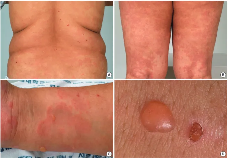
A B
C D
Fig. 1. Gross view of the skin lesion. (A-C) Generalized itchy erythematous to brownish polymorphic patches on the back, legs and arm. (C, D) Discrete tense bullae on her right arm.
A B C
Fig. 2. Histopathology of the skin lesion. (A, B) Histopathologic examination of the skin biopsy specimen showed a subepidermal blister with abundant neutrophils (H&E stain,
× 100). (C) Direct immunofluorescence examination showed linear deposition of IgG at the dermo-epidermal junction (× 100).
Seo J-Y, et al. • Bullous Systemic Lupus Erythematosus Related to Methimazole
820 http://jkms.org http://dx.doi.org/10.3346/jkms.2012.27.7.818
(ANCA) were positive. Rheumatic factor, anti-Sm, lupus antico- agulant, anti-Ro, and anti-La antibodies were negative. Com- plement levels were decreased with C3 being 70 mg/dL (RR 70- 150 mg/dL) and C4, 7 mg/dL (RR 10-35 mg/dL). The bulla on her left thigh was biopsied, and histopathologic examination showed a subepidermal blister with abundant neutrophils (Fig. 2). Direct immunofluorescence examination showed linear deposition of lgG, lgA, C3, fibrinogen, and C1q at the dermo-epidermal junc- tion (Fig. 2). There was no evidence of vasculitis.
Because bullous SLE was suspected, 40 mg/d prednisolone and 300 mg/d hydroxychloroquine were started on September 28, which alleviated the fever, and relief provided relief from the annular skin and patch lesions. The bullous skin lesions, which were refractory to prednisolone, were partially relieved with the introduction of 125 mg bid dapsone on October 1. Methotrexate at 15 mg/week was required for complete relief of the bullous lesions. The redness of the eyes disappeared slowly, and her voice returned to normal. After discharge, at 12 months, lupus nephritis occurred and she under the treatment with predniso- lone, mycophenolate mofetil.
DISCUSSION
In this case, SLE developed after the administration of methim- azole, with multiple oral ulcerations, arthritis, hemolytic ane- mia, low complement levels, and anti-nuclear, anti-cardiolipin, and anti-ds DNA antibodies. Anti-histone antibodies were also present. In addition to fulfilling the classification criteria for SLE (3), this case satisfied the diagnostic criteria for bullous SLE pro- posed by Yell: 1) subepidermal blistering in SLE, and 2) IgG, IgA, C3, fibrinogen, and C1q deposits at the basement membrane zone on direct immunofluorescence examination (4).
DILE is a lupus-like disease caused by exposure to drugs. Like idiopathic SLE, DILE can be classified into systemic, subacute, and chronic forms depending on the characteristics of the cuta- neous manifestations. The drugs frequently implicated in sys- temic DILE are hydralazine, procainamide, isoniazid, methyl- dopa, chlorpromazine, quinidine, and minocycline (5). For sub- acute forms, calcium channel blockers, angiotensin-converting enzyme inhibitors (6), docetaxel (7) and thiazide diuretics are reported to be frequent triggers. Antithyroid drugs such as pro- pylthiouracil and methimazole have also been reported to cause systemic DILE (8-12). Among the 8 cases of methimazole-in- duced SLE reported to date, none had bullous SLE (13). One case in which vesiculobullous skin lesions developed after ad- ministration of methimazole was due to antineutrophil cytoplas- mic antibody-positive vasculitis, and did not satisfy the criteria for SLE (14). Drug-induced bullous SLE has been reported for hydralazine and penicillamine (15, 16).
Our case showed peculiar autoantibody profiles. Drug-in- duced lupus is generally characterized by anti-histone antibod-
ies, but subacute forms, which include annular papulosqua- mous and bullous forms, are reported to show anti-Ro and anti- La antibodies without anti-histone antibodies (8). However, our patient showed anti-dsDNA and anti-histone antibodies with- out anti-Ro or anti-La antibody. These autoantibody profiles suggest that the disease status in our patients was between the systemic and subacute forms of lupus. At the time of initial pre- sentation, drug-induced bullous SLE was the more favored diag- nosis than idiopathic SLE, based on the reasons that follow. First, it developed 1 month after the use of methimazole. This is con- sistent with a previous report that DILE develops 2 weeks to 3.2 yr after use of a drug (8, 17). Second, the rash was distributed over the woman’s whole body, including the lower extremities. In idiopathic SLE, rash usually develops on the sun-exposed up- per body, but it can develop over the whole body, including the lower extremities, in DILE cases (5, 18). Third, systemic signs were not prominent in our case. There was a fever, but other common systemic symptoms in DILE, such as arthritis and se- rositis, were absent (19). But, after 1 yr, she presented with lupus nephritis, which means methimazole triggered SLE later.
For bullous SLE, dapsone is the cornerstone treatment; it in- duces a dramatic response within a week (8). In some cases where an adequate response is not achieved with dapsone, oth- er immunosuppressants, such as prednisolone, methotrexate, and azathioprine, can be tried (20). In our case, dapsone alone was not sufficient to control the symptoms, and prednisolone, methotrexate, and hydroxychloroquine were also required for control of the symptoms. The combined features of systemic and subacute forms in our case could have necessitated com- bined treatment.
In conclusion, we report the first case of bullous SLE triggered by methimazole. The findings in our case suggest that SLE should be considered as a differential diagnosis when bullous skin le- sions develop in patients treated with methimazole for hyper- thyroidism.
REFERENCES
1. Hess E. Drug-related lupus. N Engl J Med 1988; 318: 1460-2.
2. Vassileva S. Bullous systemic lupus erythematosus. Clin Dermatol 2004;
22: 129-38.
3. Tan EM, Cohen AS, Fries JF, Masi AT, McShane DJ, Rothfield NF, Schaller JG, Talan N, Winchester RJ. The 1982 revised criteria for the classification of systemic lupus erythematosus. Arthritis Rheum 1982; 25: 1271-7.
4. Yell JA, Allen J, Wojnarowska F, Kirtschig G, Burge SM. Bullous systemic lupus erythematosus: revised criteria for diagnosis. Br J Dermatol 1995;
132: 921-8.
5. Marzano AV, Vezzoli P, Crosti C. Drug-induced lupus: an update on its dermatologic aspects. Lupus 2009; 18: 935-40.
6. Kim BS, Hong YM, Park SM, Park IW, Lee BH, Jeong JH, Lee CW. A case of angiotensin converting enzyme inhibitor-induced systemic lupus ery- thematosus. J Rheum Dis 2011; 18: 288-91.
Seo J-Y, et al. • Bullous Systemic Lupus Erythematosus Related to Methimazole
http://jkms.org 821
http://dx.doi.org/10.3346/jkms.2012.27.7.818
7. Shin JA, Huh CW, Kwon JE, Kim HJ, Ahn CM, Chang YS. A case of docetax- el induced subacute cutaneous lupus erythematosus. Tuberc Respir Dis 2009; 66: 380-4.
8. Sontheimer RD, Henderson CL, Grau RH. Drug-induced subacute cuta- neous lupus erythematosus: a paradigm for bedside-to-bench patient- oriented translational clinical investigation. Arch Dermatol Res 2009;
301: 65-70.
9. Sakata S, Nakamura S, Nagai K, Komaki T, Kawade M, Niwa T, Miura K.
Two cases of systemic lupus erythematosus associated with hyperthyroid- ism. Jpn J Med 1987; 26: 373-6.
10. Park CO, Goo J, Ahn SK. A case of propylthiouracil-induced lupus ery- thematosus. Korean J Dermatol 2006; 44: 467-9.
11. Kim JW, Kim JS. A case of propylthiouracil-induced lupus erythematosus accompanied by antineutrophil cytoplasmic antibody-positive vasculitis.
Korean J Dermatol 2005; 43: 496-500.
12. Lee J, Yoo B, Lim YJ, Kim SH, Lim M, Cho YS, Shong YK, Moon HB. A case of propylthiouracil-induced lupus. J Korean Rheum Assoc 1999; 6:
75-8.
13. Wang LC, Tsai WY, Yang YH, Chiang BL. Methimazole-induced lupus erythematosus: a case report. J Microbiol Immunol Infect 2003; 36: 278-81.
14. Thong HY, Chu CY, Chiu HC. Methimazole-induced antineutrophil cyto- plasmic antibody (ANCA)-associated vasculitis and lupus-like syndrome with a cutaneous feature of vesiculo-bullous systemic lupus erythemato- sus. Acta Derm Venereol 2002; 82: 206-8.
15. Condon C, Phelan M, Lyons JF. Penicillamine-induced type II bullous systemic lupus erythematosus. Br J Dermatol 1997; 136: 474-5.
16. Fleming MG, Bergfeld WF, Tomecki KJ, Tuthill RJ, Norris M, Benedetto EA, Weber LA. Bullous systemic lupus erythematosus. Int J Dermatol 1989; 28: 321-6.
17. Srivastava M, Rencic A, Diglio G, Santana H, Bonitz P, Watson R, Ha E, Anhalt GJ, Provost TT, Nousari CH. Drug-induced, Ro/SSA-positive cu- taneous lupus erythematosus. Arch Dermatol 2003; 139: 45-9.
18. Bonsmann G, Schiller M, Luger TA, Ständer S. Terbinafine-induced sub- acute cutaneous lupus erythematosus. J Am Acad Dermatol 2001; 44:
925-31.
19. Antonov D, Kazandjieva J, Etugov D, Gospodinov D, Tsankov N. Drug- induced lupus erythematosus. Clin Dermatol 2004; 22: 157-66.
20. Patrício P, Ferreira C, Gomes MM, Filipe P. Autoimmune bullous derma- toses: a review. Ann N Y Acad Sci 2009; 1173: 203-10.