• 검색 결과가 없습니다.

Brain meningioma in a patient with systemic lupus erythematosus

N/A
N/A
Protected

Academic year: 2021

Share "Brain meningioma in a patient with systemic lupus erythematosus"

Copied!
3
0
0

로드 중.... (전체 텍스트 보기)

전체 글

(1)

CASE REPORT eISSN 2384-0293 https://doi.org/10.12701/yujm.2016.33.2.159

Yeungnam Univ J Med 2016;33(2):159-161

YUJM VOLUME 33, NUMBER 2, DECEMBER 2016 159

Brain meningioma in a patient with systemic lupus erythematosus

Byung-Woo Yoo

1

, Sung-Soo Ahn

1

, Jung Yoon Pyo

1

, Se-Jin Byun

1

, Jason Jungsik Song

1

, Yong-Beom Park

1

, Soo-Kon Lee

1

, Jung-Soo Song

2

, Sang-Won Lee

1

1

Division of Rheumatology, Department of Internal Medicine, Yonsei University College of Medicine;

2

Department of Rheumatology, Chung-Ang University College of Medicine, Seoul, Korea

Brain meningioma, the most common benign brain tumor, has been reported to account for 13-26% of all intracranial tumors, with a crude incidence rate of 2.3 per 100,000 persons for all types of meningiomas.

The prevalence of neuropsychiatric lupus erythematosus is 15-91% and its clinical manifestations are diverse:

from mild cognitive dysfunction to serious neurological or psychiatric symptoms. Here, we report the first Korean patient with brain meningioma and systemic lupus erythematosus who had undergone surgical tumor resection.

Keywords: Systemic lupus erythematosus; Meningioma; Headache

Received: June 11, 2015, Revised: July 30, 2015 Accepted: August 07, 2015

Corresponding Author: Sang-Won Lee, Division of Rheuma- tology, Department of Internal Medicine, Yonsei University College of Medicine, 50 Yonsei-ro, Seodaemun-gu, Seoul 03722, Korea

Tel: +82-2-2228-1947, Fax: +82-2-393-6884 E-mail: sangwonlee@yuhs.ac

Copyright ©2016 Yeungnam University College of Medicine

This is an Open Access article distributed under the terms of the Creative Commons Attribution Non-Commercial License (http://creative- commons.org/licenses/by-nc/4.0/) which permits unrestricted non-commercial use, distribution, and reproduction in any medium, provided the original work is properly cited.

INTRODUCTION

Brain meningioma is a common benign brain tumor origi- nating from non-neuroepithelial progenitor cells [1]. Menin- giomas account for 13-26% of all intracranial tumors, with a crude incidence rate of 2.3 per 100,000 persons [2]. The incidence increases in the 6th and 7th decades of life, and meningiomas are predominantly found in women, with a gen- der ratio of 3:2 or 2:1 [1]. A positive association of meningi- oma with pre-existing diseases has been reported, including diabetes mellitus and pulmonary hypertension, and a negative association with rheumatoid arthritis [3].

Systemic lupus erythematosus (SLE) is a prototypic infla- mmatory autoimmune disease with multi-organ involvement, varied manifestations, and an unpredictable course. The pre-

valence of neuropsychiatric SLE ranges from 15-91% of lupus, and its clinical manifestations are diverse: from mild cogni- tive dysfunction to serious neurological or psychiatric symp- toms [4,5]. Brain involvement is a significant cause of mor- bidity and mortality, and the primary cause of death in 4-8%

of patients with SLE [6]. Early detection and proper treat- ment of brain involvement in SLE with neuropsychiatric fea- tures might decisively influence prognosis.

Few occurrences of brain meningioma in patients with SLE have been reported [7,8]. To the best of our knowledge, no SLE patients with brain meningioma have yet been reported from Eastern Asia. In this report, we describe a Korean pa- tient with SLE and concurrent brain meningioma.

CASE

We report on a 49-year-old female patient diagnosed with

SLE who presented with a brain meningioma. She was re-

ferred to our hospital for recurrent oral ulcers, malar rash,

and arthritis. On physical examination, several ulcers were

observed on the lower lips and bilateral buccal mucosa, and

a maculopapular rash affected the bilateral malar area. There

(2)

Byung-Woo Yoo et al.

160 YUJM VOLUME 33, NUMBER 2, DECEMBER 2016

Fig. 1. A 13×18 mm-sized dural-based extra-axial mass is shown in the left parieto-occipital area as hyperintensity on an axial T2-weighted image (A), hypointensity on an axial T1-weighted image (B), and homogeneous enhancement on an axial enhanced T1-weighted image (C).

Fig. 2. Microscopic findings demonstrating tightly whorled clusters of tumor cells, suggesting meningioma of the meningothelial type (H&E stain, A×100; B×400).

was mild effusion in the right knee joint. Laboratory findings showed a hemoglobin concentration 9.2 g/dL, white blood cell 3,450/mm

3

(lymphocytes, 750/mm

3

), platelet 187,000/mm

3

, erythrocyte sedimentation rate of 45 mm/h, and C-reactive protein 2.3 mg/L. Blood urea nitrogen, creatinine, total pro- tein, albumin, aspartate aminotransferase, alanine aminotrans- ferase, and lactate dehydrogenase were within the normal range. Anti-nuclear antibodies were detected, with a titer 1:640. Reactions for anti-dsDNA, anti-Sm, anti-SSA/Ro, and anti-SSB/La antibodies were also positive. Complement C3 and C4 levels were decreased to 56 mg/dL and 6.3 mg/dL, respectively. Anti-cardiolipin and anti-beta2 glycoprotein 1 antibodies and lupus anticoagulant were not detected, and no proteinuria or red blood cell casts were detected using urine test strips.

The patient was diagnosed with SLE, and treated with low-dose prednisolone (10 mg/day) and hydroxychloroquine (300 mg/day). One week later, she visited the hospital with a severe headache of 3-day duration. On neurological exami- nation, no decreased motor strength or sensory changes were detected, and her mental status was alert. To exclude central nervous system (CNS) involvement in SLE, brain magnetic

resonance imaging (MRI) was performed, which demonstrated an extra-axial tumor in the left parietal region, based on the parietal convexity with a dural tail and showing homogeneous signal hypo-intensity on T1-weighted images and hyper-inten- sity on T2-weighted images (Fig. 1). The tumor measured 13×

18 mm in size. The radiological features were suggestive of a brain meningioma. Because her meningioma was large enough to provoke a persistent headache, complete surgical resection was indicated and performed. Histopathological findings from the resected tumor revealed a brain meningioma of the menin- gothelial type (Fig. 2). No neurological complications were observed. Follow-up brain MRI performed 4 months later showed obliteration of the sulcal spaces in the left parietal lobe without evidence of residual or recurrent tumor. The patient has since remained free of symptoms.

DISCUSSION

In the Korean population, the overall crude rate of CNS tumors is 11.69 per 100,000 person-years, and meningioma is the most common primary CNS tumor (31.2%). The female- to-male ratio has been reported as 3:2 or 2:1 and the mean age of patients at diagnosis is 57.6 years old. Most meningio- mas are benign [1,9].

Most meningiomas are discovered incidentally during exa- mination for unrelated symptoms. If symptoms present, they are determined by the location and size of the mass. Common manifestations of intracranial meningiomas include seizure disorders, cranial nerve dysfunctions, and visual disturbances [10]. Although symptoms of both lupus and meningioma vary and are not specific, imaging study is required for patients who complain of recurrent and intractable symptoms among patients with suspected neuropsychiatric lupus.

Castellino et al. reported three cases of meningioma with

lupus erythematosus [8]. The prevalence of meningioma in

patients with lupus erythematosus was 1.65% (3 among 181

patients). All three patients were women complaining of neu-

ropsychiatric symptoms characterized by headache, mild cog-

nitive dysfunction, anxiety, depression, and panic attacks, whic

h were unrelated to the location of meningioma. One of the

patients had undergone surgical removal of meningioma, but

showed no improvement of symptoms. The authors con-

cluded that their findings were pure coincidence. Richardson

and Cohen reported on a 44-year-old female patient who

(3)

Brain meningioma in SLE

YUJM VOLUME 33, NUMBER 2, DECEMBER 2016 161

developed a meningioma after the onset of subacute cuta- neous lupus erythematosus [11].

In previous studies, the risk factors for development of brain meningioma included deletions in the neurofibromato- sis type-2 gene, ionizing radiation, head injury, and defective hormonal receptors [1]. Among the risk factors, the role of hormones is expected to have most association between men- ingioma and SLE as SLE is more prevalent in women and the disease activity of SLE is often associated with oral contra- ceptive use or pregnancy. Estrogen receptors are particularly associated with aggressive meningiomas. Estrogens are also regarded as potent accelerators of disease in both male and female murine models of SLE [12].

The association between meningioma and autoimmune dis- ease is not yet well established. Meningioma may be more prevalent in patients with lupus erythematosus than in the general population. Although further studies using more ex- tensive data are required, we should be alert to the possibility of meningioma in patients with SLE who present with neuro- psychiatric symptoms and use of imaging modalities like MRI should be considered for these patients.

REFERENCES

1. Marosi C, Hassler M, Roessler K, Reni M, Sant M, Mazza E, et al. Meningioma. Crit Rev Oncol Hematol 2008;67:153- 71.

2. Rohringer M, Sutherland GR, Louw DF, Sima AA. Incidence and clinicopathological features of meningioma. J Neurosurg 1989;71:665-72.

3. Schneider B, Pülhorn H, Röhrig B, Rainov NG. Predisposing conditions and risk factors for development of symptomatic meningioma in adults. Cancer Detect Prev 2005;29:440-7.

4. Huizinga TW, Diamond B. Lupus and the central nervous system. Lupus 2008;17:376-9.

5. Borchers AT, Aoki CA, Naguwa SM, Keen CL, Shoenfeld Y, Gershwin ME. Neuropsychiatric features of systemic lupus erythematosus. Autoimmun Rev 2005;4:329-44.

6. Jönsen A, Bengtsson AA, Nived O, Ryberg B, Sturfelt G. Out- come of neuropsychiatric systemic lupus erythematosus with- in a defined Swedish population: increased morbidity but low mortality. Rheumatology (Oxford) 2002;41:1308-12.

7. Williams HM, Lundberg GA Jr. Systemic lupus erythmatosus, meningioma and carcinoma of the cervix--a case report. 1956.

Conn Med 2006;70:453-6.

8. Castellino G, Rizzo N, Bernardi S, Trotta F, Govoni M. Menin- gioma and systemic lupus erythematosus: a matter of pure coincidence? Lupus 2009;18:650-4.

9. Lee CH, Jung KW, Yoo H, Park S, Lee SH. Epidemiology of primary brain and central nervous system tumors in Korea.

J Korean Neurosurg Soc 2010;48:145-52.

10. Whittle IR, Smith C, Navoo P, Collie D. Meningiomas. Lancet 2004;363(9420):1535-43.

11. Richardson TT, Cohen PR. Subacute cutaneous lupus eryth- ematosus: report of a patient who subsequently developed a meningioma and whose skin lesions were treated with isotretinoin. Cutis 2000;66:183-8.

12. Greenstein B, Roa R, Dhaher Y, Nunn E, Greenstein A,

Khamashta M, et al. Estrogen and progesterone receptors in

murine models of systemic lupus erythematosus. Int Immuno-

pharmacol 2001;1:1025-35.

수치

Fig. 2. Microscopic findings demonstrating tightly whorled clusters of tumor cells, suggesting meningioma of the meningothelial type (H&E stain, A×100; B×400)

참조

관련 문서

• A predictor of patient’s survival and technical survival - high solute transport: mortality risk↑. - less UF, high protein

Post-surgical use of radioiodine (131I) in patients with papillary and follicular thyroid cancer and the issue of remnant ablation: a consensus

Here, we found that TAMR-MCF-7 cells had undergone EMT, evidenced by mesenchymal-like cell shape, down-regulation of the basal E-cadherin expression

Patient had laparoscopic surgery on the adnexal tumor and excised tissue was removed through Douglas pouch incision by single surgeon.. Results: The mean age

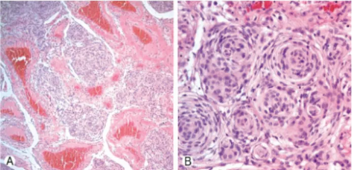
Purpose: Giant cell tumor of the tendon sheath are the most common tumors after ganglionic cysts in benign soft tissue tumors which could be recurred after surgical

Conclusion: Aromatherapy with neroli essential oil had a difference in brain wave and brain utilization ability according to the MBTI personality type

Sometimes called a direct neural interface or a brain-machine interface, is a direct communication pathway between a brain (or brain cell culture) and an external device..

Conclusions : No significant corelation of the patency rate was shown in the study on the effects of local irrigation and systemic application of heparin