INTRODUCTION
Systemic lupus erythematosus (SLE) is one of the most un- predictable systemic connective tissue diseases. Headache is a common symptom in SLE patients (1-3) but whether there is a unique syndrome attributable to SLE is not clear. Dif- ferent types of headache may occur in SLE due to various un- derlying conditions. Pseudotumor cerebri (PC) is an uncommon syndrome of neuropsychiatric SLE and one of the causes of headache in SLE patients. In 1968 Bettman et al. (4) first reported a patient with concomitant SLE and PC. Since then PC has been reported in a few sporadic cases in adult patients with SLE (5, 6). However, recurrent PC in patients with SLE, which has been reported rarely (7-11), has not been report- ed in Korea.
Here, we describe a 30-yr-old female patient with SLE who was afflicted with recurrent PC. This case illustrates that the possibility of PC should be taken into account in the differ- ential diagnosis of headache during the course of SLE.
CASE REPORT
A 30-yr-old woman was referred to our department due to intractable headache, dizziness, fatigue, nausea and vom- iting. She had been well until two weeks before admission,
when she experienced severe frontal headache. The severity of her headache increased over one week prior to admission.
She was diagnosed with SLE five year earlier in accord with the ACR criteria (12): malar rash, arthritis, photosensitivity, thrombocytopenia (85,000/ L), and positive results for anti- nuclear antibody (1:320, speckled pattern), anti-dsDNA, and anti-Ro antibodies. She had symptoms of elevated intracranial pressure: headache, diplopia, nausea and vomiting. Ophthal- mologic examination showed bilateral papilledema and the findings of brain CT scan was normal. A lumbar puncture produced clear acellular cerebrospinal fluid (CSF) with an opening pressure above 300 mmH2O. Treatment with pred- nisolone 60 mg/day was started on the basis of a diagnosis of PC associated with SLE. After 3 months of treatment, relief of the signs and symptoms with normal CSF opening pres- sure. She had been well for the next five year with low dose prednisolone and hydroxychloroquine (300 mg/day) until her headache recurred.
At the current admission, her vital signs were body tem- perature of 37.7℃, blood pressure of 100/60 mmHg, pulse rate of 79/min, and respiration rate of 17/min. A faint and pink maculopapular rash was noted over the face. No palpa- ble lymph nodes were noted. An ophthalmologic examina- tion revealed bilateral papilledema but visual acuity and visual fields were normal. Neurological examination was normal.
Wan Hee Yoo, Ji Hyun Park, Hyun Kag Kim, Tae Sun Park, Hong Sun Baek
Department of Internal Medicine, Medical School, Chonbuk National University and Research Institute of Clinical Medicine, Chonju, Korea
Received : 13 November 2000 Accepted : 1 February 2001
Address for correspondence Wan Hee Yoo, M.D.
Division of Rheumatology, Department of Internal Medicine, Medical School, Chonbuk National University and Research Institute of Clinical Medicine, 634-18, Keumam-dong, Duckjin-gu, Chonju 561-712, Korea
Tel : +82.63-254-1609, Fax : +82.63-250-1672 E-mail : [email protected]
805 J Korean Med Sci 2001; 16: 805-8
ISSN 1011-8934
Copyright � The Korean Academy of Medical Sciences
Recurrent Pseudotumor Cerebri in Systemic Lupus Erythematosus
: A Case Report
Pseudotumor cerebri is an uncommon manifestation of neuropsychiatric sys- temic lupus erythematosus (SLE), and is characterized by an elevated intracra- nial pressure, papilledema with occasional abducens nerve paresis, absence of a space-occupying Iesion or ventricular enlargement, and normal cerebrospinal fluid chemical and hematological constituents. Pseudotumor cerebri has been reported in a few sporadic cases in patients with systemic lupus erythematosus.
However, the recurrent pseudotumor cerebri in patients with systemic lupus ery- thematosus which has been rarely reported, has not been reported in Korea.
We experienced a 30-yr-old female patient with SLE who was presented with second attack of severe intractable headache. She was diagnosed pseudotu- mor cerebri twice and successfully treated with corticosteroid. Headache is the common symptom in patients with neuropsychiatric SLE and attributable to vari- ous causes. We suggest that it is important to define the cause of headache in patients with SLE and pseudotumor cerebri should be included in the spectrum of clinical manifestations during the course of SLE as a cause of headache.
Key Words : Pseudotumor Cerebri; Lupus Erythematosus, Systemic
806 W.H. Yoo, J.H. Park, H.K. Kim, et al.
Laboratory results showed a white blood cell count of 3,200/ L, hemoglobin of 9.1 g/dL, reticulocytosis of 1.1%, and platelet count of 235,000/ L. Erythrocyte sedimenta- tion rate (ESR) was 80 mm/hr. Serum electrolytes, liver and kidney function test results were normal. Coagulation test results were normal, with prothrombin time of 100% (INR 1.0), activated partial thromboplastin time of 38.0 sec (nor- mal, 29.8-41.8). Immunoglobulin profiles demonstrated an elevated level of IgG upto 1,810 mg/dL (normal, 751-1,560) and normal levels of IgA and IgM. Antinuclear antibody titer was 1:640 (speckled pattern) and anti-dsDNA anti- body was 114 IU/mL (normal, <7). Anti-Ro, anti-RNP tests were both positive. Complement levels were decreased:
C3, 60.9 mg/dL (normal, 65-125) and C4, 7.19 mg/dL (nor- mal, 12-43). Tests for anticardiolipin antibody (IgG, IgM) and lupus anticoagulant were negative. Hormonal profile showed normal levels of TSH, free T3, T4, FSH, LH, estro- gens, progesterone, prolactin, and cortisol.
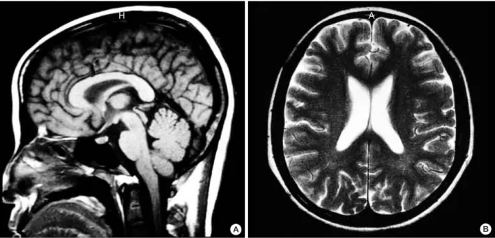
A lumbar puncture showed clear acellular CSF with an opening pressure of above 280 mmH2O (normal, <60-160 mmH2O) and glucose and protein concentrations of 30.6 mg/dL (normal, 15-45) and 62 mg/dL (normal, 50-75), re- spectively. CSF bacteria cultures and tests for viral antibody of varicella-zoster, herpes simplex, influenza A, B, cytome- galovirus, adenovirus, and Epstein-Barr virus were all nega- tive. The findings of brain magnetic resonance images were normal (Fig. 1).
The patient was diagnosed with recurrent PC represent- ing as a flare-up of neuropsychiatric SLE. Treatment with prednisolone 60 mg daily and azathioprine 100 mg/day was started, and subsequently the prednisolone was tapered by
dose to 10 mg/day over 8 weeks. After three months of fol- low-up, the patient’s signs and symptoms were noted to be relieved.
DISCUSSION
Neuropsychiatric SLE includes the neurologic syndromes of the central, peripheral, and autonomic nervous system and psychiatric syndromes observed in patients with SLE after excluding other causes. Since the first report of stupor and coma in SLE by Hera and Kaposi (13) in 1875, a mul- titude of neuropsychiatric syndromes have been reported in SLE patients. Neuropsychiatric affliction in SLE has become well established with a frequency of as high as 14 to 75%
(14), and PC has been reported in a few cases of adult patients with neuropsychiatric SLE (5, 6).
Diagnosis of PC requires increased intracranial pressure (CSF pressure >200 mmH2O) without clinical, laboratory, or radiological evidence of a space-occupying lesion or hydro- cephalus. This patient fulfilled all of these criteria for PC.
The patient manifested the symptoms and signs suggesting increased intracranial pressure as intractable headache, nausea, vomiting and bilateral papilledema. The increased intracra- nial pressure was determined by lumbar puncture during which opening pressure of CSF showed above 280 mmH2O.
The chemical and hematological compositions of the CSF were normal. The last criterion was satisfied by the absence of radiological abnormalities in findings of MRI scan.
Although PC is known to be associated with various con- ditions, the etiology of the disease is unknown. In addition,
A B
Fig. 1.Brain MRI images (T1-weighted sagittal and T2-weighted axial) show normal sized ventricle and subarachnoid space without space-occupying lesion.
A H
Pseudotumor Cerebri in SLE 807
diverse therapeutic agents, including vitamin A, tetracycline, nalidixic nitrofurantoin, sulfa derivatives, lithium, pheny- toin and indomethacin, have been used in this disorder. The systemic conditions of PC are primarily endocrinological abnormalities such as hypo- and hyperthyroidism, hypopara- thyroidism, adrenal insufficiency, Cushing’s syndrome and oral contraceptives. Pregnancy, obesity (particularly among women), anemia, hypertension and rare cases of sarcoidosis were reported to be associated with PC (15). SLE is one of the enlisted diseases underlying PC, albeit not common (5- 11, 14-19).
The following conditions rendered it reasonable think that SLE was attributable to PC in this patient: she did not have menstrual irregularities or endocrinopathies; she was not on contraceptive pills, antibiotics or vitamins, which are known to induce PC. Corticosteroid therapy could not be implicat- ed because this patient was receiving only low dose of pred- nisolone at the time of diagnosis of PC.
The pathophysiologic mechanism of PC in SLE remains unknown. It is not clear whether the overproduction of CSF, relative absorptive failure of CSF, or increased cerebral blood flow is associated with the pathogenesis of PC in SLE (16).
On the other hand, PC in SLE has been attributed to cere- bral venous thrombosis in some cases. The patients in these cases had lupus anticoagulant or anticardiolipin antibodies that were assumed to induce a hypercoagulable state that eventuated in cerebral venous thrombosis (6, 9, 17). Thro- mbotic obliteration of the arteriolar or venous vascular bed might have affected the arachnoid villi and eventually have caused PC in this patient. However, vasculitis, immune com- plex precipitation or even direct antibody injury may all result in a similar dysfunction (11). In this patient, the test for serum anticardiolipin antibody and lupus anticoagulant test were negative and evidence of sagittal or other venous or arterial thromboses was not demonstrated in MRI scans.
The laboratory and radiological findings along with the grad- ual onset and progression of PC-related symptoms in this patient favored the latter hypothesis as a causative condition.
SLE-associated PC usually indicates a favorable outcome.
The most critical sequela of PC is the permanent visual im- pairment. In a recent prospective study (18), a large propor- tion of patients were found to have some visual loss includ- ing blind spot enlargement in at least one eye on follow-up.
Furthermore, recurrent PC (7-11), chronic low-grade papil- ledema, and death from progressive renal disease have been observed in some patients (19). Corticosteroid is currently the mainstay of the treatment of PC in patients with SLE (19), and acetazolamide, intravenous mannitol and furosemide are also used. In addition, plasmapheresis, azathioprine, cyclo- phosphamide and other immunosuppressive agents can be used in the treatment of such patients.
In conclusion, we described a 30-yr-old female patient with SLE who presented with second attack of severe intractable headache. She was diagnosed recurrent PC, as a neuropsy-
chiatric SLE. Headache is the commonest symptom encoun- tered in patients with neuropsychiatric SLE and is attributable to different syndromes. It is important to define the cause of headache in patients with SLE, since there is no typical pattern in the benign headache of lupus. Although the underlying mechanisms of the cerebral manifestations are not always readily identifiable, we suggest that PC should be included in the spectrum of neuropsychiatric manifestations during the course of SLE.
REFERENCES
1. Isenberg DA, Meyrick-Thomas D, Snaith ML, McKeran RO, Roys- ton JP. A study of migraine in systemic lupus erythematosus. Ann Rheum Dis 1982; 41: 30-2.
2. Kovacs JA, Urowitz MB, Gladman DD. Dilemmas in neuropsychi- atric lupus. Rheum Dis Clin North Am 1993; 19: 795-814.
3. Amit M, Molad Y, Levy O, Wysenbeek AJ. Headache in systemic lupus erythematosus and its relation to other disease manifestations.
Clin Exp Rheumatol 1999; 17: 467-70.
4. Bettman JW Jr, Daroff RB, Sanders MD, Joyt WF. Papilledema and asymptomatic intracranial hypertension in systemic lupus erythe- matosus. A fluorescein angiographic study of resolving papillede- ma. Arch Ophthalmol 1968; 80: 189-93.
5. Edmund K, Patrick C. Pseudotumor cerebri in systemic lupus ery- thematosus. J Rheumatol 1989; 16: 113-5.
6. Kaplan R, Springate G, Feld L, Cohen M. Pseudotumor cerebri asso- ciated with cerebral venous sinus thrombosis, internal jugular vein thrombosis, and systemic lupus erythematosus. J Pediatr 1985; 107:
266-8.
7. Silberg DH, Laties SS. Increased pressure in disseminated lupus erythematosus. Arch Neurol 1973; 29: 88-90.
8. Parnass SM, Goodwin JA, Patel DV, Levinson DJ, Reinhard JD.
Dural sinus thrombosis: a mechanism for pseudotumor cerebri in systemic lupus erythematosus. J Rheumatol 1987; 14: 152-5.
9. Chevalier X, de Bandt M, Bourgeois P, Kahn MF. Primary Sjogren’s syndrome preceding the presentation of systemic lupus erythemato- sus as a benign intracranial hypertension syndrome. Ann Rheum Dis 1992; 51: 808-9.
10. Ogura N, Atsumi T, Sagawa A, Jodo S, Amasaki Y, Nakabayashi T, Watanabe I, Mukai M, Fujisaku A, Nakagawa S. Systemic lupus erythematosus associated with benign intracranial hypertension: a case report. Ryumachi 1992; 32: 66-72.
11. Horoshovski D, Amital H, Katz M, Shoenfeld Y. Pseudotumour cerebri in SLE. Clin Rheumatol 1995; 14: 708-10.
12. Tan EM, Cohen AS, Fries JF, Masi AT, McShane DJ, Rothfield NF, Schaller JG, Talal N, Winchester RJ. The 1982 revised criteria for the classification of systemic lupus erythematosus. Arthritis Rheum 1982; 25: 1271-7.
13. Hebra F, Kaposi M. On diseases of the skin including the exanthe- mata. Vol. IV. Tay W, editor/translator. London: The New Synde- ham Society 1875: 14-47.
14. Ashereson RA, Denburg SD, Denburg JA, Carbotte RM, Futrell N.
. .
808 W.H. Yoo, J.H. Park, H.K. Kim, et al.
Current concepts of neuropsychiatric systemic lupus erythematosus.
Postgrad Med J 1993; 69: 602-8.
15. Ahlskog JE, O’Neill BP. Pseudotumor cerebri. Ann Intern Med 1982;
97: 249-56.
16. Li EK, Ho PCP. Pseudotumor cerebri in systemic lupus erythemato- sus. J Rheumatol 1989; 16: 113-6.
17. Nampoory MR, Johny KV, Gupta RK, Constandi JN, Nair MP, al-
Muzeiri I. Treatable intracranial hypertension in patients with lupus nephritis. Lupus 1997; 6: 597-602.
18. Wall M, George D. Idiopathic intracranial hypertension. A prospec- tive study of 50 patients. Brain 1991; 114: 155-80.
19. Green L, Vinker S, Amital H, Amir T, Bar-Dayan Y, Levi Y, Schoen- feld Y. Pseudotumor cerebri in systemic lupus erythematosus. Semin Arthritis Rheum 1995; 25: 103-8.