Identification of Genes Differentially Expressed in the MCF-7 Cells Treated with Mitogenic Estrogens
Myeong Sook Cheon, Taesook Yoon, Do Yeon Lee, Goya Choi, AYeong Lee, Byung Kil Choo, and Ho Kyoung Kim*
Deptartment of Herbal Resources Research, Korea Institute of Oriental Medicine, Yuseong-gu, Daejeon 305-811, Korea
Received October 8, 2007; Accepted February 4, 2008
Estrogens, a group of steroid compounds functioning as the primary female sex hormone, play an important role in the development and progression of breast cancer. In this study, using a novel annealing control primer-based GeneFishing PCR technology, five differentially expressed genes (DEGs), expressed using 10 nM mitogenic estrogens, 17β-estradiol (E2) and 16α-hydroxyestrone (16α-OHE1), were selected from the estrogen receptor (ER)-positive MCF-7 human breast cancer cells. The DEGs, MRPL42, TUBA1B, SSBP1, KNCT2, and RUVBL1, were identified by comparison with the known genes via direct sequencing and sequence homology search in BLAST.
Quantitative real-time PCR data showed that two DEGs, tubulin α1b and kinetochore associated 2, were greater than 2-fold upregulated by E2 or 16α-OHE1. Both genes could be new biomarkers for the treatment and prognosis of cancers, and further study may provide insights into the molecular mechanisms underlying development and progression of breast cancer.
Key words : differentially expressed genes, 17β-estradiol, estrogen receptor-positive MCF-7 human breast cancer cells, 16α-hydroxyestrone, kinetochore associated 2, tubulin α1b
Estrogens are a group of steroid compounds functioning as the primary female sex hormone. Menopause is the physiological cessation of the menstrual cycle associated with aging in women, resulting in the decrease of the estrogen production. Estrogen deficiency leads to the post-menopausal symptoms such as flashes, insomnia, depression and osteoporosis. Post-menopausal women are at an increased risk of osteoporosis, and many studies have reported that one reason for this risk is the dramatic change in the level of female sex hormones, especially estrogens. HRT can increase the bone mineral density and, as a result, decrease the skeletal fragility [Delmas, 1997]. Unfortunately, HRT increases cardiovascular events in the postmenopausal women [Grady et al., 2002]
and a long-term compliance to HRT is limited by side effects such as breast cancer, the risk of which increases after prolonged treatments.
Estrogens play an important role in the development and progression of breast cancer [McGuire et al., 1976;
Clemons and Goss, 2001]. Despite the considerable efforts to identify the genes involved in the molecular mechanisms underlying the estrogen-mediated breast cancer, conventional differential display-methods are labor-intensive and lead to a high degree of false positives. A novel ACP-based differential display-RT- PCR technology regulated by an ACP has been used to identify the DEGs [Ryu et al., 2007; Hwang et al., 2004].
The basis of the ACP technology is the unique tripartite structure of a specific oligonucleotide primer, which contains distinct 3'-end and 5'-end regions separated by a regulator, as well as the interactions of each portion of this primer during the two-stage PCR [Kim et al., 2004].
The ACP-based PCR system facilitates the identification of DEGs from the samples displaying low mRNA levels without generating false positives [Hwang et al., 2004].
Recently, Kim et al. [2005] reported that two mitogenic estrogens, 17β-estradiol (E2) and 16α-hydroxyestrone (16α-OHE1), induced the proliferation of the estrogen
*Corresponding author
Phone: +82-42-868-9502; Fax: +82-42-863-9434 E-mail: [email protected]
Abbreviations: ACP, annealing control primer; DC, dextran/
charcoal; DEG, differentially expressed gene; DMEM, Dulbecco’s Modified Eagle’s Medium; E2, 17β-estradiol; ER, estrogen receptor; FBS, fetal bovine serum; GAPDH, glyceraldehydes-3- phosphate dehydrogenase; HDAC, histone deacetylase; HRT, hormone replacement therapy; 2-ME, 2-methoxyestradiol; 16α- OHE1, 16α-hydroxyestrone; PR, progesterone receptor.
receptor (ER)-positive MCF-7 human breast cancer cells at 10 nM with greater than 20-fold induction of the PR transcript, which is a classical example for an ER- mediated gene. This suggests that the mitogenic mechanisms of E2 and 16α-OHE1 could stem from a direct genomic action via the activation of ER. Therefore, in the present study, to identify the genes selectively expressed in MCF- 7 cells by the mitogenic estrogens, E2 and 16α-OHE1, the gene expression patterns were examined using the ACP-based GeneFishing PCR technology.
Materials and Methods
Cell culture. Human MCF-7 breast cancer cells were maintained in a humidified atmosphere of 5% CO2 at 37oC in DMEM (Life Technologies, Gaithersburg, MD) supplemented with 10% FBS (Hyclone Laboratories, South Logan, UT), 100 U/mL of penicillin, and 100 mg/
mL streptomycin with a change of the medium every 3 days.
Estrogens treatment. The MCF-7 cells were plated into 96-well plates at a density of 4,000 cells per well in DMEM with 10% FBS and incubated for 24 h. The medium was replaced with a phenol red-free DMEM containing 5% DC-treated FBS to remove the endogenous steroids in the serum and exclude the weak estrogen- agonist activity of the phenol red [Darbre et al., 1983;
Ernst et al., 1989]. After 24 h, the cells were incubated with 10 nM of each of the E2 metabolites (Steraloids Inc., Wilton, NH).
Isolation of total RNA. Using the TRIzol reagent (Life technologies), total RNA was isolated from the MCF-7 cells treated with each E2 metabolite for 24 h. The homogenized samples were incubated for 5 min at room temperature to permit the complete dissociation of the nucleoprotein complexes. After the addition of 0.2 volume of chloroform, the samples were shaken vigorously for 15 s, incubated for 3 min, and centrifuged at 12,000×g for 15 min at 4oC. The total RNA remaining in the upper aqueous phase was precipitated by mixing with an equal volume of isopropanol. The mixtures were incubated for 10 min and centrifuged at 12,000×g for 10 min at 4oC. The total RNA pellet was washed with 75%
ethanol, dried, and dissolved in the RNase-free water. The concentration and purity of total RNA were calculated based on the difference in the absorbance at 260 and 280 nm.First-strand cDNA Synthesis. Reverse transcriptase was used to synthesize the first-strand cDNAs from total RNAs . Reverse transcription was performed for 1.5 h at 42ºC in a final reaction volume of 20µL containing 3µg of the purified total RNA, 4µL of 5×reaction buffer
(Promega, Madison, WI), 5µL of dNTPs (each 2 mM), 2
µL of 10µM dT-ACP1 (5'-CGTGAATGCTGCGACTA CGATIIIIIT(18)-3'), 0.5µL of RNasin RNase Inhibitor (40 U/µL; Promega), and 1µL of Moloney murine leukemia virus reverse transcriptase (200 U/µL; Promega).
First-strand cDNAs were diluted by the addition of 80µL of the ultra-purified water for the GeneFishingTM PCR, and stored at −20oC until use.
ACP-based GeneFishingTM PCR. DEGs were screened by the ACP-based PCR method [Kim et al., 2004] using the GeneFishingTM DEG kits (Seegene, Seoul, Korea).
Briefly, the second-strand cDNA synthesis was conducted at 50ºC during one cycle of the first-stage PCR in a final reaction volume of 20µL containing 3-5µL (about 50 ng) of the diluted first-strand cDNA, 1µL of dT-ACP2 (10µM), 1µL of 10µM arbitrary ACP, and 10µL of 2
×Master Mix (Seegene). The PCR protocol for the second-strand synthesis was one cycle at 94ºC for 1 min, followed by 50ºC for 3 min, and 72ºC for 1 min. After completion of the second-strand DNA synthesis, the second-stage PCR amplification protocol was carried out as follows; 40 cycles at 94ºC for 40 s, followed by 65ºC for 40 s, 72ºC for 40 s, and a 5-min final extension at 72ºC. The amplified PCR products were separated in the 2% agarose gel stained with ethidium bromide.
Direct Sequencing. The differentially expressed bands were re-amplified and extracted from the gel using the GENCLEAN II Kit (Q-BIO gene, Carlsbad, CA), and directly sequenced with the ABI PRISM 3100-Avant Genetic Analyzer (Applied Biosystems, Foster City, CA).
Complete sequences were determined by searching for similarities using a BLAST program (http://www.ncbi.
nlm.nih.gov).
Primer design and real-time PCR. To set the most suitable PCR amplification conditions, the sequences of the primers were determined by an on-line primer design program [Rozen and Skaletsky, 2000]. The primer sets used in this study are shown in Table 1. First-strand cDNA was synthesized with 1µg of total RNAs and 1µM of oligo-dT15 primer using the Omniscript Reverse Transcriptase (Qiagen, Valencia, CA). SYBR Green- based quantitative PCR amplification was performed using the Stratagene Mx3000P Real-Time PCR system and Brilliant SYBR Green Master Mix (Stratagene, La Jolla, CA) with the 1 : 50 diluted first-strand cDNA and 20 pmole of the primers according to the manufacturer’s protocols. The PCR reaction consisted of initial denaturation at 94oC for 3 min, 3-step cycling (40 cycles) at 94oC for 40 s, 60oC for 40 s, and 72oC for 1 min, and a final extension at 72oC for 5 min. All reactions were run in triplicates, and the data were analyzed using the 2-∆∆CT method [Livak and Schmittgen, 2001]. To assess the
differential transcript expression among the groups, GAPDH was used as the control gene. Significance was determined by Student’s t-test of Microsoft Excel with a GAPDH-normalized 2-∆∆CT value, and the expression differences were considered significant when P<0.05.
Results
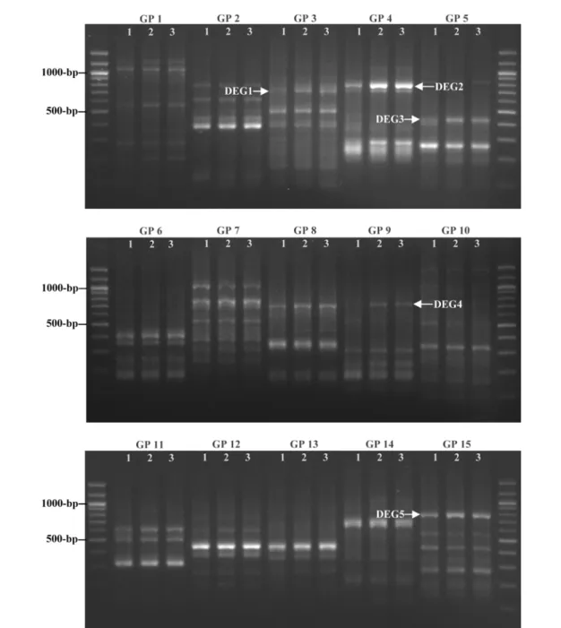
Identification of up-regulated genes by the treatment of E2 or 16α-OHE1 in MCF-7 cells. To identify the estrogen-dependently expressed genes, the MCF-7 cells were cultured in a phenol red-free DMEM with 5% DC-treated FBS and treated with 10 nM of E2 or 16α-OHE1 under the serum-free condition. After the incubation for 24 h, the DEGs were observed using the ACP-based GeneFishing PCR technology. From 20 GPs, 5 DEGs that were up-regulated by E2 or 16α-OHE1 were identified (Fig. 1). Between GP 16 and GP 20, no DEGs were found (data not shown).
Sequence homology search for DEGs. Five differentially expressed transcripts, MRPL42, TUBA1B, SSBP1, KNCT2, and RUVBL1, were identified by comparison with the known genes via direct sequencing and sequence homology search in BLAST. The sequence similarities and characterizations of these five DEGs are summarized in Table 2.
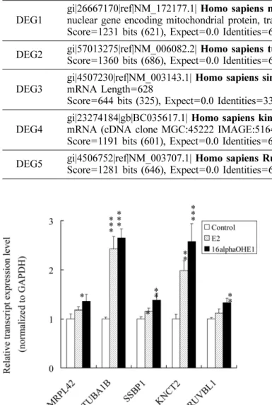
Confirmation of DEGs expression levels by quantitative real-time PCR. To confirm the efficacy of the ACP system and further determine the estrogen- dependent expression patterns of the DEGs identified, SYBR Green-based quantitative real-time PCR was performed as described in Materials and Methods. Under the serum-free condition, TUBA1B, SSBP1, and KNTC2 were significantly up-regulated by E2 or 16α-OHE1, consistent with the results of ACP-based PCR; however,
MRPL42 and RUVBL1 were significantly up-regulated only in the cells treated with 16α-OHE1 (Fig. 2).
Interestingly, the upregulation of the TUBA1B and
KNTC2 by E2 or 16α-OHE1 were greater than 2-fold.
Discussion
E2 is the most biologically active estrogen in the breast tissue and its administration in the rodent models has shown to be carcinogenic. Interestingly, its hydroxylated metabolites showing the estrogenic activities are also carcinogenic. The estrogenic activity of 16α-OHE1 has been proposed to be more potent than that of E2 [Fishman
et al., 1980]. In a population of women with breast cancer, abnormal 16α-hydroxylation of E2 has been observed [Schneider et al., 1982], and the activity of 16α- hydroxylase, the enzyme involved in the formation of 16α-OHE1, increased [Fishman et al., 1984]. Since 16α- OHE1 with the potential to induce the proliferation of the MCF-7 human breast cancer cells has been shown to covalently bind to ER, the covalent modification of ER by 16α-OHE1 was suggested be one mechanism of the malignant transformation in the estrogen target tissues [Swaneck and Fishman, 1988; Gupta et al., 1998; Lewis
et al., 2001].
Recently, Kim et al. [2005] showed that E2 and 16α- OHE1 have highly mitogenic estrogen activities at a low concentration (10 nM), suggesting these mitogenic mechanisms of E2 and 16α-OHE1 could stem from their direct genomic action via the activation of ER. Therefore, in the present study, the genes selectively expressed by E2 or 16α-OHE1 in the ER-positive MCF-7 cells were identified using a novel ACP-based GeneFishing PCR technology. Five DEGs, MRPL42, TUBA1B, SSBP1, KNCT2, and RUVBL1, were identified by comparison with the known genes, and their transcript expression patterns were confirmed by SYBR Green-based quantitative real-time PCR. Under the serum-free condition, TUBA1B and KNTC2 were up-regulated greater than 2-fold by 10 nM of E2 or 16α-OHE1 in the MCF-7 cells per se.
Tubulin, the structural protein of microtubules, is a heterodimer composed of two 50-kD acidic monomers,
α- and β-tubulin, that polymerize to form microtubules, a component of the cytoskeletal system. A recent study Table 1. Primer sequences used in this study
Gene Forward Reverse
MRPL42 5'-atg cta cca ccc ttc tgt gg-3' 5'-tgt gat acc gtc cat gag ga-3' TUBA1B 5'-gcc cta caa ctc cat cct ca-3' 5'-gtc aac att cag ggc tcc at-3' SSBP1 5'-aag atc cct gaa tcg tgt gc-3' 5'-gtt gct tgt cgc ctc aca t-3' KNTC2 5'-agt tta atc ccg agg ctg gt-3' 5'-cag gtg ctt gtg ttt ctc ca-3' RUVBL1 5'-gcc agc taa tga agc caa ag-3' 5'-cct cag tgc ctc tga tga ca-3' GAPDH 5'-GTCAGTGGT GGACCTGACCT-3' 5'-agg ggt cta cat ggc aac tg-3'
showed that the expression level of HDAC 6 mRNA/
protein and the cell motility increased in the MCF-7 cells treated with E2 [Saji et al., 2005]. In addition, the accumulation of HDAC also caused the deacetylation of
α-tubulin. Together with the up-regulation of TUBA1B by the mitogenic estrogens, this result suggests that E2 can regulate the expressions of HDAC6 and tubulin isotype(s), and consequently affect the survival of the breast cancer cells. Moreover, the treatment of the prostate cancer cells with diethylstilbestrol and 2-ME suppressed the levels of the transcripts and the protein for β-tubulin isotype IVa [Montgomery et al. 2005]. In addition, upon the 2-ME
treatment, the levels of acetylated and detyrosinated tubulins decreased in the wild type of the human breast cancer cells, while remaining unchanged in the 2-ME- resistant cells [Goekmen-Polar et al. 2005]. Interestingly, anti-cancer drug-resistant MCF-7 cells contain significantly higher amounts of tyrosinated α-tubulin than do the wild- type cells, suggesting that the level of certain isotype of tubulin becomes elevated in the drug-resistant cells [Banerjee, 2002]. In other words, the regulations of the tubulin gene expression and the microtubule dynamics by targeting (or binding) the tubulins could be promising ways to treat cancers [Sengupta and Thomas, 2006].
Fig. 1. Identification of mitogenic estrogen-regulated DEGs in MCF-7 cells. The mRNA expression profiling in MCF-7 cells (lane 1) was compared with that of cells treated with either 10 nM of E2 (lane 2) or 16α-OHE1 (lane 3). Between GP 1 and 15, 5 DEGs (DEG1 to 5) were identified to be up-regulated by both estrogens.
KNTC2 (also referred to as highly expressed in cancer 1, HEC1) is one of kinetochore protein directly involved in the kinetochore microtubule interactions, the chromosome congression, and the spindle checkpoint signaling [Martin-Lluesma et al., 2002; DeLuca et al., 2006]. High levels of KNTC2 were observed in the majority of the lung cancers of various histologic types, and the elevated expressions were associated with the poorer prognosis of the non-small cell lung carcinoma patients [Hayama et
al., 2006]. Considering that this simultaneous up-regulation is a frequent and important feature of the cell growth and survival of the lung cancer, selective suppression of the
KNTC2 activity could be a promising therapeutic target for the treatment of lung cancers. Moreover, the mitotic checkpoint is an important determinant for the efficacy of the microtubule-targeting drugs in eradicating the cancer cells [Lee et al., 2004]. Interestingly, the depletion of KNTC2 protein by the cytotoxic recombinant adeno- associated virus that expresses short hairpin resulted in the persistent spindle checkpoint activation, followed by the death of the tumor cells [Li et al., 2007].
In conclusion, TUBA1B and KNTC2, the mitogenic estrogen-dependently expressed genes, were identified in the breast cancer cells. The elucidation of the estrogen effects on the gene expression could be helpful in understanding the action mechanism of the estrogens, because the estrogens are highly involved in the progress of breast cancer. Furthermore, both genes could be promising therapeutic and prognostic biomarkers for the breast cancers.
References
Banerjee A (2002) Increased levels of tyrosinated α-, β(III)-, and β(IV)-tubulin isotypes in paclitaxel-resistant MCF-7 breast cancer cells. Biochem Biophys Res Commun 293, 598-601.
Clemons M and Goss P (2001) Estrogen and the risk of breast cancer. N Engl J Med344, 276-285.
Darbre P, Yates J, Curtis S, and King RJ (1983) Effect of estradiol on human breast cancer cells in culture. Can- cer Res43, 349-354.
Delmas PD (1997) Hormone replacement therapy in the pre- vention and treatment of osteoporosis. Osteoporos Int
Suppl 1, S3-7.
DeLuca JG, Gall WE, Ciferri C, Cimini D, Musacchio A, and Salmon ED (2006) Kinetochore microtubule dynam- ics and attachment stability are regulated by Hec1. Cell
Table 2. Sequence homology search for DEGs
DEG No. Sequence homology search
DEG1 gi|26667170|ref|NM_172177.1| Homo sapiens mitochondrial ribosomal protein L42 (MRPL42), nuclear gene encoding mitochondrial protein, transcript variant 2, mRNA Length=2073
Score=1231 bits (621), Expect=0.0 Identities=627/629 (99%), Gaps=0/629 (0%) Strand=Plus/Plus DEG2 gi|57013275|ref|NM_006082.2| Homo sapiens tubulin, alpha 1b (TUBA1B), mRNA Length=1771
Score=1360 bits (686), Expect=0.0 Identities=688/689 (99%), Gaps=0/689 (0%) Strand=Plus/Plus DEG3 gi|4507230|ref|NM_003143.1| Homo sapiens single-stranded DNA binding protein 1 (SSBP1),
mRNA Length=628
Score=644 bits (325), Expect=0.0 Identities=334/336 (99%), Gaps=1/336 (0%) Strand=Plus/Plus DEG4 gi|23274184|gb|BC035617.1| Homo sapiens kinetochore associated 2 (KNTC2),
mRNA (cDNA clone MGC:45222 IMAGE:5164424), complete cds Length=2140
Score=1191 bits (601), Expect=0.0 Identities=622/622 (100%), Gaps=0/622 (0%) Strand=Plus/Plus DEG5 gi|4506752|ref|NM_003707.1| Homo sapiens RuvB-like 1 (E. coli) (RUVBL1), mRNA Length=1750
Score=1281 bits (646), Expect=0.0 Identities=646/646 (100%), Gaps=0/646 (0%) Strand=Plus/Plus
Fig. 2. Evaluation of transctript expression levesl by quantitative real-time PCR. The effect of each estrogen on transctript expression was evaluated by quantitative real-time PCR. The GAPDH-normalized fold changes are expressed as the mean±SD from three independent experiments. *P<0.05; **P<0.01; ***P<0.001 (compared to control).
127, 969-982.
Ernst M, Schmid C, and Froesch ER (1989) Phenol red mimics biological actions of estradiol: enhancement of osteoblast proliferation in vitro and of type I collagen gene expression in bone and uterus of rats in vivo. Ste- roid Biochem33, 907-914.
Fishman J and Martucci C (1980) Biological properties of 16α-hydroxyestrone: implications in estrogen physiol- ogy and pathophysiology. Clin Endocrinol Metab 51, 611-615.
Fishman J, Schneider J, Hershcope RJ, and Bradlow HL (1984) Increased estrogen-16α-hydroxylase activity in women with breast and endometrial cancer. Steroid Bio- chem20, 1077-1081.
Grady D, Herrington D, Bittner V, Blumenthal R, Davidson M, Hlatky M, Hsia J, Hulley S, Herd A, Khan S, Newby LK, Waters D, Vittinghoff E, and Wenger N;
HERS Research Group (2002) Cardiovascular disease outcomes during 6.8 years of hormone therapy: Heart and estrogen/progestin replacement study follow-up (HERS II). JAMA288, 49-57.
Gupta M, McDougal A, and Safe S (1998) Estrogenic and antiestrogenic activities of 16α- and 2-hydroxy metabo- lites of 17β-estradiol in MCF-7 and T47D human breast cancer cells. J Steroid Biochem Mol Biol67, 413-419.
Hayama S, Daigo Y, Kato T, Ishikawa N, Yamabuki T, Miyamoto M, Ito T, Tsuchiya E, Kondo S, and Naka- mura Y (2006) Activation of CDCA1-KNTC2, mem- bers of centromere protein complex, involved in pulmonary carcinogenesis. Cancer Res66, 10339-10348.
Hwang KC, Cui XS, Park SP, Shin MR, Park SY, Kim EY, and Kim NH (2004) Identification of differentially regu- lated genes in bovine blastocysts using an annealing con- trol primer system. Mol Reprod Dev69, 43-51.
Kim SH, Lee SU, Kim MH, Kim BT, and Min YK (2005) Mitogenic estrogen metabolites alter the expression of 17β-estradiol-regulated proteins including heat shock proteins in human MCF-7 breast cancer cells. Mol Cells
20, 378-384.
Kim YJ, Kwak CI, Gu YY, Hwang IT, and Chun JY (2004) Annealing control primer system for identification of dif- ferentially expressed genes on agarose gels. BioTech- niques36,424-426, 428, 430 passim.
Lee EA, Keutmann MK, Dowling ML, Harris E, Chan G, and Kao GD (2004) Inactivation of the mitotic check- point as a determinant of the efficacy of microtubule-tar- geted drugs in killing human cancer cells. Mol Cancer
Ther3, 661-669.
Lewis JS, Thomas TJ, Klinge CM, Gallo MA, and Thomas T (2001) Regulation of cell cycle and cyclins by 16α- hydroxyestrone in MCF-7 breast cancer cells. J Mol Endocrinol27, 293-307.
Li L, Yang L, Scudiero DA, Miller SA, Yu ZX, Stukenberg PT, Shoemaker RH, and Kotin RM (2007) Development of recombinant adeno-associated virus vectors carrying small interfering RNA (shHec1)-mediated depletion of kinetochore Hec1 protein in tumor cells. Gene Ther 14, 817-824.
Livak KJ and Schmittgen TD (2001) Analysis of relative gene expression data using real-time quantitative PCR and the 2-∆∆CT method. Methods25, 402-408.
Martin-Lluesma S, Stucke VM, and Nigg EA (2002) Role of Hec1 in spindle checkpoint signaling and kinetochore recruitment of Mad1/Mad2. Science297, 2267-2270.
McGuire WL, Horwitz KB, Chamness GC, and Zava DT (1976) A physiological role for estrogen and progester- one in breast cancer. J Steroid Biochem7, 875-882.
Rozen S and Skaletsky HJ (2000) Primer3 on the WWW for general users and for biologist programmers. In Bio- informatics Methods and Protocols: Methods in Molecu- lar Biology, Krawetz S and Misener S. pp. 365-386, Humana Press. Totowa, NJ.
Ryu JY, Lee BM, Kacew S, and Kim HS (2007) Identifica- tion of differentially expressed genes in the testis of Sprague-Dawley rats treated with di(n-butyl) phthalate.
Toxicology234, 103-112.
Saji S, Kawakami M, Hayashi S, Yoshida N, Hirose M, Horiguchi S, Itoh A, Funata N, Schreiber SL, Yoshida M, and Toi M (2005) Significance of HDAC6 regula- tion via estrogen signaling for cell motility and progno- sis in estrogen receptor-positive breast cancer. Oncogene
24, 4531-4539.
Schneider J. Kinne D, Fracchia A, Pierce V, Anderson KE, Bradlow HL, and Fishman J (1982) Abnormal oxidative metabolism of estradiol in women with breast cancer.
Proc Natl Acad Sci USA79, 3047-3051.
Sengupta S and Thomas SA (2006) Drug target interaction of tubulin-binding drugs in cancer therapy. Expert Rev Anticancer Ther6, 1433-1447.
Swaneck GE and Fishman J (1988) Covalent binding of the endogenous estrogen 16α-hydroxyestrone to estradiol receptor in human breast cancer cells: characterization and intranuclear localization. Proc Natl Acad Sci USA
85, 7831-7835.