• 검색 결과가 없습니다.

Identification of differentially expressed genes using an annealing control primer system in periodontitis

N/A
N/A
Protected

Academic year: 2021

Share "Identification of differentially expressed genes using an annealing control primer system in periodontitis"

Copied!
6
0
0

로드 중.... (전체 텍스트 보기)

전체 글

(1)

*Correspondence to: Jin Chung, Department of Oral

Microbiology, School of Dentistry, Pusan National University, Address: Mool Keum, Beomoe Ri, Yangsan, 626-810, Korea, TEL: 82-51-510-8247, FAX: 82-51-510-8246,

E-mail: [email protected]

Identification of differentially expressed genes using an annealing control primer system in periodontitis

Hee Sam Na, Ji S. Kim, and Jin Chung*

Department of Oral Microbiology, School of Dentistry, Pusan National University, Yangsan, Korea (received August 22, 2012 ; revised September 6, 2012 ; accepted September 12, 2012)

In the gingival tissues of patients with periodontitis, inflammatory responses are mediated by a wide variety of genes. In this study, we screened for differentially exp- ressed genes (DEGs) in periodontitis compared with normal tissue using an annealing control primer (ACP) system. By ACP RT-PCR analysis, we obtained about 160 amplicons, 8 of which were found to be differentially expressed. DEGs in patients with periodontitis were thus successfully and reliably identified by the ACP-based RT PCR technique.

The DEGs identified in the screen may also enhance our understanding of the pathogenesis of periodontitis.

KEY WORDS: periodontitis ACP-PCR, differentially ex- pressed genes

INTRODUCTION

Periodontal disease consists of a group of infections that leads to the inflammation of gingival tissues and destruc- tion of periodontal tissues and, in severe cases, is accom- panied by the loss of alveolar bonewith eventual teeth lose [1]. Mechanisms of host response in the periodontal tissues are complex and involve numerous systems of interactions.

The interaction of bacterial antigen with the host immune

system is believed to be the basis for the destructive inflam- matory response found in periodontal disease. Specific pathogens such as Porphyromonas gingivalis (P. gingivalis) and Aggreagtibacter actinomycetemcomitans (A. actinomycetem- comitans) associated with plaque biofilm have been known to induce strong systemic and mucosal immune responses in the oral cavity [2,3]. Therefore, in the gingival tissues of patients with periodontitis, a wide variety of genes are either up- or down-regulated to mediate the inflammatory response.

Among several reverse transcription-polymerase chain reac- tion (RT-PCR) methods, differential display methods, which are based on PCR using short arbitrary primers, are simple and fast but produce high rates of false positives. Annea- ling control primer (ACP) system is a highly accurate and sensitive PCR technology that can be used to amplify the minute amounts of material available in tissues [4,5]. This primer anneals specifically to the template and allows only genuine product to be amplified, thus eliminating false- positives. In this study, differentially expressed genes (DEGs) in a normal gingiva vs. in an inflammatory gingiva were identified by using an ACP. In addition, the sequences of DEGs were analyzed.

MATERIALS AND METHODS

Tissue sampling

Patients from Pusan National University Dental Hospital

109

(2)

Table 1. List of primers used in ACP-based PCR

Primer name Sequnece

cDNA synthesis primer

dT-ACP1 5'-CTGTGAATGCTGCGACTACGATIIII(T)18-3'

Reverse primer

dT-ACP2 5'-CTGTGAATGCTGCTGCGACTACGATIIIII(T)15-3'

Arbitrary primer (forward primer)

ACP1 5'-GTCTACCAGGCATTCGCTTCATIIIIIGCCATCGACC-3'

ACP2 5'-GTCTACCAGGCATTCGCTTCATIIIIIAGGCGATGCC-3'

ACP3 5'-GTCTACCAGGCATTCGCTTCATIIIIICCGGAGGATG-3'

ACP4 5'-GTCTACCAGGCATTCGCTTCATIIIIIGCTGCTCGCG-3'

ACP5 5'-GTCTACCAGGCATTCGCTTCATIIIIIAGTGCGCTCG-3'

ACP6 5'-GTCTACCAGGCATTCGCTTCATIIIIIGGCCACATCG-3'

ACP7 5'-GTCTACCAGGCATTCGCTTCATIIIIICTGCGGATCG-3'

ACP8 5'-GTCTACCAGGCATTCGCTTCATIIIIIGGTCACGGAG-3'

ACP9 5'-GTCTACCAGGCATTCGCTTCATIIIIIGATGCCGCTG-3'

ACP10 5'-GTCTACCAGGCATTCGCTTCATIIIIITGGTCGTGCC-3'

ACP11 5'-GTCTACCAGGCATTCGCTTCATIIIIICTGCAGGACC-3'

ACP12 5'-GTCTACCAGGCATTCGCTTCATIIIIIACCGTGGACG-3'

ACP13 5'-GTCTACCAGGCATTCGCTTCATIIIIIGCTTCACCGC-3'

ACP14 5'-GTCTACCAGGCATTCGCTTCATIIIIIGCAAGTCGGC-3'

ACP15 5'-GTCTACCAGGCATTCGCTTCATIIIIICCACCGTGTG-3'

ACP16 5'-GTCTACCAGGCATTCGCTTCATIIIIIGTCGACGGTG-3'

ACP17 5'-GTCTACCAGGCATTCGCTTCATIIIIICAAGCCCACG-3'

ACP18 5'-GTCTACCAGGCATTCGCTTCATIIIIICGGAGCATCC-3'

ACP19 5'-GTCTACCAGGCATTCGCTTCATIIIIICTCTGCGAGC-3'

ACP20 5'-GTCTACCAGGCATTCGCTTCATIIIIIGACGTTGGCG-3'

with and without adult periodontitis were used as donor.

Criteria for patient selection were: (1) absence of systema- tic diseases, (2) no antibiotics taken for at least 6 months before sampling, and (3) non-smokers. Informed consent was obtained from all donors and their rights were protected according to the protocol reviewed and approved by the institutional review board of Pusan National University Dental Hospital. Sites with no overt signs of gingival inflammation and with a probing depth of < 3mm were defined as clinically healthy; sites with obvious alveolar bone loss detected radiographically and a probing depth > 3were defined as exhibiting signs of periodontitis. Gingival samp- les were collected from both normal and periodontitis sites after infiltration with an appropriate local anesthetic. Samp- les comprised the epithelial lining and a portion of the under- lying connective tissue. The gingival tissue specimens obtained were thoroughly rinsed with sterile normal saline

solution, transferred into Eppendorf tubes, and stored -70℃

until use. The histolopathological diagnoses were determined and one sample from each group was used in the experiment.

RNA isolation and cDNA synthesis

Total RNA was isolated from both normal gingiva and inflammatory gingiva with the Trizol reagent (Invitrogen, Carlsbad, CA). Further purification of the extracted RNA was achieved by the use of the RNeasy total RNA isolation kit (Qiagen, Valencia, CA) according to the manufacturer’s instructions. The purified RNA was quantified spectropho- tometrically.

Reverse transcription was performed for 1.5 hr at 42℃ in

a final reaction volume of 20 μl containing the purified

mRNA, 4 ml of 5 × reaction buffer (Promega, Madison,

WI), 5 ml of dNTP (each 2mM), 2 ml of 10 mM cDNA

synthesis primer dT-ACP1 (Table 1), 0.5 ml of RNasin

(3)

Plus RNase Inhibitor (Promega, Madison, WI) and 1 ml of Superscript II reverse transcriptase (200 U/μl) (Invitrogen, Carlsbad, CA). First-strand cDNAs were diluted with 120 ml of sterile water.

ACP-based PCR

The cDNAs are subjected to second-strand cDNA syn- thesis by random PCR amplification using dT-ACP2 and one of 20 arbitrary ACPs (Table 1). Second-strand cDNA syn- thesis was carried out at 50℃ during one cycle of first- stage PCR in a final reaction volume of 49.5 ml contai- ning 1 ml of the diluted first-strand cDNA, 5 ml of 10 × PCR reaction buffer (Roche Applied Science, Mannheim, Germany), 5 ml of dNTP (each 2mM), 1 ml of 10 mM dT- ACP2, and 1 ml of 10 mM arbitrary primer. The tube containing the reaction mixture was held at 94℃ while 0.5 ml of Taq DNA polymerase (5U/ul) (Roche Applied Science, Mannheim, Germany) was added to the reaction mixture.

The PCR protocol for second-strand synthesis was one cycle at 94℃ for 1 min, followed by 50℃ for 3 min, and 72℃

for 1 min. After second-strand DNA synthesis was comp- leted, 40 amplification cycles wereperformed. Each cycle involved denaturation at 94℃for 40 sec, annealing at 65℃

for 40 sec, and extension at 72℃was performed to comp- lete the PCR. The amplified PCR products were then sub- jected to electrophoresis on 2% agarose gels and stained with 1 mg/ml ethidium bromide.

Identification of differentially expressed genes

The differentially expressed bands were extracted and cloned into a TOPO vector of the TA cloning kit (Invit- rogen, Carlsbad, CA) and used to transform competent TOP10 Escherichia coli cells. The colonies were grown for 18 hrs at 37℃ on Luria broth agar plates containing ampi- cillin, X-gal (5-bromo 4-chloro 3-indoyl-β-D-galactopyra- noside), and isopropyl- β-D-thiogalactopyranoside for blue/

white colony selection. In order to verify the identity of inserted DNA, isolated plasmids were sequenced automa- tically (Applied Biosystems, Foster city, CA). Complete sequences were analyzed by searching for similarities using BLASTN or BLASTX search program at the National Center for Biotechnology Information (NCBI) GenBank.

RESULTS

To identify genes which were differentially expressed in

Fig. 1. Differentially expressed genes in gingiva from healthy and periodontitis patient. Band patterns for differential expression between a normal gingiva and a inflammatory gingiva after annealing control primer-polymerase chain reaction (ACP-PCR) using ACP1 to ACP10 (A) and ACP 11 to ACP 20 (B). Total RNA was isolated from both tissues and subjected to reverse transcription-polymerase chain reaction (RT-PCR) using ACP. The PCR products were separated by electrophoresis on 2% agarose gels. M, lane containing a 100-bp DNA ladder.

gingiva from healthy and periodontitis, the mRNAs from both types of gingiva were extracted and subjected to ACP RT-PCR analysis using a combination of 20 arbitrary primers and two anchored oligo(dT) primers (dT-ACP 1 and dT-ACP 2). The analysis produced about 160 amplicons, 8 of which were differentially expressed (Fig. 1). We have found 7 genes that were markedly up-regulated in a gingivafrom patient with periodontitis, while only one was enhanced in a healthy gingiva. The sequence of a total 8 DEGs analyzed by BLAST search program were determined (Table 2). One DEG up-regulated in a normal gingiva was for the constant region of Homo sapiens immunoglobulin kappa chain, while the expressions of two DEGs coding for the variable region of kappa chain and lamda constant were enhanced in an inflammatory gingiva.

Other DEGs increased in an inflammatory gingiva were for nuclear ribonucleoprotein, hydroxyacyl glutathione hydro- lase, and plasminogen activator inhibitor-1 mRNA-binding protein (PAI-RBP1).

DISCUSSION

In periodontal tissues, a number of genes are up- or

(4)

Table 2. List of genes identified by ACP system

DEG No. GenBank Accession No. Sequence homology search

DEG 1 BC036082 Homo sapiens,Similar to HSPC159 protein, clone IMAGE : 5301908, mRNA

DEG 2 BC090336 (BlastN) Homo sapiens immunoglobulin kappa constant, mRNA (cDNA clone MGC : 88341 IMAGE : 30330282), complete cds

BAC01760 (BlastX) Immunoglobulin kappa light chain VLJ region [Homo sapiens].

DEG 3

CR609605 (BlastN) Full-length cDNA clone CS0CAP007YC20 of thymus of Homo sapiens (human).

BC073792 (BlastN) Homo sapiens immunoglobulin kappa variable 1-5, mRNA (cDNA clone MGC : 88810 IMAGE : 6280971), complete cds

BAC01738 (BlastX) immunoglobulin kappa light chain VLJ region [Homo sapiens].

DEG 4 AK129814 (BlastN) Homo sapiens cDNA FLJ26303 fis, clone DMC07819,

highly similar to HETEROGENEOUS NUCLEAR REBONUCLEOPROTEIN A1 AAH52296 (BlastX) Heterogeneous nuclear ribonucleoprotein A1, isoform

DEG 5 NM_15640 Homo sapiens PAI-1 mRNA-binding protein (PAI-RBP1), mRNA

DEG 6

HS429E7 (BlastN) Human DNA sequence from clone LA16-429E7 on chromosome 16 Contains the HAGH (hydroxyacyl glutathione hydrolase) gene, ESTs, STSs, GSSs and CpG Islands, complete sequence

NM_005326 (BlastN) Homo sapiens hydroxyacylglutathione Hydrolase (HAGH), mRNA NP_005317 (BlastX) Hydroxyacyl glutathione hydrolase [Homo sapiens].

DEG 7 BC034142 Homo sapiens immunoglobulin kappa variable 1-5, mRNA (cDNA clone MGC : 32715 IMAGE : 4694346), complete cds

DEG 8 BC071804 Homo sapiens immunoglobulin lambda constant 2 (Kern-Oz-marker), mRNA (cDNA clone MGC : 88451 IMAGE : 4717660),

complete cds

down-regulated by subgingival plaque biofilm comprised of facultative or anaerobic bacteria such as P. gingivalis, Tan- nerella forsythia and A. actinomycetemcomitans [1]. The primary mediators, such as IL-1b and TNF-αhave been shown to contribute to several events that are essential for the inflammatory response. Moreover, various inflam- matory mediators including cytokines, chemokines and their receptors are expressed by gingival fibroblast, epithelial cells, endothelial cells and inflammatory cells in perio- dontally disease tissues [6-8]. In this study, we have searc- hed for the genes expressed differentially in a gingiva from periodontitis patients compared to a healthy gingiva.

We used the ACP system to search DEGs in gingival tissues. The ACP system is based on two principles: the unique tripartite structure of the primers, which have distinct 3’- and 5’- end regions that are separated by a polydeoxyinosine [poly(dI)] linker, and the interaction of each region during two-stage PCR [4,5]. For first-strand cDNA synthesis, the reaction was carried out with dT- ACP1, wherein the 3’- end core portion comprises a hybri- dizing sequence complementary to a poly A region of

mRNA transcripts. Polymerase chain reaction was con- ducted by using arbitrary ACPs to synthesize second-strand cDNAs under annealing conditions such that the 3’-end core portion of the dT-ACP2 is prevented from annealing to first-strand cDNAs and only the 3’-end core portion of the arbitrary ACP anneals to the first-strand cDNA. Arbitrary ACPs contain random sequence 10-mers as the 3’- end core sequences, and only those ACPs that are sufficiently complementary to a region of a first-strand cDNA anneal.

Thus, the ACP system allows only real products to be amplified and eliminates false-positive results.

In present study, using ACP system, 8 DEGs were detected (Fig 1, Table 2). Seven DEGs including the va- riable region of kappa chain, nuclear ribonucleoprotein, hydroxyacyl glutathione hydrolase, and PAI-RBP1 were identified in an inflammatory gingiva. Among them, the increased expression of PAI-RBP1 is especially noticeable.

PAI-RBP1 has been reported to be increased in non-small cell lung cancer with different tumor metastatic potential and ovarian cancer associated with tumor progression [9,10].

PAI-RBP1 has been suggested to be involved in tumor

(5)

development orinvasion and metastasis that are mediated via angiogenesis and cell migration. PAI-RBP1 is a binding protein for plasminogen activator inhibitor-1 (PAI-1). A major role of PAI-1 in immune response is the regulation of cell migration, thereby suggesting that PAI-1 may also modulate cellular recruitment during the acute inflammatory process [11-13]. PAI-1 expression is regulated by growth factors, cytokines, and hormones, including agents that regu- late cellular cAMP levels [14-16].

The initialstages of periodontal inflammation are accom- panied by vascular hyperpermeability and LPS from Porphy- romonas gingivalis has been reported to induce vascular permeability [17]. PAI-1 can mediate vascular permeability [18] suggesting the possibility of regulating host response in periodontal disease. In relation to periodontal disease, it was reported that there was a significant increase of plasminogen activator activity-2and t-PA antigen in samples from inflamed gingival tissue compared with normal gingi- val tissue [19-21]. It has been suggested that t–PA and PAI-2 may play a significant role in the periodontal tissue destruction and tissue remodeling because of the increased concentration of t–PA and PAI-2 in inflamed gingiva and gingival crevicular fluid [22-24]. Deppe et al. reported that urokinase-type plasminogen and its inhibitor PAI-1 antigen was detected in normal or inflamed periodontal tissues [25].

In this study, 8 DEGs were identified in a normal gingiva vs. in an inflammatory gingiva using 20 ACPs.

However, inflammatory cytokines and chemokines that are frequently reported in inflammatory diseases were not detected in this study, which limit our study. It is ex- pected that more DEGs will be detected if more ACPs will be used. In future, further studies will be needed to detect more new DEGs serving as the targets for control or therapy of periodontal disease and to elucidate the relation between PAI-1 or PAI-RBP1 and periodontitis.

ACKNOW LEDGMENT

This work was supported by the Bio-Scientific Research Grant funded by the Pusan National University (PNU, Bio-Scientific Research Grant)(PNU-2010-101-257).

REFERENCES

1. Darveau RP. Periodontitis: a polymicrobial disruption of host homeostasis. Nat Rev Microbiol 2010;8:481-490.

2. Katz J, Michalek SM. Effect of immune T cells derived from mucosal or systemic tissue on host responses to Porphyromonas gingivalis. Oral Microbiol Immunol 1998;

13:73-80.

3. Kinane DF, Mooney J, Ebersole JL. Humoral immune response to Actinobacillus actinomycetemcomitans and Porphyromonas gingivalis in periodontal disease. Perio- dontol 2000 1999;20:289-340.

4. Hwang IT, Kim YJ, Kim SH, Kwak CI, Gu YY, Chun JY.

Annealing control primer system for improving specificity of PCR amplification. Biotechniques 2003;35:1180-1184.

5. Kim YJ, Kwak CI, Gu YY, Hwang IT, Chun JY. Annealing control primer system for identification of differentially expressed genes on agarose gels. Biotechniques 2004;

36:424-426, 428, 430 passim.

6. Eskan MA, Benakanakere MR, Rose BG, Zhang P, Zhao J, Stathopoulou P, et al. Interleukin-1beta modulates pro- inflammatory cytokine production in human epithelial cells. Infect Immun 2008;76:2080-2089.

7. Gaibani P, Caroli F, Nucci C, Sambri V. Major surface protein complex of Treponema denticola induces the production of tumor necrosis factor alpha, interleukin-1 beta, interleukin-6 and matrix metalloproteinase 9 by primary human peripheral blood monocytes. J Periodontal Res 2010;45:361-366.

8. Jonsson D, Nebel D, Bratthall G, Nilsson BO. The human periodontal ligament cell: a fibroblast-like cell acting as an immune cell. J Periodontal Res 2010;46:153-157.

9. Koensgen D, Mustea A, Klaman I, Sun P, Zafrakas M, Lichtenegger W, et al. Expression analysis and RNA localization of PAI-RBP1 (SERBP1) in epithelial ovarian cancer: association with tumor progression. Gynecol Oncol 2007;107:266-273.

10. Sun W, Guo C, Meng X, Yu Y, Jin Y, Tong D, et al.

Differential expression of PAI-RBP1, C1orf142, and COTL1 in non-small cell lung cancer cell lines with differenttumor metastatic potential. J Investig Med 2012;

60:689-694.

11. Czekay RP, Aertgeerts K, Curriden SA, Loskutoff DJ.

Plasminogen activator inhibitor-1 detaches cells from extracellular matrices by inactivating integrins. J Cell Biol 2003;160:781-791.

12. Czekay RP, Wilkins-Port CE, Higgins SP, Freytag J, Overstreet JM, Klein RM, et al. PAI-1: An Integrator of Cell Signaling and Migration. Int J Cell Biol 2011;

2011:562481.

13. McMahon GA, Petitclerc E, Stefansson S, Smith E, Wong

MK, Westrick RJ, et al. Plasminogen activator inhibitor-1

regulates tumor growth and angiogenesis. J Biol Chem

2001;276:33964-33968.

(6)

14. Kaneko T, Fujii S, Matsumoto A, Goto D, Ishimori N, Watano K, et al. Induction of plasminogen activator inhibitor-1 in endothelial cells by basic fibroblast growth factor and its modulation by fibric acid. Arterioscler Thromb Vasc Biol 2002;22:855-860.

15. Ma Z, Kwong KY, Tovar JP, Paek D. Cyclic adenosine monophosphate induces plasminogen activator inhibitor-1 expression in human mast cells. Biochem Biophys Res Commun 2010;400:569-574.

16. Pandey M, Tuncman G, Hotamisligil GS, Samad F.

Divergent roles for p55 and p75 TNF-alpha receptors in the induction of plasminogen activator inhibitor-1. Am J Pathol 2003;162:933-941.

17. Kim SR, Jeong SK, Kim WS, Jeon HJ, Park HJ, Kim MK, et al. The Lipopolysaccharide from Porphyromonas gingivalis induces vascular permeability. International J Oral Biology 2011;36:6.

18. Dohgu S, Takata F, Matsumoto J, Oda M, Harada E, Watanabe T, et al. Autocrine and paracrine up-regulation of blood-brain barrier function by plasminogen activator inhibitor-1. Microvasc Res 2011;81:103-107.

19. Buduneli N, Becerik S, Buduneli E, Baylas H, Kinnby B.

Gingival status, crevicular fluid tissue-type plasminogen activator, plasminogen activator inhibitor-2 levels in pregnancy versus post-partum. Aust Dent J 2010;

55:292-297.

20. Kardesler L, Buduneli N, Cetinkalp S, Lappin D, Kinane DF. Gingival crevicular fluid IL-6, tPA, PAI-2, albumin levels following initial periodontal treatment in chronic periodontitis patients with or without type 2 diabetes.

Inflamm Res 2010;60:143-151.

21. Olofsson A, Matsson L, Kinnby B. Plasminogen activating capacity in gingival fluid from deteriorating and stable periodontal pockets. J Periodontal Res 2002;37:60-65.

22. Kinnby B. The plasminogen activating system in periodontal health and disease. Biol Chem 2002;383:

85-92.

23. Kucharewicz I, Kowal K, Buczko W, Bodzenta-Lukaszyk A. The plasmin system in airway remodeling. Thromb Res 2003;112:1-7.

24. Ullbro C, Kinnby B, Lindberg P, Matsson L. Tissue plasminogen activator (t-PA) and placental plasminogen activator inhibitor (PAI-2) in gingival crevicular fluid from patients with Papillon-Lefevre syndrome. J Clin Periodon- tol 2004;31:708-712.

25. Deppe H, Hohlweg-Majert B, Holzle F, Kesting MR,

Wagenpfeil S, Wolff KD, et al. Content of urokinase-type

plasminogen activator (uPA) and its inhibitor PAI-1 in oral

mucosa and inflamed periodontal tissue. Quintessence Int

2010;41:165-171.

수치

Table 1. List of primers used in ACP-based PCR
Fig. 1. Differentially expressed genes in gingiva from healthy  and periodontitis patient
Table 2. List of genes identified by ACP system

참조

관련 문서

Comparing the gene expression profiles be- tween fungal infection and control silkworm, we detected 10 genes that were differentially expressed in fungal induction

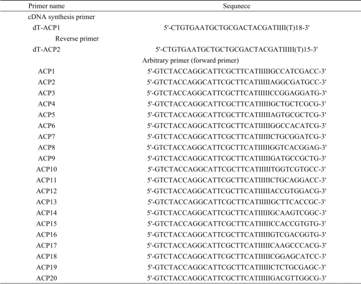
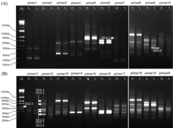
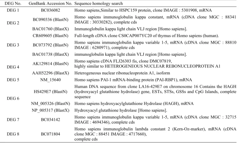
In this study, we applied annealing control primer (ACP)-based reverse transcriptase PCR (polymerase chain reaction) technique to identify differentially expressed

By using different gene fishing primer we identified several Cu- induced genes including UDP-glucuronic acid decarboxylase (DEG 4), transmembrane protein (DEG 10), small heat