—118—
INTRODUCTION
Myostatin (MSTN) belongs to the TGF-β superfamily and acts as a negative regulator of skeletal muscle devel- opment and growth. It is primarily expressed in skeletal muscles in mice (McPherron et al., 1997). MSTN-muta- tions of cattle and mice result in an increase of skeletal muscle mass with both hyperplasia and hypertrophy (Kambadur et al., 1997; McPherron et al., 1997). In re- viewed by Bradley et al., 2008, several studies have re- ported the regulation methods of the myostatin activity to provide possible therapeutic candidates for human’s muscle diseases such as muscular dystrophy and motor neuron disease, and to promote muscle growth for mam- malian and marine organisms; Anti-myostatin monoclo- nal antibodies such as RK35 and JA16, myostatin prodo- main, Follistatin, histone deacetylase (HDAC) inhibitors,
myostatin peptide and soluble activin IIB receptor. Among them, myostatin prodomain has bound myostatin prevent- ing the release of mature myostatin, and then increased body weight and muscle mass caused by hypertrophy and/or hyperplasia (Bogdanovich et al., 2005). There have been several studies about the functions of myosta- tin prodomain in mammalian and fish models. In mam- malian, overexpression of prodomain in transgenic mice (Lee and McPherron, 2001; Yang et al., 2001; Pirottin et al., 2005) and delivery of myostatin prodomain by an adeno-associated virus vector system in dystrophic mice (Bartoli et al., 2007; Qiao et al., 2008) and dogs (Qiao et al., 2009) were increased in skeletal muscle mass up to two-fold. In fish, transgenic zebrafish overexpressing MSTN prodomain was observed significantly increase in fiber number (Xu et al., 2003) and MSTN prodomain of the marine fish Sparus aurata suppressed myostatin activity in vitro (Rebhan and Funkenstein, 2008). Recent- ly Lee et al. (2010) first reported to the production and expression of soluble fish (Paralichthys olivaceus) myo-
Identification of Differentially Expressed Genes in Improved Rainbow Trout Growth by Treatment with a Fish Myostatin Prodomain Using the Annealing Control Primer System
By Sang Beum Lee and Hyung-Joo Jin*
Department of Marine Molecular Biotechnology, Gangneung-Wonju National University, Gangneung 210-702, Korea
ABSTRACT The present study was conducted to investigate different gene expression profile between treated poMSTNpro and non-treated in rainbow trout and to identify those genes that are specifically or predominantly expressed in treated poMSTNpro by employing annealing control primer (ACP)-based GeneFishing polymerase chain reaction (PCR). We isolated total RNAs in muscle tissues from the treated poMSTNpro fish by immersion bath technique with fish myostatin prodomain (Paralichthys olivaceus, poMSTNpro) for one month and the other was non-treated poMSTNpro, and synthesized cDNA using annealing control primers (ACP, Seegene, Korea). Using 20 different ACPs for PCR, were cloned sequ- enced, and analyzed identities of 2 differentially expressed genes (DEGs). According to BLAST analysis, sequences of 2 clones significantly matched database entries and confirmed by semi-quantitative RT- PCR. The functional roles of one up-regulated gene, cytochrome P450 mono-oxygenases 2K1v2 (CYP2K1v2), and one down-regulated gene was Profilin-1 were identified. We identified distinctive gene expression profiles in improved rainbow trout growth by treatment with a fish myostatin prodomain using ACP-based GeneFishing.
Key words : Myostatin, myostatin prodomain, Annealing Control Primer (ACP), cytochrome P450 mono-oxy- genase, profilin, Oncorhynchus mykiss
*Corresponding author: Hyung-Joo Jin Tel: 82-33-640-2349, Fax: 82-33-640-2340, E-mail: [email protected]
ISSN: 1225-8598
Accepted: April 29, 2012
http://www.fishkorea.or.kr
statin prodomain (poMSTNpro) in E. coli system to im- prove muscle growth through blocking mature myostatin.
In an in vitro gene reporter assay system, the anti-myo- statin activity of the soluble myostatin prodomain is sim- ilar to one of commercial mouse myostatin prodomain.
In vivo assay, rainbow trout (Oncorhynchus mykiss) has improved the growth in a dose-dependent manner with the maximum growth increase up to 42% by immersion bath method. Therefore, the prodomain of myostatin can be increased a muscle mass of organisms through block- ing myostatin to bind it’s receptor.
In general, the function, signal pathway and downstream target genes by myostatin like TGF-β superfamily mem- bers known as well-established compared to those of myostatin’s prodomain. Yang et al. (2005) reported the responded genes were identified from myostatin-stimu- lated myoblasts to understand the pathway of myostatin.
However, myostatin prodomain only has been known as a function of binding the mature myostatin to blocking the signal transduction. Elucidation of the functional mechanisms of myostatin prodomain is very important to its application in medicine and animal breeding.
The aim of this study was to find differentially express- ed genes in treated poMSTNpro and non-treated rainbow trouts for one month by Annealing Control Primer (ACP)- based RT-PCR.
MATERIALS AND METHODS
1. Rainbow trout sample
We examined two groups of the rainbow trouts; one group was the poMSTNpro treated fish by immersion bath technique with fish myostatin prodomain (Parali- chthys olivaceus, poMSTNpro) for one month and the other was non-treated poMSTNpro. The muscle samples of 10 fishes at each group were immediately frozen in liquid nitrogen and then stored at -70�C before use.
Rainbow trout was selected as our model fish because it was easily available at the time of our experiment.
2. Genefishing reverse transcription polymerase chain reaction
To identify poMSTNpro-related genes in rainbow trout, the mRNAs from the muscle of both samples, poMSTNpro and non-treated ones, were extracted and applied to ACP RT-PCR analysis using 20 arbitrary primers (from ACP1 to ACP20). Total RNA was isolated from the muscle of non- and poMSTNpro treated-rainbow trout with TRIzol Reagent (Invitrogen, USA) following the manufacturer’s.
The RNA quality and quantity were verified using spec- trophotometry (Ultrosepc 3100pro, Amersham Biosci- ences). The RNAs were used for the first-strand cDNA synthesis by reverse transcription. Reverse transcription
was carried using GeneFishingTM DEG kits (Seegene, Korea). Reverse transcription was performed for 90 min at 42�C in a final reaction volume of 20μL containing 3 μg of total RNA, 4 μL of 5 x RT buffer (Invitorgen, USA), 5μL of 2 mM dNTP, 2 μL of 10 μM dT-ACP1 (5′-CGT GAATGCTGCGACTACGATIIIIIT (18)-3′), 0.5 μL of RNase inhibitor (40 U/μL, Promega, USA) and 1 μL of MMLV reverse transcriptase (200 U/μL, Invitorgen). First- strand Cdnas were diluted with 80μL of RNase-free water for the GeneFishingTMPCR and stored at -20�C until use.
The differentially expressed genes were screened by ACP (Annealing Control Primer)-based PCR method using the GeneFishingTM DEG kits (Seegene, Korea).
The second-strand cDNA was synthesized at 50�C dur- ing one cycle of first-stage PCR in a final reaction volume of 20μL containing 3 μL (about 50 ng) of first-strand cDNA, 1μL of 10 μM dT-ACP2 (5′-CGTGAATGCTG CGACTACGATIIIIIT (15)-3′), 2 μL of 5 μM arbitrary ACP and 10μL 2 x master mix (Seegene). The PCR protocol for the second-strand synthesis was one cycle at 94�C for 5 min, followed by 50�C for 3 min and 72�C for 1 min. After the second-strand DNA synthesis was completed, the second-stage PCR amplification protocol was 40 cycles of 94�C for 40 sec, followed by 65�C for 40 sec, 72�C for 40 sec and final extension at 72�C for 5 min.
3. Cloning and sequencing
The PCR product bands showing differential expres- sion on 1.5% agarose gel between poMSTNpro treated and non-treated rainbow trout samples were excised and purified by agarose gel extraction kit (Bioneer, Korea) and cloned into a pGEM-T easy vector (Promega) for sequencing. The DNA sequences were identified by BLAST search program at the National Center for Bio- technology Information (NCBI) GenBank.
4. Semi-quantitative reverse transcription polymerase chain reaction analysis
The differential expression of DEGs was confirmed by reverse transcriptase polymerase chain reaction (RT- PCR) using each gene specific primer pair. The primer sets and annealing temperatures of two genes are shown in Table 1. The cDNA was amplified using primers deriv- ed from the sequence of the DEGs and β-actin gene as a control reference. The PCR reaction was conducted in a final volume of 25μL containing 3 μL (about 50 ng) of diluted first-strand cDNA, 2.5μL of 10X reaction buffer, 1.5μL of MgCl2(25 mM), 0.5μL of dNTP (10 μM), 0.5 μL of PCR primers (10 μM) and 0.2 μL of Taq DNA polymerase (5 unit). The PCR amplification protocol was an initial 3 min denaturation at 94�C, followed by 20~25 cycles of 94�C for 30 s, 55�C for 30 s, 72�C for
30 s, and a 7 min final extension at 72�C. The amplified PCR products were separated by electrophoresis on a 1.5% agarose gel. The bands were photographed using Polaroid film under ultraviolet light after ethidium bro- mide staining. The optical density of each band was ana- lysied by densitometry. The relative amount of mRNA was determined by calculating the ratio of the amount of each mRNA relative to the amount of β-actin.
RESULTS AND DISCUSSION
Previously works, we expressed the soluble fish myo- statin prodomain (poMSTNpro) in E. coli system. The expressed poMSTNpro showed the activity of myostatin inhibition both in vitro and in vivo assays. Especially, in vivo test, the growth of the poMSTNpro-treated rainbow trout was improved in a dose-dependent manner with the maximum growth increased up to 42% for one month compared to non-treated one (Lee et al., 2010). Fig. 1 showed two differentially expressed genes between two groups on the basis of the differential expression levels of the mRNA fragments observed on the agarose gel. The expression levels of one (cytochrome P450 mono-oxy- genase2K1v2; CYP2K1v2) were found to be markedly up-regulated gene in poMSTNpro-treated rainbow trout, while the other band (profilin-1) was down-regulated.
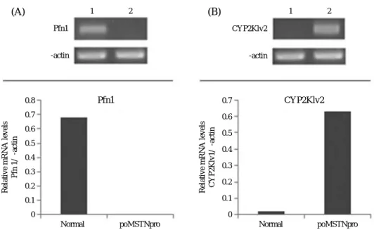
We confirmed different expressions of two genes using sequence-specific primers designed to amplify products with lengths ranging from 100 to 200 bp (Table 1). Quan- titative expression patterns of two differentially express- ed genes that CYP2K1v2 was up-regulated and profilin- 1 was down-regulated in poMSTNpro-treated rainbow trouts.
Understanding the molecular mechanisms of poMSTNpro requires investigation of differentially ex- pressed genes. In this study, we used a ACP that specifi- cally targets sequence hybridization to a template via a polydeoxyinosine linker using poMSTNpro-treated rain- bow trout samples. The structure of ACP comprises a 3′ end region with a target core nucleotide sequence that substantially complements the template nucleic acid for hybridization, a 5′ end region with a non-target universal nucleotide sequence, and a polydeoxyinosine linker bridg- ing the 3′end and 5′ end sequences. Using dT-ACP2
(reverse primer) and 20 arbitrary ACPs (forward primer) for PCR amplification, we were able to display two of differentially expressed genes. The genes were sequenced and screened by a BLAST search. For comparative analy- sis, their expression was quantified by semi-quantitative RT-PCR.
We found that Cytochrome P450 mono-oxygenases 2K1v2 (CYP2K1v2) was expressed significantly more highly in treated samples than in non-treated samples (Fig. 2). Cytochrome P450 mono-oxygenases (CYPs) catalyze oxidation of a wide range of drugs and other xenobiotics and are responsible for the metabolism of endogenous compounds, including steroids (breakdown including estrogen and testosterone synthesis and meta- bolism) (Thum and Borlak, 2000; Borlak and Thum,
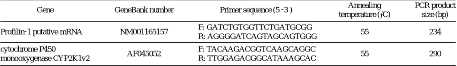
Table 1. Sequence of primers used in the cloning and RT-PCR
Gene GeneBank number Primer sequence (5′-3′) Annealing PCR product
temperature (�C) size (bp) Profilin-1 putative mRNA NM001165157 F: GATCTGTGGTTCTGATGCGG
55 234
R: AGGGGATCAGTAGCAGTGGG cytochrome P450
AF045052 F: TACAAGACGGTCAAGCAGGC
55 290
monooxygenase CYP2K1v2 R: TTGGAGACGGCATAAAGCAC
Findicates forward primer and R indicates reverse primer.
ACP19
1 2
Fig. 1. Annealing control primer (ACP)-based polymerase chain reac- tion (PCR) for the identification of differentially expressed genes from treated poMSTNpro and non-treated in rainbow trout. mRNA from treated poMSTNpro and normal tissues was used for the synthe- sis of first strand cDNA using dT-ACP1. Using a combination of dT- ACP2 (reverse primer) and 20 arbitrary ACPs (forward primer), second- strand cDNA sequences were amplified during second-stage PCR, separated for differentially expressed genes on 1.5% standard agarose gels, and stained with ethidium bromide for visualisation. Bands were excised from the gel for further cloning and sequencing. 1, mRNA from non-treated muscle tissue; 2, treated poMSTNpro muscle tissue.
Arrows indicate differential cDNA bands and ACP19 indicate the ACP number.
2001), cholesterol, bile, fatty acids (Simpson, 1997; Cap- devila et al., 2000), bradykinin (Fulton et al., 1995), and certain vitamins (Omdahl, 2001). In mammals, proteins belonging to the first families (CYP1-CYP4) are highly associated with drug activation and metabolism. However, studies on CYP function and modulation in non-mamma- lian vertebrate systems are much less defined, and the function is often inferred from mammalian data, assum- ing similar function across vertebrate taxa. CYP families and subfamilies, with the exception of the CYP2 subfa- milies, are nearly identical in vertebrate taxa and the total numbers of CYP genes in vertebrate species is similar (Thomas, 2007). The mammalian CYP2 family is regulat- ed through a number of receptors, including the retinoic acid receptor (RAR), the constitutive androstane receptor (CAR) and hepatocyte nuclear factor 4 (Honkakoski and Negishi, 2000; Lewis et al. 2002). Similar induction pathways exist across vertebrate classes and with the exception of CAR, all of these nuclear receptors have been identified in fish (Moore et al., 2002, 2003; Maglich et al., 2003). Thum and Borlak (2002) show metabolism of testosterone to be increased in hypertrophic hearts and induction of CYP mono-oxygenases to be linked to enhanced production of certain testosterone metabolites.
Androgens seem to have strong negative impact on myo- statin expression, which might be a key factor in the weight regulation of LA muscle (Mendler, 2007). From the higher expression of CYP2K1v2 in treated samples in this study, it was suggested that CYP2K1v2 might
play an important role in mediating the muscle growth of treated rainbow trout.
The level of profilin-1 (Pfn1) was expressed signifi- cantly down-regulated in treated samples (Fig. 2). Pro- filins (Pfns) belong to a class of small G-actin-binding proteins comprising of four members identified to date:
Pfn1 (ubiquitously expressed in almost all cell types), Pfn2 (mainly expressed in nervous system in vertebrates), Pfn3 and 4 (expression restricted to kidney and testis).
Besides binding to actin, Pfns also interact with a multi- tude of other ligands including various phosphoinositides and proteins containing proline-rich motifs that are in- volved in actin cytoskeletal regulation, endocytosis, and gene transcription. Pfn1, the founding member of the protein family, promotes actin polymerization in cells by virtue of its ability to (i) catalyze nucleotide exchange factor (ADP to ATP) on G-actin, (ii) shuttle G-actin to the barbed ends of actin filaments, and (iii) interact with almost all major protein families that are known to be involved in nucleation and/or elongation of actin fila- ments (Witke, 2004; Jockusch et al., 2007). Cell-cycle progression is tightly regulated by coordinated activities of cyclin/cyclin-dependent kinase (CDK) complexes.
The interactions of cyclins with their partner CDKs are negatively regulated by CDK inhibitors (CDKIs). Two families of CDKIs, namely the CIP/KIP (p21Cip1/Waf1 (p21), p27Kip1(p27), and p57Kip2(p57)) and INK4 (p16INK4a, p15INK4b, p18INK4c, and p19INK4d) cause cell- cycle arrest at GI phase (Sherr, 1994; Sherr and Roberts,
1 2 1 2
Relative mRNA levels Pfn 1/β-actin Relative mRNA levels CYP2Klv1/β-actin
0.8 0.7 0.6 0.5 0.4 0.3 0.2 0.1 0
0.7 0.6 0.5 0.4 0.3 0.2 0.1 0
Pfn1 CYP2Klv2
β-actin β-actin
Pfn1 CYP2Klv2
Normal poMSTNpro Normal poMSTNpro
(A) (B)
Fig. 2. Confirmation by semi-quantitative reverse transcription-polymerase chain reaction (RT-PCR) of the differential mRNA expression pattern of two genes that were identified by sequence-specific primers as being differentially expressed genes. RT-PCR and densitometric analysis. The comparison of the expression patterns of the two differentially expressed genes (1: normal and 2: treated-poMSTNpro) was determined by semi- quantitative RT-PCR. The amplified DNA products were separated on 1.5% agarose gels and stained with ethidium bromide. Quantitative analyses of expression patterns of bands were carried out. β-actin was used as a control to confirm the integrity of the mRNA samples and each band was scaled to the intensity of beta actin in the respective lane. (A) Profilin-1 (Pfn1) gene expression pattern. (B) Cytochrome P450 mono-oxygenase 2K1v2 (CYP2K1v2) gene expression pattern.
1995). Myostatin functions by binding to two different type of serine/threonine kinase receptors, which leads to the phosphorylation of Smad2 and Smad3 (Lee and Mc- Pherron, 2001; Rebbapragada et al., 2003). Phosphor- ylated Smad2 and Smad3 form a complex with Smad4 that translocates into the nucleus, where it is involved in regulating the transcription of target genes (Shi and Mas- sague, 2003). In vitro, an addition of MSTN to muscle cell culture inhibits cell proliferation by up-regulation of p21 and Cdk2, which control the cell cycle progres- sion process (Thomas et al. 2000; Joulia et al. 2003).
Profilin-1 overexpression inhibits proliferation of MDA- MB-231 breast cancer cells partly through p27kip1upregu- lation (Zou et al., 2010). Decrease Profilin-1 might play a role in the muscle growth that was the hallmark of treat- ed samples.
In conclusion, we found differential expression of two genes (CYP2K1v2 and Pfn1) in relation to drug, steroids metabolism and proliferation in treated poMSTNpro rainbow trouts using the GeneFishingTMPCR technique.
Although the detailed function of these genes remain to be determined, they could be important and deserve fur- ther investigation, and their identification in this study provides preliminary data for further study of the mole- cular mechanism underlying treated poMSTNpro in rain- bow trout.
ACKNOWLEDGMENTS
This research was supported by Basic Science Research Program through the National Research Foundation of Korea (NRF) funded by the Ministry of Education, Sci- ence and Technology (2010-0023056) and the Regional Technology Innovation Program (RTI05-01-02).
REFERENCES
Bartoli, M., J. Poupiot, A. Vulin, F. Fougerousse, L. Arandel, N. Daniele, C. Roudaut, F. Noulet, L. Garcia, O. Danos and I. Richard. 2007. AAV-mediated delivery of a mutated myostatin propeptide ameliorates calpain 3 but not alpha-sarcoglycan deficiency. Gene Ther., 14:
733-740.
Bogdanovich, S., K.J. Perkins, T.O. Krag, L.A. Whittemore and T.S. Khurana. 2005. Myostatin propeptide-medi- ated amelioration of dystrophic pathophysiology.
FASEB J., 19: 543-549.
Borlak, J. and T. Thum. 2001. Induction of nuclear transcrip- tion factors, cytochrome P450 monooxygenases, and glutathione S-transferase a gene expression in Aroclor 1254-treated rat hepatocyte cultures. Biochem. Phar- macol., 61: 145-153.
Bradley, L., P.J. Yaworsky and F.S. Walsh. 2008. Myostatin as a therapeutic target for musculoskeletal disease.
Cell. Mol. Life Sci., 65: 2119-2124.
Capdevila, J.H., J.R. Falck and R.C. Harris. 2000. Cytochrome P450 and arachidonic acid bioactivation. Molecular and functional properties of the arachidonate mono- oxygenase. J. Lipid Res., 41: 163-181.
Fulton, D., K. Mahboubi, J.C. McGiff and J. Quilley. 1995.
Cytochrome P450-dependent effects of bradykinin in the rat heart. Br. J. Pharmacol., 114: 99-102.
Honkakoski, P. and M. Negishi. 2000. Regulation of cyto- chrome P450 (CYP) genes by nuclear receptors. Bio- chem. J., 347: 321-337.
Jockusch, B.M., K. Murk and M. Rothkegel. 2007. The pro- file of profilins. Rev. Physiol. Biochem. Pharmacol., 159: 131-149.
Joulia, D., H. Bernardi, V. Garandel, F. Rabenoelina, B. Ver- nus and G. Cabello. 2003. Mechanisms involved in the inhibition of myoblast proliferation and differen- tiation by myostatin. Exp. Cell Res., 286: 263-275.
Kambadur, R., M. Sharma, T.P. Smith and J.J. Bass. 1997.
Mutations in myostatin (GDF8) in double-muscled Belgian blue and piedmontese cattle. Genome Res., 7: 910-916.
Lee, S.B., Y.S. Kim, M.Y. Oh, I.H. Jeong, K.B. Seong and H.J. Jin. 2010. Improving rainbow trout (Oncorhyn- chus mykiss) growth by treatment with a fish (Para- lichthys olivaceus) myostatin prodomain expressed in soluble forms in E. coli. Aquaculture, 302: 270- 278.
Lee, S.J. and A.C. McPherron. 2001. Regulation of myostatin activity and muscle growth. Proc. Natl. Acad. Sci.
USA., 98: 9306-9311.
Lewis, D.F., M.N. Jacobs, M. Dickins and B.G. Lake. 2002.
Quantitative structure-activity relationships for indu- cers of cytochromes P450 and nuclear receptor ligands involved in P450 regulation within the CYP1, CYP2, CYP3 and CYP4 families. Toxicology, 176: 51-57.
Maglich, J.M., J.A. Caravella, M.H. Lambert, T.M. Willson, J.T. Moore and L. Ramamurthy. 2003. The first com- pleted genome sequence from a teleost fish (Fugu rubripes) adds significant diversity to the nuclear re- ceptor superfamily. Nucleic. Acids Res., 31: 4051- 4058.
McPherron, A.C., A.M. Lawler and S.J. Lee. 1997. Regulation of skeletal muscle mass in mice by a new TGF-beta superfamily member. Nature, 387: 83-90.
Mendler, L., Z. Baka, A. Kovacs-Simon and L. Dux. 2007.
Androgens negatively regulate myostatin expression in an androgen-dependent skeletal muscle. Biochem.
Bioph. Res. Co., 361: 237-242.
Moore, J.T., L.B. Moore, J.M. Maglich and S.A. Kliewer.
2003. Functional and structural comparison of PXR and CAR. Biochimica. Et. Biophysica. Acta, 1619:
235-238.
Moore, L.B., J.M. Maglich, D.D. McKee, B. Wisely, T.M.
Willson, S.A. Kliewer, M.H. Lambert and J.T. Moore.
2002. Pregnane X receptor (PXR), constitutive andro- stane receptor (CAR), and benzoate X receptor (BXR) define three pharmacologically distinct classes of nu- clear receptors. Mol. Endocrinol., 16: 977-986.
Omdahl, J.L., E.A. Bobrovnikova, S. Choe, P.P. Dwivedi and B.K. May. 2001. Overview of regulatory cyto- chrome P450 enzymes of the vitamin D pathway.
Steroids, 66: 381-389.
Pirottin, D., L. Grobet, A. Adamantidis, F. Farnir, C. Herens, H. DaaSchrøder and M. Georges. 2005. Transgenic engineering of male-specific muscular hypertrophy.
Proc. Natl. Acad. Sci. USA., 102: 6413-6418.
Qiao, C., J. Li, J. Jiang, X. Zhu, B. Wang, J. Li and X. Xiao.
2008. Myostatin propeptide gene delivery by adeno- associated virus serotype 8 vectors enhances muscle growth and ameliorates dystrophic phenotypes in mdx mice. Hum. Gene. Ther., 19: 241-254.
Qiao, C., J. Li, H. Zheng, J. Bogan, J. Li, Z. Yuan, C. Zhang, D. Bogan, J. Kornegay and X. Xiao. 2009. Hydro- dynamic limb vein injection of AAV8 canine myosta- tin propeptide gene in normal dogs enhances muscle growth. Hum. Gene Ther., 20: 1-10.
Rebbapragada, A., H. Benchabane, J.L. Wrana, A.J. Celeste and L. Attisano. 2003. Myostatin Signals through a Transforming Growth Factor β-Like Signaling Path- way To Block Adipogenesis. Mol. Cell. Biol., 23:
7230-7242.
Rebhan, Y. and B. Funkenstein. 2008. Inhibition of fish myostatin activity by recombinant fish follistatin and myostatin prodomain: Potential implications for en- hancing muscle growth in farmed fish. Aquaculture, 284: 231-238.
Sherr, C.J. 1994. G1 phase progression: Cycling on cue. Cell, 79: 551-555.
Sherr, C.J. and J.M. Roberts. 1995. Inhibitors of mammalian
G1 cyclin-dependent kinases. Genes Dev., 9: 1149- 1163.
Shi, Y. and J. Massague. 2003. Mechanisms of TGF-beta signaling from cell membrane to the nucleus. Cell, 113: 685-700.
Simpson, A.E. 1997. The cytochrome P450 4 (CYP4) family.
Gen. Pharmacol., 28: 351-359.
Thomas, J.H. 2007. Rapid birth-death evolution specific to xenobiotic cytochrome P450 genes in vertebrates.
PLoS Genet., 3: e67.
Thomas, M., B. Langley, C. Berry, M. Sharma, S. Kirk, J.
Bass and R. Kambadur. 2000. Myostatin a negative regulator of muscle growth, functions by inhibiting myoblast proliferation. J. Biol. Chem., 275: 40235- 40243.
Thum, T. and J. Borlak. 2000. Cytochrome P450 monooxy- genase gene expression and protein activity in cultures of adult cardiomyocytes of the rat. Br. J. Pharmacol., 130: 1745-1752.
Thum, T. and J. Borlak. 2002. Testosterone, cytochrome P450, and cardiac hypertrophy. FASEB J., 16: 1537- 1549.
Witke, W. 2004. The role of profilin complexes in cell moti- lity and other cellular processes. Trends Cell Biol., 14: 461-469.
Xu, C., G. Wu, Y. Zohar and S.J. Du. 2003. Analysis of myostatin gene structure, expression and function in zebrafish. J. Exp. Biol., 206: 4067-4079.
Yang, J., T. Ratovitski, J.P. Brady, M.B. Solomon, K.D.
Wells and R.J. Wall. 2001. Expression of myostatin pro domain results in muscular transgenic mice. Mol.
Reprod. Dev., 60: 51-61.
Yang, W., Y. Zhang, G. Ma, X. Zhao, Y. Chen and D. Zhu.
2005. Identification of gene expression modifications in myostatin-stimulated myoblasts. Biochem. Bioph.
Res. Co., 326: 660-666.
Zou, L., Z. Ding and P. Roy. 2010. Profilin-1 overexpression inhibits proliferation of MDA-MB-231 breast cancer cells partly through p27kip1 upregulation. J. Cell Phy- siol., 223: 623-629.
Annealing control primer system을 이용한 어류 재조합 myostatin prodomain 단백질에 의해 성장이 증가된 무지개송어의 특이적 발현 유전자 탐색
이상범∙진형주
강릉원주대학교 생명과학대학 해양분자생명공학과
요 약 :이전 연구에서 넙치유래 재조합 마이오스타틴 프로도메인을 무지개송어에 한달간 침지법을 통하여 처리한 결과 대조군에 비하여 무게가 최대 약 42% 증가되었다. 따라서 본 연구는 재조합 마이오스타틴 프로도 메인을 침지법에 의해 처리된 무지개송어와 대조군의 근육으로부터 발현되는 cDNA를 제작하여 마이오스타틴 프로도메인에 의해서 유도된 특정유전자를 선발하기 위하여 ACP (annealing control primer)를 이용한 DDRT법 을 통하여 분석하였다. 총 20가지의 ACP를 이용한 결과 2개의 특정 유전자를 분석하였으며, NCBI BLAST분석 결과 Cytochrome P450 mono oxygenase와 Profilin으로 판명되었다. 이 중 Cytochrome P450 mono oxygenase는 대조군보다 발현량이 증가하였으며, Profilin는 대조군에 비해서 발현량이 감소하였다. 이러한 결과를 재확인하기 위하여 두 유전자의 primer를 각각 제작하여 semi-quantitative RT-PCR를 시행한 결과 DDRT법에 의한 분석과 동일하였다. 본 결과는 어류의 성장에서 마이오스타틴 프로도메인의 기능 및 메카니즘에 대한 연구에 유용한 자 료가 될 것으로 사료된다.
찾아보기 낱말 :마이오스타틴, 마이오스타틴 프로도메인, Annealing control primer, cytochrome P450 mono- oxygenase, profilin, 무지개송어