915
V-erbA Related EAR-3 유전자의 발현분석
Analysis of Human V-erbA Related EAR-3 Gene Expression between Transitional Cell Carcinoma and Normal Tissue in Bladder Cancer
Won Sik Ham, Joo Hyoung Lee, Ho Song Yu, Young Deuk Choi From the Department of Urology and Urological Science Institute, Yonsei Univer- sity College of Medicine, Seoul, Korea
Purpose: The prognosis of bladder cancer is related to tumor grade and stage. Because these pathological changes are preceded by molecular alterations, new molecular markers are needed in early diagnosis. New target molecular biomarkers can be differentially expressed genes (DEGs) between normal and cancer tissues. We tried to find a new DEG and demonstrated that it may be related to the development of the bladder cancer.
Materials and Methods: Cancer tissues were obtained from 39 patients with urothelial cell carcinoma, treated by transurethral resection of tumor (TURB) since 2002. Normal bladder tissues were obtained from the same patients during TURB. We compared the mRNA profiles between normal and cancer tissues using annealing control primer (ACP)-based Genefish- ingTM PCR to identify the DEGs in normal and cancer tissues of one same patient. To validate the result of ACP-based GeneFishingTM PCR, reverse transcription-polymerase chain reaction (RT-PCR) was performed on those of 39 patients.
Results: According to the result of ACP-based GenefishingTM PCR, EAR-3 gene was only present or markedly upregulated in normal tissue, com- pared with cancer tissues. The expression pattern that EAR-3 gene was downregulated in cancer tissues, irrespective of the clinicopathologic para- meters was confirmed by RT-PCR in 39 patients.
Conclusions: EAR-3 gene was downregulated in cancer tissues, irrespective of clinicopathologic parameters, compared with normal tissues in the bladder of the same patient. Therefore, we suggested that EAR-3 gene may be also play a role in bladder cancer development. (Korean J Urol 2007;48:915-920)
Key Words: Bladder cancer, ACP-based GeneFishingTM PCR, Reverse transcriptase polymerase chain reaction, EAR-3 gene
대한비뇨기과학회지 제 48 권 제 9 호 2007
연세대학교 의과대학
비뇨기과학교실, 비뇨의과학연구소
함원식ㆍ이주형ㆍ유호송ㆍ최영득
접수일자:2007년 6월 29일 채택일자:2007년 7월 24일
교신저자: 최영득
세브란스병원 비뇨기과 서울시 서대문구 성산로 250 (신촌동 134)
120-752
TEL: 02-2228-2317 FAX: 02-312-2538 E-mail: [email protected] 본 연구는 2005년도 연세대학교 학술 연구비의 일부 지원으로 수행되었음.
서 론
방광암은 전 세계적으로 9번째로 흔한 악성종양으로 모든 방광암의 95%는 이행상피세포암이다. 방광암으로 진단받 은 환자의 70%는 진단 당시 표재성 (Ta 또는 T1)으로 발견 되어 경요도방광종양절제술로 치료한다. 그러나 술 후 70%
의 환자에서 재발하고 15%는 침윤성으로 진행하기 때문에 이러한 표재성 방광암환자들에서 조기 진단과 함께 질환의 진행에 대한 정확한 예측이 가능하다면 적절한 치료를 선 택하는 데 도움이 될 수 있다.1
방광경검사는 방광 내 악성 병변을 발견하는 데 기본이 되는 검사방법이나 침습적이며, 상피내암종 (carcinoma in situ) 같은 편평한 병변들은 발견하지 못하는 단점이 있다.1
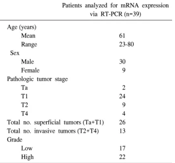
Table 1. Patients' characteristics
Patients analyzed for mRNA expression via RT-PCR (n=39)
Age (years)
Mean 61
Range 23-80
Sex
Male 30
Female 9
Pathologic tumor stage
Ta 2
T1 24
T2 9
T4 4
Total no. superficial tumors (Ta+T1) 26 Total no. invasive tumors (T2+T4) 13 Grade
Low 17
High 22
RT-PCR: reverse transcription-polymerase chain reaction 요세포검사는 비침습적이며, 방광경검사를 보완하기 위해
흔히 임상에서 사용하는 검사법으로 상피내암종 같은 높은 세포분화도의 병변 발견에는 특이도가 높으나, 판독의의 주관적인 판단에 따라 진단이 달라지고 상대적으로 낮은 감수성 때문에 진단적인 가치는 제한적이다.2
현재까지 방광암 환자의 추적관찰과 치료과정에 대한 중 요 결정 요인은 종양의 병기와 세포분화도이다. 그러나 병 리적인 판단에 의존한 종양의 병기 및 세포분화도의 결정 은 판독에 따라 달라지는 경향이 있고, 이러한 변수들은 아 직까지 질환의 진행경로를 충분히 정확히 예측하지 못하고 있다.3 분자학적 변화들이 이러한 병리적인 변화들과 임상 적인 진행을 선행하기 때문에, 이러한 분자학적 변화들을 파악할 수 있다면 예후에 관한 정보들을 조기에 얻을 수 있다. DNA ploidy와 같은 분자의 거대한 변화들과 여러 염 색체들의 변화들이 방광암의 침윤성 병변으로의 진행과 연 관이 있다.4,5 DNA의 변화들 외에도 p53과 RB gene과 같은 경우에서와 같이 mRNA 발현정도의 차이가 방광암의 진행 과 연관이 있다.6,7 그러나 이런 표지자들의 감수성이 낮고, 방광암의 임상적인 진행경로를 정확히 예측하지 못하기 때 문에 새로운 표지자들이 필요하다.
새로운 분자학적 표지자의 대상으로는 정상조직과 종양 조직사이에 다르게 발현되는 유전자들 (differentially expre- ssed genes; DEGs) 또는 그로 인한 단백질산물들이 될 수 있다. 이에 저자들은 두 조직 간에 다르게 발현되는 새로운 유전자를 찾아 그 임상적인 의미를 찾고자 하였다.
대상 및 방법
2002년 6월에서 2004년 11월까지 본원에서 방광암으로 진단되어 경요도방광종양절제술를 시행받은 환자 중 방광 암조직과 정상방광조직이 동시에 존재하는 39명의 환자를 대상으로 하였다. 같은 환자의 방광암조직과 정상방광조직 을 구별한 후, Hwang 등8-11의 genefishing 방법에 따라 annealing control primer (ACP)-based GenefishingTM polyme- rase chain reaction (PCR)를 이용하여 두 조직에서의 mRNA 의 발현차이를 보이는 유전자들을 탐색하였다.
우선 방광암 환자에서 경요도방광종양절제술을 시행 후 생리식염수로 방광 내부를 충분히 세척한 후에 방광암 부 위에서 3cm 이상 먼 방광암 의심 병변이 없는 부위에서 정상요로상피에 대한 조직을 얻었다. 모든 조직표본들은 80oC로 동결 보관하여 사용하였다. 표재성 방광암 환자 26 명 중 22명에서 경요도방광종양절제술 후 추가적인 방광 내 약물주입요법을 시행받았다. 침윤성 방광암 환자 (n=13) 중 경요도방광종양절제술 후 9명에서 근치적 방광적출술
을 시행받았으며, 4명은 술 후 항암화학요법를 시행받았다 (Table 1). 방광암의 병리학적 소견은 모두 이행상피세포암 이었으며, 정상방광조직은 병리의사에 의해 정상방광조직 생검표본 내에 종양세포가 없음을 확인하였다. 방광암의 세포분화도결정을 위해 WHO와 ISUP 기준을 사용하였고, 병기결정을 위해 TNM 분류기준을 사용하여 임상병리적인 변수들을 평가하였다.12 RNA 추출 전에 모든 방광암조직표 본들에서의 종양세포의 비율을 병리의사가 평가하였다.
종양조직과 정상조직에서 총 RNA를 추출하였다. 모든 조직표본들은 무균의 막자 (sterile pestle)를 이용하여 1ml의 Ultraspec II (Biotecx Lab., Houston, USA)에 넣어 분쇄하였 다. 제조사의 포로토콜에 따라 추출한 RNA를 260nm 흡광 도에서 정량하고, 일부는 Agilent 2100 bioanalyzer (Agilent, Palo Alto, USA)를 이용하여 ‘Lab On a Chip’ (developed by Agilent Technologies, Waldbronn, Germany, in co-operation with Caliper Technologies, Mountain View, USA)법으로 정량 분석하여 RNA비율이 1 이상인 mRNA만을 취하여 사용하 였다.
DEGs의 가능성이 있는 유전자들을 파악하기 위해 ACP- based GenefishingTM PCR을 시행하여 같은 환자의 정상조직 과 종양조직 간의 mRNA양상을 비교하였다. First strand cDNA 합성을 위해 역전사는 3μg 정제된 total RNA, 4μl 5x reaction buffer, 5μl dNTPs (각각 2mM), 2μl 10μM cDNA 합성 primer dT-ACP1 (5’-CGTGAATGCTGCGACTACGATIIIII (T)18-3’), 0.5μl RNasin RNase Inhibitor (40U/μl), 그리고 1μl
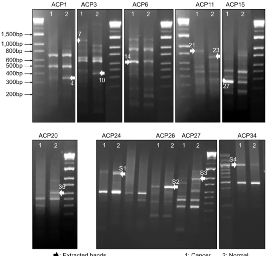
Fig. 1. Results of annealing control primer (ACP)-based polymerase chain reaction (PCR) for identification of differentially expressed genes (DEGs) from normal and cancer tissue.
Arrowheads indicate 12 differential cDNA bands, excised from the gel for further cloning and sequencing and 35 band is found to be EAR-3 gene according to the sequence homology search.
Moloney Murine Leukemia Virus reverse transcriptase (200U/
μl; Promega, Madison, USA)를 초기 용량 20μl로 42oC에서 1.5시간 동안 시행하였다. 역전사 후 first strand cDNAs는 GeneFishingTM PCR용 ultra-purified water 80μl로 희석하고 사용시까지 20oC에 보관하였다.
Second strand cDNA 합성과 일련의 PCR 반응은 단일 튜 브 내에서 시행되었다. Second strand cDNA 합성은 50oC에 서 3-5μl (약 50ng) 희석된 first strand cDNA, 10μl 2x Master Mix (씨젠, 서울, 한국), 1μl 10μM dT-ACP2, 1μl 10μM 임 의의 ACP를 25μl의 용량으로 반응하였다. PCR 반응은 94oC에서 1분, 50oC에서 3분, 그리고 72oC에서 1분간 진행 된 후 40회의 증폭 cycles이 시행되었다. 각각의 cycle은 94oC에서 40초간의 denaturation 과정, 65oC에서 40초간의 annealing 과정, 그리고 72oC에서 40초간의 extension 과정을 거친 후, 72oC에서의 5분간의 최종 extension 과정을 통해 PCR 반응을 완료하였다. PCR 산물들은 2% agarose gels에 서 전기영동 후 ethidium bromide로 염색하였다.
Gel 위에 전기영동하여 발현차이가 있는 다수의 cDNA bands 중에 GENCLEAN II Kit (Q-BIO gene, Carlsbad, USA)를
이용하여 12개의 DEGs를 추출한 후 제조사의 포로토콜대 로 TOPO TA cloning vector로 바로 클로닝하였다. 클로닝된 plasmids는 ABI PRISM 3100 Genetic Analyzer (Applied Bio- systems, Foster City, USA)로 automatic sequencing을 수행하 였고 National Center for Biotechnology Information (NCBI) GenBank의 BLASTX 탐색프로그램을 이용하여 분석하였 다. Sequence 분석결과를 토대로 하여 이들 중 종양조직에 서 발현이 저하되는 EAR-3 유전자를 선별하여 reverse tran- scription-polymerase chain reaction (RT-PCR)의 대상으로 선 정하였다.
ACP-based GenefishingTM PCR의 결과를 입증하기 위해 EAR-3 유전자에 대한 RT-PCR oligodeoxynucleotides로 설계 하였고 (forward, 5'-CGG CAC GGC GGG GGA CAA GGG, reverse, 5'-TGC GTA CTG GCC TGG ATT GGG), RT-PCR를 39명의 환자들의 두 조직들에 대해 시행하였다. First strand cDNA는 human β-actin 유전자로 정상화하고, 정상화된 cDNA를 template로 사용하였다. PCR반응은 2-4μl (약 50ng) 희석된 first strand cDNA, 10μl 2x Master Mix (씨젠, 서울, 한국), 1μl 10μM primer 5’, 1μl 10μM primer 3’의 25μl로
Fig. 2. Reverse transcription-poly- merase chain reaction (RT-PCR) pro- duct analysis by agarose gel electro- phoresis and ethidium bromide stain- ing showing the intensity of EAR-3 gene expression in bladder cancer tissues (C) and normal bladder tissue (N) for each patient. The second line represents internal control β-actin expression.
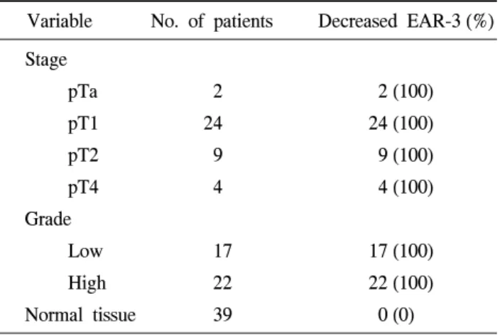
Table 2. Incidence of lower EAR-3mRNA expression among superficial and invasive tumor types
Variable No. of patients Decreased EAR-3 (%) Stage
pTa 2 2 (100)
pT1 24 24 (100)
pT2 9 9 (100)
pT4 4 4 (100)
Grade
Low 17 17 (100)
High 22 22 (100)
Normal tissue 39 0 (0) 하였다. PCR 반응 후 PCR 산물들은 2% agarose gels위에서
전기영동 후 ethidium bromide로 염색하였다.
결 과
방광이행상피세포암조직과 정상방광조직이 함께 동결 보관된 39명의 환자들을 대상으로 정상조직 및 종양조직에 서 추출된 mRNA에 대해 ACP-based GeneFishingTM PCR을 시행하였다 (Fig. 1). 정상조직과 종양조직에서 유전자 발현 의 차이를 보인 다수의 cDNA band들 중 12개의 DEGs에 대 해 클로닝과 sequencing 과정을 거친 후 NCBI GenBank의 BLASTX 검색프로그램을 이용하여 분석하였다. Sequence 분석 결과를 토대로 하여 종양조직에 비해 정상조직에서만 존재하거나 발현이 증가하는 것으로 나타난 EAR-3 유전자 를 RT-PCR의 대상으로 선정하였다.
방광암 환자 39명에서 정상조직과 종양조직에 대해 EAR-3 유전자의 상태를 RT-PCR로 확인하였다. ACP-based GeneFishingTM PCR의 결과와 같이 RT-PCR에서도 39명의 환자 모두에서 정상조직에서만 EAR-3 유전자가 발현되고 종양조직에서는 발현되지 않거나 매우 약하게 나타났다 (Fig. 2). 이러한 EAR-3 유전자는 방광암의 병기와 세포분화 도 차이에 상관없이 모든 종양조직에서 발현이 저하되는 것으로 나타났다 (Table 2).
고 찰
방광암의 형성, 진행, 재발 및 예후척도에 대해 많은 인자 들이 있으나, 조기 진단을 좀 더 정밀히 예측하기 위한 새로 운 분자학적 표지자에 대한 연구는 계속되고 있다. 본 저자 들의 연구에서는 종양의 병기, 세포분화도와는 상관없이 모든 종양조직에서 정상조직에 비해 EAR-3 유전자의 발현 이 저하되어 있었다.
EAR-3 유전자는 1988년 Miyajima 등13에 의한 cDNA cloning을 통한 분석에 의해 밝혀진 human v-erbA related
genes 중의 하나이다. 다른 연구진들에 의해서는 chicken ovalbumin promoter 내의 direct repeat regulatory element에 결 합하는 homodimer로 발견되어 chicken ovalbumin upstream promoter-transcription factor I (COUP-TFI)로 명칭되었고, 그 동안 두가지 명칭으로 각각 불리며 연구되어 왔다. 이후에 이어 COUP-TFII도 발견되었는데 그것 또한 다른 연구진들 에 의해 apolipoprotein regulating protein 1 (ARP-1)으로 독립 적으로 clone되었다.14,15
Orphan receptor는 리간드 (ligand)가 아직까지 밝혀지지 않은 nuclear receptor를 의미하는 것으로 COUP-TFs는 그동 안 가장 많은 연구가 이루어져 온 orphan receptors 중의 하 나로, sequence를 분석한 결과 steroid/thyroid receptor (TR) superfamily의 member에 속한다. COUP-TFs는 homodimer를 형성하거나 retinoid X receptor (RXR)와 몇몇의 다른 nuclear receptors와 heterodimer를 형성하여 다양한 spacings의 불완 전한 AGGTCA direct 또는 inverted repeats를 포함한 다양한 response elements에 결합한다.16 일반적으로는 retinoid acid receptor (RAR), TR, vitamin D receptor (VDR), peroxisome proliferator-activated receptor (PPAR), hepatocyte nuclear factor 4 (HNF4) 등과 같은 다른 nuclear hormone receptors에
대한 전사억제물질 (transcriptional repressor)로 알려지고 있 으나,17 상황에 따라 이러한 억제기능 외에도 많은 다른 genes에 대해 양성적인 조절인자들로 작용할 수도 있다. 아 직까지 이러한 기능적인 양면성의 기전에 대해서는 정확히 알려져 있지 않으나 일부 기전은 COUP-TFs과 작용하는 공 동조절인자 단백질 (coregulator protein)들의 종류에 따라 달 라질 수 있고, 또한 대상 유전자들의 촉진자 (promoter) 환경 에 따라서도 달라질 수 있다.18
레티노이드 (retinoid)는 방광암을 포함한 여러 악성종양 들에 대해 항암효과를 나타낸다.19 최근의 연구들은 여러 다 른 종류의 종양세포들에 대한 레티노이드의 성장억제효과 를 매개하는 데 RARβ가 중요한 역할을 하며, 이러한 RARβ 의 소실이 종양세포들이 정상적인 성장조절을 벗어나는 중 요 요인이 되어 종양발생에 기여할 것이라고 추정하고 있
다.20-22 어떻게 RARβ 발현이 조절되고, 종양세포에서 소실
되는지는 아직까지 알려지지 않았다. Wu 등23은 COUP- TFs 의 발현이 폐암세포주에서 RARβ 유도와 레티노이드에 의 한 성장억제와 양성적으로 연관된다고 보고하였다. Lin 등
24은 레티노이드가 암세포에서의 RARβ 발현, 성장억제, 세 포고사를 유도하는 데 COUP-TFs가 필요하다고 보고하였 다. 레티노이드에 저항성이 있는 종양세포주들의 대부분에 서 COUP-TFs가 발현되지 않아 COUP-TFs의 소실이 종양세 포들에서의 레티노이드에 대한 저항성을 가지게 되는 중요 기전이 될 것이라고 추정하였다. 또한 어떻게 COUP-TFs의 발현이 소실되는지 이해하는 것이 종양세포들에서 레티노 이드와 연관된 결손을 이해하는 것에 핵심적일 것이라고 예상하였다. RARβ의 소실이 유방암 발생의 조기단계에 발 생한다는 사실은 COUP-TFs가 종양 발생에 역할을 할 수도 있다는 것을 의미한다.25
이러한 여러 연구들에도 불구하고 아직까지 COUP-TFs 의 체내 기능에 대해서는 정확히 알려진 것이 거의 없다.
본 연구에서 저자들은 EAR-3/COUP-TFI mRNA가 종양의 병기, 분화도에 상관없이 종양조직에서는 발현이 저하되는 것을 발견하여 이러한 EAR-3/COUP-TFI 유전자의 발현저 하가 방광이행상피세포암의 발생에 역할을 할 것이라고 추 정하였다. 이러한 종양조직들은 레티노이드에 대한 저항성 을 나타낼 것이고, 만약 이러한 종양조직에 EAR-3/COUP- TFI 유전자를 첨가하여 준다면 레티노이드의 항암효과를 증가시키는 데 도움이 될 수 있다. 본 저자들의 연구에서는 EAR-3/COUP-TFI mRNA와 단백질에 대한 양적인 측정을 하지 않아, 향후 real time quantitative RT-PCR과 western blot 분석을 이용한 추가적인 연구를 계획 중이며 이러한 연구 들은 궁극적으로 EAR-3/COUP-TFI 유전자의 생리적인 기 능과 그들의 고유한 리간드를 밝히는 데 도움이 될 것이다.
결 론
정상방광조직과 종양조직 간에 다르게 발현되는 새로운 유전자를 확인함으로써 방광암에 대한 새로운 표지자의 대 상을 찾고자 하는 연구를 한 결과 EAR-3/COUP-TFI 유전자 가 종양의 병기, 분화도에 상관없이 같은 환자의 정상조직 에 비해 종양조직에서 발현이 저하되었다. EAR-3/COUP- TFI 유전자가 방광이행상피세포암의 발생과정에 역할을 할 수도 있다.
REFERENCES
1. Scher HI, Shipley WU, Herr HW. Cancer of the bladder. In:
Devita VT, Hellman S, Rosenberg SA, editors. Cancer:
principles and practice of oncology. 5th ed. Philadelphia:
Lippincott-Raven; 1997;1300-22
2. Matzkin H, Moinuddin SM, Soloway MS. Value of urine cytology versus bladder washing in bladder cancer. Urology 1992;39:201-3
3. Ooms EC, Anerson WA, Alons CL, Boon ME, Veldhuizen RW. Analysis of the performance of pathologists in the grading of bladder tumors. Hum Pathol 1983;14:140-3 4. Dorkin TJ, Robson CN, Neal DE. The molecular pathology
of urological malignancies. J Pathol 1997;183:380-7 5. Cordon-Cardo C. Molecular alterations in bladder cancer.
Cancer Surv 1998;32:115-31
6. Esrig D, Elmajian D, Groshen S, Freeman JA, Stein JP, Chen SC, et al. Accumulation of nuclear p53 and tumor progression in bladder cancer. N Engl J Med 1994;331:1259-64 7. Cairns P, Proctor AJ, Knowles MA. Loss of heterozygosity at
the RB locus is frequent and correlates with muscle invasion in bladder carcinoma. Oncogene 1991;6:2305-9
8. Hwang KC, Cui XS, Park SP, Shin MR, Park SY, Kim EY, et al. Identification of differentially regulated genes in bovine blastocysts using an annealing control primer system. Mol Reprod Dev 2004;69:43-51
9. Hwang KC, Lee HY, Cui XS, Kim JH, Kim NH. Identification of maternal mRNAs in porcine parthenotes at the 2-cell stage:
a comparison with the blastocyst stage. Mol Reprod Dev 2005;70:314-23
10. Hwang IT, Kim YJ, Kim SH, Kwak CI, Gu YY, Chun JY.
Annealing control primer system for improving specificity of PCR amplification. Biotechniques 2003;35:1180-4
11. Kim YJ, Kwak CI, Gu YY, Hwang IT, Chun JY. Annealing control primer system for identification of differentially expressed genes on agarose gels. Biotechniques 2004;36:424- 34
12. Epstein JI, Amin MB, Reuter VR, Mostofi FK. The World Health Organization/International Society of Urological Path-
ology consensus classification of urothelial (transitional cell) neoplasms of the urinary bladder. Bladder Consensus Confer- ence Committee. Am J Surg Pathol 1998;22:1435-48 13. Miyajima N, Kadowaki Y, Fukushige S, Shimizu S, Semba K,
Yamanashi Y, et al. Identification of two novel members of erbA superfamily by molecular cloning: the gene products of the two are highly related to each other. Nucleic Acids Res 1988;16:11057-74
14. Wang LH, Tsai SY, Cook RG, Beattie WG, Tsai MJ, O'Malley BW. COUP tanscription factor is a member of the steroid receptor superfamily. Nature 1989;340:163-6 15. Wang LH, Ing NH, Tsai SY, O'Malley BW, Tsai MJ. The
COUP-TFs compose a family of functionally related transcrip- tion factors. Gene Expr 1991;1:207-16
16. Tsai SY, Tsai MJ. Chick ovalbumin upstream promoter- transcription factors (COUP-TFs): coming of age. Endocr Rev 1997;18:229-40
17. Leng X, Cooney AJ, Tsai SY, Tsai MJ. Molecular mechanisms of COUP-TF-mediated transcriptional repression: evidence for transrepression and active repression. Mol Cell Biol 1996;16:
2332-40
18. Park JI, Tsai SY, Tsai MJ. Molecular mechanism of chicken ovalbumin upstream promoter-transcription factor (COUP-TF) actions. Keio J Med 2003;52:174-81
19. Kelloff GJ. Perspectives on cancer chemoprevention research
and drug development. Adv Cancer Res 2000;78:199-334 20. Li XS, Shao ZM, Sheikh MS, Eiseman JL, Sentz D, Jetten
AM, et al. Retinoic acid nuclear receptor beta inhibits breast carcinoma anchorage independent growth. J Cell Physiol 1995;
165:449-58
21. Li Y, Dawson MI, Agadir A, Lee MO, Jong L, Hobbs PD, et al. Regulation of RAR beta expression by RAR- and RXR-selective retinoids in human lung cancer cell lines: effect on growth inhibition and apoptosis induction. Int J Cancer 1998;75:88-95
22. Minna JD, Mangelsdorf DJ. Retinoic acid receptor expression abnormalities in lung cancer: important clues or major obstacles? J Natl Cancer Inst 1997;89:602-4
23. Wu Q, Li Y, Liu R, Agadir A, Lee MO, Liu Y, et al. Mo- dulation of retinoic acid sensitivity in lung cancer cells through dynamic balance of orphan receptors nur77 and COUP-TF and their heterodimerization. EMBO J 1997;16:1656-69
24. Lin B, Chen GQ, Xiao D, Kolluri SK, Cao X, Su H, et al.
Orphan receptor COUP-TF is required for induction of retinoic acid receptor β, growth inhibition, and apoptosis by retinoic acid in cancer cells. Mol Cell Biol 2000;20:957-70
25. Xu XC, Sneige N, Liu X, Nandagiri R, Lee JJ, Lukmanji F, et al. Progressive decrease in nuclear retinoic acid receptor β messenger RNA level during breast carcinogenesis. Cancer Res 1997;57:4992-6