in Brain PET/CT
Hong-Jae Lee, Yong-Ho Do, Jin-Eui Kim
Department of Nuclear Medicine, Seoul National University Hospital, Seoul, Korea
Purpose The purpose of this study was to evaluate image quality change by truncated region in field of view (FOV) of attenuation correction computed tomography (AC-CT) in brain PET/CT.
Materials and Methods Biograph Truepoint 40 with TrueV (Siemens) was used as a scanner.
68
Ge phantom scan was performed with and without applying brain holder using brain PET/CT protocol. PET attenuation correction factor (ACF) was evaluated according to existence of pallet in FOV of AC-CT. FBP, OSEM-3D and PSF methods were applied for PET reconstruction. Parameters of iteration 4, subsets 21 and gaussian 2 ㎜ filter were applied for iterative reconstruction methods. Window level 2900, width 6000 and level 4, 200, width 1000 were set for visual evaluation of PET AC images. Vertical profiles of 5 slices and 20 slices summation images applied gaussian 5㎜ filter were produced for evaluating integral uniformity.
Results Patient pallet was not covered in FOV of AC-CT when without applying brain holder because of small size of FOV. It resulted in defect of ACF sinogram by truncated region in ACF evaluation. When without applying brain holder, defect was appeared in lower part of transverse image on condition of window level 4200, width 1000 in PET AC image evaluation. With and without applying brain holder, integral uniformities of 5 slices and 20 slices summation images were 7.2%, 6.7% and 11.7%, 6.7%.
Conclusion Truncated region by small FOV results in count defect in occipital lobe of brain in clinical or research studies.
It is necessary to understand effect of truncated region and apply appropriate accessory for brain PET/CT.
Key Words bain PET/CT, truncated region, AC-CT, FOV, brain holder
3)
서 론
PET 는 1994년 한국에 도입된 이후 brain을 위주로 발전하 였다. 간질을 진단하고 노인성 질환에서 진단하던 기존에 방 법에서 PET/CT (positron emission tomography/computed tomography) 가 도입되어 종양 특히 암 분야에 생화학적인 영
∙Received: October 02. 2015 Accepted: October 13. 2015
∙Corresponding author: Hong-Jae Lee
∙Department of Nuclear Medicine, Seoul National University Hospital, 101 Daehangno, Jongno-gu, Seoul, 110-744, Korea Tel.: +82-2-2072-3804, Fax.: +82-2-747-0208
E-mail: [email protected]
상과 형태학적으로 결합된 영상의 발전은 급속도로 종양 분
야에 발전을 가져왔다. 특히 2006년 18 F-FDG PET 가 급여화
됨에 따라 PET 건수는 급격히 증가하였다. 암을 진단하는 영
상의 흐름을 전체적으로 변경하는 계기가 되어 핵의학의 중
흥기를 이끌었고 또한 핵의학 발전의 획을 그었다. 2014년
30 만 건 이상 시행되었고 2014년 12월 1일부로 고시가 변경
되어 FDG PET 검사에 대한 제한이 되어 다시 감소세로 들어
왔다. 이제 핵의학의 발전은 새로운 방사성의약품으로 인한
또 다른 도약과 더불어 특히 21세기 노인성 질환으로 인한
brain 환자가 늘어날 예상이다. 이에 brain 검사 시 중요한 것
중 하나가 액서서리인 brain holder를 사용하지 않는 경우 CT
Fig. 1. Phantom Holder (L-bracket)
Fig. 2. Patient table
Fig. 3. Siemens Biograph Truepoint 40 with TrueV PET/CT scanner was used for acquisition
Fig. 4. 68 Ge-uniform phantom
의 small FOV 의하여 whole pallet이 AC-CT에 cover되지 않 으며 본 논문에서는 brain holder를 사용하지 않았을 경우 truncated region 에 의한 영상의 질을 평가하고 한다.
실험재료 및 방법
1. 연구 대상68 Ge 팬텀을 이용하여 brain holder를 사용하여 검사하고 (Fig. 1), pallet 위에 68 Ge 팬텀을 놓고(Fig. 2) 영상을 획득하여 비교 평가하였다.
2. 실험 장비 및 선원
검사 시 이용된 장비는 Biograph Truepoint 40 with TrueV (Siemens medical system, Germany) 이며(Fig. 3) 68 Ge-uniform phantom (Fig. 4) 를 이용하였다.
3. 검사 방법
1) 영상획득 방법
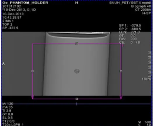
68 Ge-uniform phantom 를 patient table위에 올려놓고 topogram 을 실시하고(Fig. 5), AC-CT를 검사 후 사각 박스에 서 보듯 coverage를 나타내며 holder를 사용하지 않는 경우 pallet 이 AC-CT의 FOV에 포함되지 않았다(Fig. 6).
2) PET 영상 재구성 방법
영상 획득 후 FBP, OSEM, TrueX를 재구성하여 parameters 는 iteration : 4, subsets : 21, gaussian filer : 2 ㎜, 5 ㎜로 설정하였 다. WL : -4200, WW : 1000으로 하여 영상의 균일도를 평가하 였다. 또한 vertical profile를 이용하여 비교하였고 5장과 20장 을 합하여 integral uniformity를 비교하였다.
Fig. 5. Image acquisition after CT Topogram
Fig. 6. Not including pallet of FOV by AC-CT
Fig. 7. Patient table
Fig. 8. Phantom holder
Fig. 9. Patient table
Fig. 10. Phantom holder
결 과
1. Phantom AC CT imagesAC-CT 의 영상을 통하여 holder를 사용하지 않는 경우 FOV 내에 pallet이 모두 포함되지 않음을 알 수 있다(Fig. 8).
2. Phantom PET (corrected) sinogram
PET corrected sinogram 영상에서 holder를 사용하지 않는
경우 truncated region에 의한 defected 부위를 확인할 수 있으
며(Fig. 9), holder를 사용한 경우 uniform한 영상을 확인 할 수
있다(Fig. 10).
Fig. 11. Image of used table
Fig. 12. Image of used holder
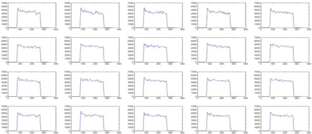
Fig. 13. Profile of used table
3. PET AC image(Table vs Holder)
Holder 유무에 따라서 AC영상을 비교하면 window level 4200, window width 1000 으로 설정 시 FBP, OSEM, TrueX recon 방법 모두에서 holder를 사용한 경우 uniform한 영상이 획득되었지만(Fig. 12), holder를 사용하지 않은 경우 영상하
단에 defect가 관찰 되었다(Fig. 11).
4. Vertical profile with different recon parameter
Vertical profile 은 table을 사용 했을 때 보다 phantom
holder 를 사용했을 시 하단부위의 count defect가 나타나지
Fig. 14. Profile of used holder
Fig. 15. Profile of used table
Fig. 16. Profile of used holder 않았다.
5. Integral uniformity