Vol. 66, No. 6, June, 2004
484
서 론
복강내탈장은 장폐색증 환자의 0.2∼0.9%를 차지하고 있 으며(1) 그 중 십이지장 주위 탈장은 복강내탈장의 30∼
53% 정도를 차지하고 있으나(2) 복강내탈장의 빈도가 매우 드물기 때문에 장폐색증 환자를 진단할 때 그 원인질환으 로 생각되는 경우가 드물다. 그러므로 대부분 증례보고로 문헌에 보고되고 있으며, 증상은 무증상부터 경한 위장장 애, 우상복부동통, 장폐색 및 장 괴사에 의한 심한 복통까지 다양하게 나타난다.(3,4) 명백한 징후나 증상이 없어 수술 전 진단이 거의 불가능할 정도로 진단이 어려우며 진단이 지연될 경우 장이 괴사되어 장절제를 해야 하는 경우가 생 기는 등 환자에게 치명적인 손상을 일으키는 경우가 있다.
이러한 복강내 탈장환자들에 대하여 임상경과를 고찰하고 급성복통을 호소하는 환자들의 진단에 있어서 드문 원인 중의 하나인 복강내 탈장을 시사하는 소견에 대하여 특징 적인 것이 있는지 확인하고 임상경과와 합병증과 관계된 인자가 있는지와 이에 대한 치료방향을 제시하고자 하였 다.
급성 장폐색을 일으킨 십이지장주위탈장의 임상분석
가톨릭대학교 의과대학 의정부성모병원 외과학교실
김기환․윤영철․서학준․김지일․안창혁․진형민․김정수․전해명․임근우
Clinical Analysis of an Acute Intestinal Obstruc- tion with a Paraduodenal Hernia
Kee Hwan Kim, M.D., Young Chul Yoon, M.D., Hak Jun Seo, M.D., Ji Il Kim, M.D., Chang Hyeok Ahn, M.D., Hyung Min Chin, M.D., Jeong Soo Kim, M.D., Hae Myung Jeon, M.D. and Keun Woo Lim, M.D.
Purpose: A paraduodenal hernia is an uncommon congenital disease that manifests as an intestinal obstruction, which may lead to strangulation and, subsequently, result in gan- grene of the intestine. In this retrospective study, 12 para- duodenal hernia cases were evaluated and the clinical prognostic factors investigated.
Methods: Paraduodenal hernias leading to intestinal obstruc- tions were noted in 12 patients. The patients were retro- spectively evaluated with respect to signs and symptoms.
The laboratory and radiological findings, type of operation, time elapsed between the onset of symptoms and laparo- tomy and postoperative complications and hospital stays were also reviewed. The relationships between clinical factors and outcomes were also statistically evaluated.
Results: In our series, postoperative short bowel syndrome was encountered in one patient (case 1) with bowel strangu- lation, but there were no mortalities. The time elapsed between the onset of symptoms and laparotomy was found to be longer in the patients with strangulation than in those without (6.8±4.5 day versus 4.1±3.6 day). Additionally, the postoperative hospital stay was longer in those patients with strangulation (24.4±11.6 day versus 15.3±7.7 day), but the relationship was not statistically significant (P=0.283 and 0.130, respectively).
Conclusion: Since the preoperative diagnosis of a paraduo- denal hernia is very difficult, due to the lack of specific signs and symptoms, the postoperative complications can only be decreased with early surgical intervention in those patients
with an acute intestinal obstruction. Although the postopera- tive morbidity and mortality were not correlated with any of the factors evaluated in this study, further study will be needed to evaluate the significance of the time elapsed between the onset of symptoms and a laparotomy as a prognostic factor. (J Korean Surg Soc 2004;66:484-489) Key Words: Intestinal obstruction, Paraduodenal hernia,
Prognostic factor
중심 단어: 장폐색, 십이지장주위 탈장, 예후인자 ꠏꠏꠏꠏꠏꠏꠏꠏꠏꠏꠏꠏꠏꠏꠏꠏꠏꠏꠏꠏꠏꠏꠏꠏꠏꠏꠏꠏꠏꠏꠏꠏꠏꠏꠏꠏꠏꠏꠏꠏꠏꠏꠏꠏꠏꠏꠏꠏꠏꠏ Department of Surgery, Uijeongbu St. Mary's Hospital, College of Medicine, The Catholic University of Korea, Uijeongbu, Korea
책임저자:김정수, 경기도 의정부시 금오동 65-1
ꂕ 480-130, 가톨릭대학교 의정부성모병원 외과 Tel: 031-820-3048, Fax: 031-847-2717
E-mail: [email protected]
접수일:2004년 1월 20일, 게재승인일:2004년 3월 22일
ꠏꠏꠏꠏꠏꠏꠏꠏꠏꠏꠏꠏꠏꠏꠏꠏꠏꠏꠏꠏꠏꠏꠏꠏꠏꠏꠏꠏꠏꠏꠏꠏꠏꠏꠏꠏꠏꠏꠏꠏꠏꠏꠏꠏꠏꠏꠏꠏꠏꠏꠏꠏꠏꠏꠏꠏꠏꠏꠏꠏꠏꠏꠏꠏꠏꠏꠏꠏꠏꠏꠏꠏꠏꠏꠏꠏꠏꠏꠏꠏꠏꠏꠏꠏꠏꠏꠏꠏꠏꠏꠏꠏꠏꠏꠏꠏꠏꠏꠏꠏꠏꠏꠏꠏꠏꠏꠏꠏꠏꠏꠏꠏꠏꠏꠏ
방 법
가톨릭대학교 의과대학 외과학교실에서 1993년 4월부터 2003년 10월까지 과거력에서 복부수술 경력이 없었던 환자 에서 소장폐색을 일으켜 수술 전이나 수술 후에 십이지장 주위탈장으로 진단된 12명의 환자를 대상으로 하였고 이들 의 임상징후와 증상, 말초혈액학적 소견, 방사선학적 소견, 증상발현 후 수술까지의 기간, 수술소견, 수술방법, 수술 후 합병증과 재원기간에 대해 의무기록을 검토하여 후향적으 로 조사하였다.
결 과
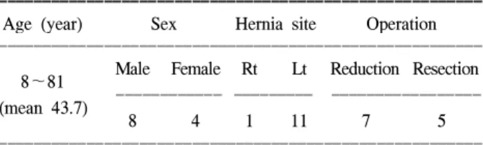
1) 임상적 분석(Table 1)
12명의 환자 중 8명(66.7%)이 남자였고 4명(33.3%)이 여자 였다. 평균연령은 43.7세(8∼81세)였고 소장절제 및 정복술 을 시행한 군은 47.8세로 수술적 정복술만 시행한 군에서의 40.8세보다 약간 나이가 더 많았으나 의의는 없었다. 발생 위치에 따라서 좌측 십이지장 주위 탈장이 11명, 우측 십이
지장 주위 탈장이 1명이었다. 환자의 평균 증상 기간은 2.0 일이었고 모든 환자가 복통 및 복부팽만을 호소하였으며 8명(66.7%)의 환자가 오심과 구토증상을 호소하였다. 오심 및 구토증세가 없이 복통증세만 있는 환자는 4명(33.3%)이 었다. 말초혈액학적 소견에서 혈색소는 평균 15.2 gm/dl (12.4∼20.6 gm/dl), 헤마토크리트는 평균 44.4% (37.2∼60.1%) 를 보였고 일부 환자에서 혈액농축(hemoconcentration) 양상 을 보였으며 그 외의 혈액학적 소견에서 특이소견은 보이 지 않았다.
2) 진단방법
8명(61.6%)의 환자에서 복부전산화단층촬영을 시행하였
Table 2. Summary of clinical data in all paraduodenal patients
ꠚꠚꠚꠚꠚꠚꠚꠚꠚꠚꠚꠚꠚꠚꠚꠚꠚꠚꠚꠚꠚꠚꠚꠚꠚꠚꠚꠚꠚꠚꠚꠚꠚꠚꠚꠚꠚꠚꠚꠚꠚꠚꠚꠚꠚꠚꠚꠚꠚꠚꠚꠚꠚꠚꠚꠚꠚꠚꠚꠚꠚꠚꠚꠚꠚꠚꠚꠚꠚꠚꠚꠚꠚꠚꠚꠚꠚꠚꠚꠚꠚꠚꠚꠚꠚꠚꠚꠚꠚꠚꠚꠚꠚꠚꠚꠚꠚꠚꠚꠚꠚꠚꠚꠚꠚꠚꠚꠚꠚꠚꠚꠚꠚꠚꠚ Time elapsed
between the onset Hospital
Case Age/sex Initial symptoms CT Operation Complication
of symptoms and stay (day)
laparotomy (day)
ꠏꠏꠏꠏꠏꠏꠏꠏꠏꠏꠏꠏꠏꠏꠏꠏꠏꠏꠏꠏꠏꠏꠏꠏꠏꠏꠏꠏꠏꠏꠏꠏꠏꠏꠏꠏꠏꠏꠏꠏꠏꠏꠏꠏꠏꠏꠏꠏꠏꠏꠏꠏꠏꠏꠏꠏꠏꠏꠏꠏꠏꠏꠏꠏꠏꠏꠏꠏꠏꠏꠏꠏꠏꠏꠏꠏꠏꠏꠏꠏꠏꠏꠏꠏꠏꠏꠏꠏꠏꠏꠏꠏꠏꠏꠏꠏꠏꠏꠏꠏꠏꠏꠏꠏꠏꠏꠏꠏꠏꠏꠏꠏꠏꠏꠏ
Abdominal clamp Short bowel
1 8/F 1 Midgut volvulus Resection 43
nausea syndrome
Abdominal clamp
2 73/M 3 Left paraduodenal Resection 11 None
nausea Abdominal clamp
3 M 10 Strangulation Resection 23 None
nausea Abdominal clamp
4 73/M 11 Not done Resection 22 None
nausea Abdominal clamp
5 31/M 9 Internal hernia Resection 23 None
nausea
ꠏꠏꠏꠏꠏꠏꠏꠏꠏꠏꠏꠏꠏꠏꠏꠏꠏꠏꠏꠏꠏꠏꠏꠏꠏꠏꠏꠏꠏꠏꠏꠏꠏꠏꠏꠏꠏꠏꠏꠏꠏꠏꠏꠏꠏꠏꠏꠏꠏꠏꠏꠏꠏꠏꠏꠏꠏꠏꠏꠏꠏꠏꠏꠏꠏꠏꠏꠏꠏꠏꠏꠏꠏꠏꠏꠏꠏꠏꠏꠏꠏꠏꠏꠏꠏꠏꠏꠏꠏꠏꠏꠏꠏꠏꠏꠏꠏꠏꠏꠏꠏꠏꠏꠏꠏꠏꠏꠏꠏꠏꠏꠏꠏꠏꠏ
6 11/M Abdominal clamp 4 Not done Reduction 10 None
Abdominal clamp
7 81/F 2 Not done Reduction 21 None
nausea
8 20/M Abdominal clamp 2 Right paraduodenal Reduction 10 None
9 28/F Abdominal clamp 11 Not done Reduction 27 None
10 57/F Abdominal clamp 2 Internal hernia Reduction 12 None
Abdominal clamp
11 45/M 7 Internal hernia Reduction 21 None
nausea Abdominal clamp
12 44/M 1 Left paraduodenal Reduction 10 None
nausea
ꠏꠏꠏꠏꠏꠏꠏꠏꠏꠏꠏꠏꠏꠏꠏꠏꠏꠏꠏꠏꠏꠏꠏꠏꠏꠏꠏꠏꠏꠏꠏꠏꠏꠏꠏꠏꠏꠏꠏꠏꠏꠏꠏꠏꠏꠏꠏꠏꠏꠏꠏꠏꠏꠏꠏꠏꠏꠏꠏꠏꠏꠏꠏꠏꠏꠏꠏꠏꠏꠏꠏꠏꠏꠏꠏꠏꠏꠏꠏꠏꠏꠏꠏꠏꠏꠏꠏꠏꠏꠏꠏꠏꠏꠏꠏꠏꠏꠏꠏꠏꠏꠏꠏꠏꠏꠏꠏꠏꠏꠏꠏꠏꠏꠏꠏ Table 1. Patients characteristics (n=12)
ꠚꠚꠚꠚꠚꠚꠚꠚꠚꠚꠚꠚꠚꠚꠚꠚꠚꠚꠚꠚꠚꠚꠚꠚꠚꠚꠚꠚꠚꠚꠚꠚꠚꠚꠚꠚꠚꠚꠚꠚꠚꠚꠚꠚꠚꠚꠚꠚꠚꠚꠚꠚꠚꠚꠚ Age (year) Sex Hernia site Operation ꠏꠏꠏꠏꠏꠏꠏꠏꠏꠏꠏꠏꠏꠏꠏꠏꠏꠏꠏꠏꠏꠏꠏꠏꠏꠏꠏꠏꠏꠏꠏꠏꠏꠏꠏꠏꠏꠏꠏꠏꠏꠏꠏꠏꠏꠏꠏꠏꠏꠏꠏꠏꠏꠏꠏ
Male Female Rt Lt Reduction Resection 8∼81 ꠏꠏꠏꠏꠏꠏꠏꠏꠏꠏꠏꠏ ꠏꠏꠏꠏꠏꠏꠏꠏꠏ ꠏꠏꠏꠏꠏꠏꠏꠏꠏꠏꠏꠏꠏꠏꠏꠏꠏ (mean 43.7)
8 4 1 11 7 5
ꠏꠏꠏꠏꠏꠏꠏꠏꠏꠏꠏꠏꠏꠏꠏꠏꠏꠏꠏꠏꠏꠏꠏꠏꠏꠏꠏꠏꠏꠏꠏꠏꠏꠏꠏꠏꠏꠏꠏꠏꠏꠏꠏꠏꠏꠏꠏꠏꠏꠏꠏꠏꠏꠏꠏ
ꠏꠏꠏꠏꠏꠏꠏꠏꠏꠏꠏꠏꠏꠏꠏꠏꠏꠏꠏꠏꠏꠏꠏꠏꠏꠏꠏꠏꠏꠏꠏꠏꠏꠏꠏꠏꠏꠏꠏꠏꠏꠏꠏꠏꠏꠏꠏꠏꠏꠏꠏꠏꠏꠏꠏꠏꠏꠏꠏꠏꠏꠏꠏꠏꠏꠏꠏꠏꠏꠏꠏꠏꠏꠏꠏꠏꠏꠏꠏꠏꠏꠏꠏꠏꠏꠏꠏꠏꠏꠏꠏꠏꠏꠏꠏꠏꠏꠏꠏꠏꠏꠏꠏꠏꠏꠏꠏꠏꠏꠏꠏꠏꠏꠏꠏ 고 이 검사에서 십이지장 주위 탈장으로 진단된 경우가 3명
(37.5%)이었으며 3명(37.5%)에서 복강내 탈장으로, 그 외 중장 염전, 소장 감돈으로 진단된 경우가 각각 1예였다. 환 자 중 2명(16.6%)에서 복부초음파를 시행하였는데 복강내 탈장을 진단한 예는 없었다. 4명(33.3%)의 환자에서 임상증 상과 복부단순촬영만으로 수술을 시행하여 십이지장주위 탈장을 진단하였다.
3) 수술방법, 증상발현 후 수술까지의 시간, 수술 후 합병증과 재원기간(Table 2)
수술을 시행한 12명의 환자 중 5명(41.6%)은 이미 소장의 일부가 괴사되어 소장절제 및 정복술을 시행하였고 나머지 7명(58.3%)에서는 수술적 정복술만을 시행하였다. 증상발 현 후 수술까지의 시간은 전체 12명의 환자에서 평균 5.2±
4.1일이 소요되었고 소장절제 및 정복술을 시행한 군에서 는 평균 6.8±4.5일, 수술적 정복술만 시행한 군에서 평균 4.1±3.6일로서 소장절제 및 정복술을 시행한 군에서 수술 까지의 기간이 길었으나 통계학적인 유의성은 없었다(P=
0.283). 소장절제 및 정복술을 시행한 군에서 1명(20%)에서 단장증후군의 합병증을 보였다. 재원일수에서 소장절제 및 정복술을 시행한 군에서는 평균재원일수 24.4±11.6일, 수 술적 정복술만 시행한 군에서의 평균재원일수는 15.3±7.7 일로서 소장절제 및 정복술을 시행한 군에서의 재원기간이 길었으나 통계학적인 유의성은 없었다(P=0.130).
4) 통계적 분석
모든 자료는 SPSS 11.0을 사용하여 분석하였다. 두 군 간 의 자료비교는 independent t-test를 이용하였다. 모든 통계는 P값이 0.05 미만인 경우를 의의있다고 판정하였다.
고 찰
십이지장 주위탈장(paraduodenal hernia)은 선천성 결장간 막 탈장(congenital mesocolic hernia), mesentericoparietal her- nia 그리고 후복막탈장(retroperitoneal hernia) 등으로 불린다.
태생기에 중장의 장회전의 이상으로 발생하는 선천성질환 으로 복강내 탈장의 30∼53%를 차지한다.(2) 이 질환은 문 헌에 의하면 남녀비가 3:1로 남자에게 주로 호발한다고 되어 있고 일반적으로 30∼50대에서 증상이 나타난다고 하 며, 일부 이전에 발견되기도 하지만 평균진단 연령은 38.5 세로 되어 있다.(5-9) 저자들의 연구 결과에서도 남자가 66.6% (8/12)로 여성에 비해 많았으며 평균 연령은 43.7세로 다른 연구 결과에 비해 많은 편이었다. 연령분포는 8세에서 81세까지 다양한 분포를 보이고 있었다. 또한 좌십이지장 주위 탈장이 우십이지장 주위 탈장에 비해 3배 정도 호발한 다고 하며,(9) 본 연구에서도 좌십이지장 주위 탈장이 12명 중 11명을 차지하였다.
발생기전에 대해서 알아보면 1923년 Andrews(10)가 처음 으로 태생기에 장 회전의 이상에 의한 것이라고 기술하였 다. 십이지장주위 탈장은 세 가지 유형으로 분류하여 좌측 십이지장주위 탈장, 우측 십이지장주위 탈장 및 횡행결장 간막 탈장으로 구별하였고,(11) 좌측 십이지장주위 탈장이 75%를 차지한다. 좌측과 우측의 탈장은 각각 발생기전이 다르며 발생학적으로 태생 5주 정도에 위장관은 배측 장간 막(dorsal mesentery)에 지지되어 있는 곧은 관으로 나타나기 시작하는데, 상장간막동맥(superior mesentery artery)의 혈액 공급을 받는 중장(midgut)은 곧 자라나서 태아의 복부 밖까 지 위치하게 된다. 중장은 동맥전분절(prearterial segment) 과 동맥후분절(postarterial segment), 두 가지 부위로 각각 나 뉘는데, 동맥전분절은 공장과 대부분의 회장으로 발생하고 동맥후분절은 회장말단, 맹장, 상행결장 및 횡행결장의 일 부로 발생한다. 태생기 동안 시계 반대방향으로 장 회전이 이루어져 맹장이 우하복부에 위치하고 상행 결장이 후복막 에 고정이 되며 소장은 상장간막동맥의 하좌측 부위에 장 간막이 후복벽에 고정되어 지지하면서 위치하게 된다. 우 측 십이지장주위 탈장은 위와 같은 태생기의 정상 장회전 과정 중 동맥후분절의 회전은 정상적으로 이루어지지만 동 맥전분절의 회전이 이루어지지 않아서 소장의 대부분이 우 측 결장장간막후방의 우측 복부에 위치하게 되어 발생하 며, 좌측 십이지장주위 탈장은 장 회전 동안 소장이 좌측결 장장간막 내에 형성된 낭(Landzert's fossa)에 포획(entrap- ment)되어 발생된다.(5,10-13) 이처럼 우측 십이지장주위 탈 장은 회전이상으로 발생하고 좌측 십이지장주위 탈장은 회 전 중에 장이 포획되어 발생한다.
환자의 증상은 오심, 구토, 간헐적인 식후 복부 불편감, 만성 복부 동통, 체중감소 등 모호한 증상부터 급성 장폐색 증상까지 다양하게 나타날 수 있다.(3,4) 문헌에 의하면 환 자가 호소하는 증상과 장폐색의 정도는 직접적인 연관성이 있다.(9) 이러한 장폐색의 진행정도에 따른 장의 손상결과 가 소장절제 여부와도 연관성이 있다고 하겠다. 저자들의 연구에서는 모든 환자들이 복통과 복부 팽만을 호소하였고 아울러 66.7% (8/12)의 환자가 오심 및 구토증세를 호소하 였다. 이 중 수년 간의 간헐적인 복부 불편감을 호소했던 환자(증례 6)도 있었고 그러한 증상없이 갑자기 복통을 주 소로 내원한 환자도 있었다. 물론 증상만으로 감별해야 할 질환으로는 담도 질환, 위염, 위식도역류 등이 있기 때문에 수술 전 감별을 위한 여러 가지 검사방법을 이용하여야 하 겠다.
진단은 복부단순촬영, 소장조영술, 복부 전산화 단층촬영 등이 있는데 복부단순촬영에서는 소장에 가스가 차 있는 확장된 분절 등의 소견이 보일 수 있고, 소장 조영술에서 정중선 좌측 혹은 우측에 국한된 소장을 발견할 수 있으며, 복부 전산화 단층 촬영에서는 좌십이지장 주위 탈장은 위 와 췌장 사이에 낭에 감싸인 소장 고리를 관찰할 수 있고
ꠏꠏꠏꠏꠏꠏꠏꠏꠏꠏꠏꠏꠏꠏꠏꠏꠏꠏꠏꠏꠏꠏꠏꠏꠏꠏꠏꠏꠏꠏꠏꠏꠏꠏꠏꠏꠏꠏꠏꠏꠏꠏꠏꠏꠏꠏꠏꠏꠏꠏꠏꠏꠏꠏꠏꠏꠏꠏꠏꠏꠏꠏꠏꠏꠏꠏꠏꠏꠏꠏꠏꠏꠏꠏꠏꠏꠏꠏꠏꠏꠏꠏꠏꠏꠏꠏꠏꠏꠏꠏꠏꠏꠏꠏꠏꠏꠏꠏꠏꠏꠏꠏꠏꠏꠏꠏꠏꠏꠏꠏꠏꠏꠏꠏꠏ (Fig. 1) 우십이지장 주위 탈장에서는 상장간막동맥 뒤쪽에
공장 동․정맥 가지의 고리 모양을 관찰할 수 있다(Fig.
2).(4,6,7,14) 이외 동맥혈관조영술 및 초음파가 이용될 수 있다. 수술 전 진단으로 복부 전산화 단층 촬영이 매우 유용 하여 우십이지장 주위 탈장의 경우에는 우측 중간복부에서 우대장정맥을 전방으로 전위시키는 낭으로 둘러싸인 소장 고리가 보이고 이 소장고리가 우결장간막의 후방에 위치하 면서 소장장간막의 기시부에서 상장간막동맥과 상장간막 정맥주위근처에서 소장고리를 형성한다. 또한 회전이상으 로 상장간막정맥의 위치가 상장간막동맥의 좌측과 복측으 로 위치하고 정상적인 평행주행하는 십이지장이 보이지 않 는다. 좌십이지장 주위 탈장의 경우에는 낭으로 둘러싸인
소장고리가 하장간막정맥을 전방으로 전위시키면서 좌결 장간막의 후방에 위치하면서 낭으로 둘러싸인 소장고리는 확장되어있는 소견을 보인다. 이 소장고리가 교액상태에 빠지면 때때로 소장장간막의 울혈을 보인다.(14) 본 연구에 서는 4명(33.3%)의 환자에서 임상증상 및 복부단순촬영만 으로 수술을 시행하여 십이지장 주위탈장으로 진단되었다.
복부 전산화 단층촬영에서 66.7% (8/12)의 환자에서 시행 후 복강내 탈장으로 진단하여 수술 후 십이지장 주위 탈장 으로 확진되었다. 복부초음파는 십이지장 주위 탈장의 진 단에는 도움을 주지 못했다. 그러나 실제적으로 수술 전 진 단은 매우 어려운 것으로 보고되고 있다.(15)
Fig. 3. Surgical photograph shows left paraduodenal hernia (A) (case 12), and surgical photograph obtained after withdrawal of the herniated bowel loops shows the hernial hiatus (B). (*)(Landzert's fossa), PJ = proximal jejunum withdrawn from the sac, T = Treitz ligament.
A B
Transverse colon
Hernia sac
PJ
T
*
Inferior mesenteric vein
Incision line
Fig. 1. Left-sided paraduodenal hernia in a 43-year-old man (case 12) with acute abdominal pain. Transverse CT scan shows a saclike mass of jejunal loops (white arrows) in the left upper quadrant interposed between the pancreas (P) and stomach (S).
S P
Fig. 2. Right-sided paraduodenal hernia in a 20-year-old man (case 8) with acute abdominal pain. Transverse CT scan shows a saclike mass of small bowel loops and diffuse dilatation of small bowel loops with abrupt narrowing of proximal ileum (arrow).
ꠏꠏꠏꠏꠏꠏꠏꠏꠏꠏꠏꠏꠏꠏꠏꠏꠏꠏꠏꠏꠏꠏꠏꠏꠏꠏꠏꠏꠏꠏꠏꠏꠏꠏꠏꠏꠏꠏꠏꠏꠏꠏꠏꠏꠏꠏꠏꠏꠏꠏꠏꠏꠏꠏꠏꠏꠏꠏꠏꠏꠏꠏꠏꠏꠏꠏꠏꠏꠏꠏꠏꠏꠏꠏꠏꠏꠏꠏꠏꠏꠏꠏꠏꠏꠏꠏꠏꠏꠏꠏꠏꠏꠏꠏꠏꠏꠏꠏꠏꠏꠏꠏꠏꠏꠏꠏꠏꠏꠏꠏꠏꠏꠏꠏꠏ
수술방법에 있어서 좌측 십이지장주위 탈장의 수술방법 은 낭의 우측에 있는 하부 장간막정맥의 우측 변연을 따라 서 절개를 하고 소장을 하부 장간막정맥의 하부로 환원시 키고 낭의 확장된 경부를 폐쇄하기 위해 정맥에 인접한 복 막을 후복막에 봉합하는 것이다(Fig. 3).(1,5,16,17) 또한 우 측 십이지장주위 탈장의 경우 탈장된 소장을 환원 정복하 기 위하여 상행결장의 측부 복막을 절개하여 중장의 발생 학적 과정 제1기 말기형태, 즉 정중선의 우측에 십이지장, 공장, 회장 등을 위치시키고, 말단회장, 맹장 및 대장 등은 정중선의 좌측에 놓이게 하는 것이다(Fig. 4).(1,5,16) 본 저 자들의 대상군에서는 41.6% (5/12)에서 개복 시 이미 소장 괴사를 동반하였기 때문에 소장절제를 시행하였다. 증상발 현 후 수술까지의 기간은 소장절제 및 정복술을 시행한 군 에서는 평균 6.8±4.5일, 수술적 정복술만 시행한 군에서 평 균 4.1±3.6일로 소장절제 및 정복술을 시행한 군에서 수술 까지의 기간이 더 지연된 경향을 보이나 통계학적인 유의 성은 없었다(P=0.283). 소장절제 및 정복술을 시행한 군이 나 수술적 정복술을 시행한 군에서 증상발현 후 수술까지 짧게는 1일에서 길게는 11일로 두 군은 동일기간 내에 있었 다. 단장증후군의 중대 합병증이 생긴 8세 여아(증례 1)는 증상발생 후 1일로 바로 응급수술을 시행하였으나 이미 장 괴사가 진행되어 대량 소장절제를 시행하였다. 또한 28세 여자환자(증례 9)는 증상발현 후 11일 경과 후 수술을 시행 하였으나 장절제 없이 수술적 정복술만을 시행하여 치료된 경우도 있어 증상발현 후 수술까지의 시간이 장절제의 중 요인자로 생각되지만 증례 1이나 증례 9에서처럼 상반된 결과를 보여 증상발현에서 수술까지의 시간 외의 다른 인
자의 가능성이 있으나 정확한 이유는 설명할 수가 없었다.
이러한 결과를 볼 때 급성 장폐색 환자에서 수술경력이 없 다고 하여 보존적 치료를 시행하여 수술이 지연될 경우 환 자에게 치명적인 손상이 올 수 있으므로 신속하게 복부전 산화단층촬영 및 반복적인 이학적검사 등을 시행하여 수술 의 적절한 시기를 선택해야 한다. 소장절제 및 정복술을 시 행한 군에서는 평균재원일수가 24.4±11.6일, 수술적 정복술 만 시행한 군에서의 평균재원일수는 15.3±7.7일로 소장절 제 및 정복술을 시행한 군에서의 재원기간이 더 길었으나 통계학적인 유의성은 보이지 않았다(P=0.130). 증상발현에 서 수술까지의 시간과 평균재원일수에서 수치상으로는 소 장절제 및 정복술을 시행한 군에서 각각의 인자에서 차이 가 있었으나 통계적으로 의의가 없는 것은 대상군의 수가 적은 것이 이유로 생각이 되며 대상군의 수를 확대하고 추 적연구를 계속해야 할 것이다. 또한 합병증을 유발할 수 있 는 상황으로 수술 당시 이 질환에 대한 경험이 없는 경우 탈장낭 절개 시 탈장 낭벽의 장간막 동맥이나 정맥을 절단 할 위험이 있고, 또한 주요 장간막 혈관을 함유한 새로운 복강내 탈장의 형성 또는 장간막 결손을 조장할 수 있기 때문에 수술 시 맹목적인 탈장낭의 절개는 피해야 한다.
Cutler와 Scott(18)은 감돈된 소장의 장절제를 시행한 군에 서의 사망률이 32%로 수술적 정복술만 시행한 군에서의 사 망률 16%에 비해 사망률이 높은 결과를 보고하면서 증상발 현 후 수술까지의 소요된 시간이 장생동성의 중요한 인자 임을 강조하였고, 초기검사 시 조기진단과 조기치료의 중 요성을 강조하였다. 또한 장폐색으로 인한 장허혈이 진행 된 경우 응급수술적 정복술을 시행하여도 50% 이상의 사망 률을 보인다.(19,20) 본 연구에서는 사망한 경우는 없었으나 1예(증례 1)에서 단장증후군의 합병증이 생겨서 고위 영양 요법을 시행하여야 하였다.
이러한 결과를 볼 때 수술경력이 없는 환자에서의 소장 폐색의 원인의 하나로 복강내탈장 특히 십이지장주위 탈장 유무를 감별해야 하며 소장폐색환자의 원인에 대한 검사방 법으로 복부전산화단층촬영이 도움이 되었다. 그러나 진단 이 모호한 경우에는 임상증상과 수술자의 판단에 의해 결 정되는 경우가 있었다. 수술 후 장절제를 시행한 군에서 재 원기간이 길었고 심한 합병증으로 단장증후군 같은 영구적 인 장애를 유발할 수 있음을 인지하여 소장폐색증 원인의 하나로서 이 질환에 대한 각별한 관심을 가져야 할 것으로 생각한다.
결 론
십이지장주위 탈장은 모호한 증상으로 합병증이 많고 또 한 진단이 지연될 경우 유병률과 사망률이 증가하는 경우 가 있는 질환의 하나이므로 초기 진단 시 복부전산화단층 촬영을 반드시 시행하고 임상증상에 대한 반복적인 이학적 Fig. 4. Surgical photograph shows Ladd's band within herniated
small bowel loops in right-sided paraduodenal hernia patient (case 8).
. Ascending colon
Ladd's bands
Incision line
Herniated small bowel
ꠏꠏꠏꠏꠏꠏꠏꠏꠏꠏꠏꠏꠏꠏꠏꠏꠏꠏꠏꠏꠏꠏꠏꠏꠏꠏꠏꠏꠏꠏꠏꠏꠏꠏꠏꠏꠏꠏꠏꠏꠏꠏꠏꠏꠏꠏꠏꠏꠏꠏꠏꠏꠏꠏꠏꠏꠏꠏꠏꠏꠏꠏꠏꠏꠏꠏꠏꠏꠏꠏꠏꠏꠏꠏꠏꠏꠏꠏꠏꠏꠏꠏꠏꠏꠏꠏꠏꠏꠏꠏꠏꠏꠏꠏꠏꠏꠏꠏꠏꠏꠏꠏꠏꠏꠏꠏꠏꠏꠏꠏꠏꠏꠏꠏꠏ 검사로서 소장폐색의 원인에 대한 신속한 진단과 수술을
시행하여 장괴사 등의 합병증을 줄임으로써 유병률과 사망 률을 감소시켜야 할 것이다.
REFERENCES
1) Bartlett MK, Wang CA, Williams WH. The surgical manage- ment of paraduodenal hernia. Ann Surg 1968;168:249-54.
2) Brigham RA, d'Avis JC. Paraduodenal hernia. In: Nyhus LM, Condon RE, eds. Hernia. 3rd Ed. Philadelphia: JB Lippincot;
1989, p.481-6.
3) Tesson JA. Left paraduodenal hernia. Am J Surg 1957;93:
470-2.
4) Blachar A, Federle MP. Internal henia: An increasingly com- mon cause of small bowel obstruction. Seminars in Ultrasond, CT, and MRI 2002;23:174-83.
5) Newsom BD, Kukora JS. Congenital and acquired internal hernias: Unusual cause of small bowel obstruction. Am J Surg 1986;152:279-85.
6) Warshauer DM, Mauro MA. CT diagnosis of paraduodenal hernia. Gastrointest Radiol 1992;17:13-5.
7) Harbin WP. Computed tomographic diagnosis of internal hernia. Radiololgy 1982;143:736-40.
8) Berardi RS. Paraduodenal hernias. Surg Gynecol Obstet 1981;152:99-110.
9) Khan MA, Lo AY, Vande Maele DM. Paraduodenal hernia.
Am Surg 1998;64:1218-22.
10) Andrews E. Duodenal hernia: A misnomer. Surg Gynecol
Obstet 1923;37:740-50.
11) Willwerth BM, Zollinger RM, Izant RJ. Congenital mesocolic (paraduodenal) hernia: Embryologic basis of repair. Am J Surg 1974;128:358-61.
12) Zimmerman LM, Laufman H. Intra-abdominal hernias due to developmental and rotational anomalies. Ann Surg 1953;138:
82-5.
13) Callander LC, Rusy GY, Nemir A. Mechanisms, symptoms and treatment of hernia into the descending mesocolon (left paraduodenal hernia). Surg Gynecol Obstet 1935;60:1052-71.
14) Okino Y, Kiyosue H, Mori H, Komatsu E, Matsumoto S, Yamada Y, et al. Root of the small-bowel mesentery: Correla- tive anatomy and CT features of pathologic conditions.
Radiographics 2001;21:1475-90.
15) Yoo HY, Mergelas J, Seibert DG. Paraduodenal hernia: A treatable cause of upper gasrointestinal tract symptoms. J Clin Gastroenterol 2000;31:226-9.
16) Brigham RA, Fallon WF, Saunders JR, Harmon JW, d'Avis JC. Paraduodenal hernia: Diagnosis and surgical management.
Surgery 1984;96:498-502.
17) Davis R. Surgery of left paraduodenal hernia. Am J Surg 1975;129:570-3.
18) Cutler GD, Scott HW. Transmesenteric hernia. Surg Gynecol Obstet 1944;79:509-15.
19) Freund H, Berlatzky Y. Small paraduodenal hernias. Arch Surg 1977;112:1180-3.
20) Cogswell HS, Thomas CA. Right paraduodenal hernia. Ann Surg 1941;114:1035-41.