pISSN: 2287-4208 / eISSN: 2287-4690
World J Mens Health 2013 December 31(3): 265-267
http://dx.doi.org/10.5534/wjmh.2013.31.3.265
Case Report
Received: Jun 19, 2013; Revised: Jul 10, 2013; Accepted: Jul 18, 2013 Correspondence to: Gilho Lee
Department of Urology, Dankook University Medical College, 119 Dandae-ro, Dongnam-gu, Cheonan 330-714, Korea.
Tel: +82-41-550-3963, Fax: +82-41-551-6630, E-mail: [email protected] Copyright © 2013 Korean Society for Sexual Medicine and Andrology
This is an Open Access article distributed under the terms of the Creative Commons Attribution Non-Commercial License (http://creativecommons.
org/licenses/by-nc/3.0) which permits unrestricted non-commercial use, distribution, and reproduction in any medium, provided the original work is properly cited.
An Unusual Presentation of Crossed Testicular Ectopia as an Incarcerated Inguinal Hernia
Yoonjoon Park1, Gilho Lee2
Departments of 1Pediatric Surgery and 2Urology, Dankook University Medical College, Cheonan, Korea
Crossed testicular ectopia (CTE) is generally defined as both testes located in the same hemiscrotum and contralateral hydrocele with the absence of a testis. However, the initial presentation of CTE in an infant as an incarcerated inguinal hernia is extremely rare. We report on a 10-month-old infant with CTE, who visited the emergency room presenting with a left incarcerated inguinal hernia. After manual reduction for an incarcerated hernia and left inguinal herniorraphy, the left testis was fixed into the left hemiscrotum and right transseptal orchiopexy was performed.
Key Words: Crossed ectopia; Hernia, inguinal; Testis
The common characteristics of crossed testicular ectopia (CTE) are ipsilateral cryptorchidism or ectopic gonads and contralateral hydrocele with the absence of a testis [1-7].
In addition, hypospadias, pseudohermaphroditism, and scrotal abnormalities can also occur together [1,6]. Even though about 100 cases have been reported in the world literature, infantile CTE cases initially presenting as an in- carcerated inguinal hernia are very rare.
CASE REPORT
A healthy 10-month-old boy was admitted to the emer- gency room with a painful swelling in the left inguinal region. Physical examination showed a tender 2-cm lump in the left inguinal canal. Inguino-scrotal ultrasonography in this patient showed a left inguinal hernia with two gonads in the left scrotal sac and an impalpable right non-scrotal testis
(Fig. 1A, 1B). In addition, we could not find evidence of per- sistent Müllerian duct syndrome, which is characterized by the presence of a uterus and fallopian tubes in abdominal ultrasonography. There was no evidence of developmental malformations of the external genitalia, such as hypo- spadias, bifid scrotum, or micropenis. After urgent manual reduction for an incarcerated inguinal hernia, left inguinal exploration was undertaken. A thin-walled inguinal hernial sac was found, containing two normal-sized and nor- mal-shaped testes that were attached to one gubernaculum from the left inguinal ring (Fig. 1C). Each had its own blood supply and had separate vasa deferentia. After left inguinal herniorrhaphy, the gubernaculum was divided to strength- en each of the spermatic cord without creating undue ten- sion on either side. The left testis was placed into the left sub- dartos space and the ectopic right testis was fixed trans- septally into the right subdartos space.
266 World J Mens Health Vol. 31, No. 3, December 2013
Fig. 1. (A) Scrotal sonography reveals the dilated bowel loop and mesenteric fat (M) in the left hemiscrotum. (B) Two testes in the left hemiscrotum. (C) Both testes are attached to one guber- naculum (**). A ligated suture (*) for the inguinal hernial sac and normally developed penis (***) are visible. T: testis, LT: left testis, RT: tight testis.
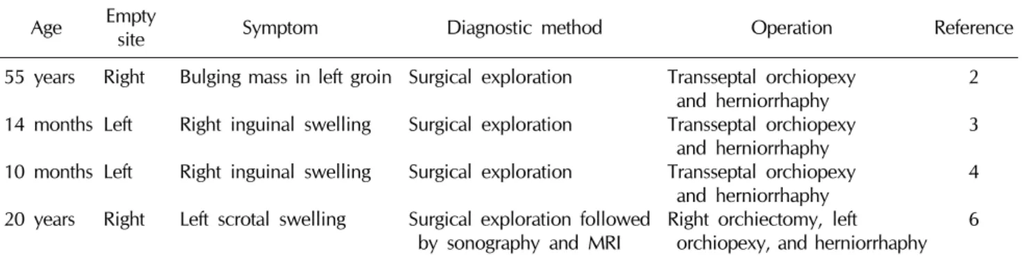
Table 1. Summary of the cases of crossed testicular ectopia as an incarcerated inguinal hernia
Age Empty
site Symptom Diagnostic method Operation Reference
55 years 14 months 10 months 20 years
Right Left Left Right
Bulging mass in left groin Right inguinal swelling Right inguinal swelling Left scrotal swelling
Surgical exploration Surgical exploration Surgical exploration
Surgical exploration followed by sonography and MRI
Transseptal orchiopexy and herniorrhaphy Transseptal orchiopexy and herniorrhaphy Transseptal orchiopexy and herniorrhaphy Right orchiectomy, left orchiopexy, and herniorrhaphy
2 3 4 6
MRI: magnetic resonance imaging.
DISCUSSION
This uncommon anomaly can occur together with per- sistent Müllerian duct syndrome or the presence of male pseudohermaphroditism, and an abnormal karyotype [1,6]. CTE has been classified into three types, based upon the presence of associated developmental abnormalities [1,2]. Type I, the most common, is associated with an in- direct inguinal hernia. Type II is associated with the pres- ence of persistent Müllerian duct structures, such as a rudi- mentary uterus or fallopian tubes. Type III is associated with other genitourinary abnormalities, such as hypo- spadias or scrotal abnormalities [1]. In this case, we could not find any ambiguity in genital phenotypes or any trace of a uterus and fallopian tubes in the pelvic cavity by ultrasonography. Furthermore, the architecture of both of the kidneys and the spleen was normal. On the basis of these findings, our case can be classified as type I.
CTE cases have rarely been reported in the English-lan-
guage literature worldwide [1-7]. Our case is particularly notable because of the unusual presentation of CTE as an incarcerated inguinal hernia. To our knowledge, the pres- ent case represents the third case of CTE diagnosed in an infant with an incarcerated inguinal hernia in the world (Table 1) [3,4].
In most reported cases, the correct diagnosis of CTE is not appropriately made preoperatively because of its rarity [5]. Furthermore, because incarcerated inguinal hernia like this case should usually be immediately managed, a complete evaluation is usually not possible [2-4,6]. For this reason, the correct diagnosis is not easily made preoperatively. In our case, ultrasonography was very helpful for the diagnosis of CTE and for deciding upon the proper management [7].
Surgical procedures for contralateral orchiopexy have been described; contralateral orchiopexy can be per- formed through the scrotal septum (transseptal orchi- opexy) [3,4] or through a suprapubic subcutaneous tunnel
Yoonjoon Park and Gilho Lee: Testicular Ectopia as an Incarcerated Hernia 267
(through their respective superficial inguinal rings) [5].
Because the incidence of testicular cancer generally in- creases in fixed testes, careful follow-ups are imperative [8].
REFERENCES
1. Esteves E, Pinus J, Maranhão RF, Abib Sde C, Pinus J.
Crossed testicular ectopia. Sao Paulo Med J 1995;113:935-40.
2. Kuwayama DP, Peterson JE. Transverse testicular ectopia in a fertile elderly male presenting with incarcerated inguinosc- rotal hernia. Hernia 2008;12:313-5.
3. Tatli D, Numanoglu KV. Transverse testicular ectopia asso- ciated with incarcerated inguinal hernia: a case report. Cases J 2008;1:200.
4. Vaos G, Zavras N. Irreducible inguinal hernia due to crossed testicular ectopia in an infant. Hernia 2004;8:397-8.
5. Pandey A, Gupta DK, Gangopadhyay AN, Sharma SP. Misdi- agnosed transverse testicular ectopia: a rare entity. Hernia 2009;13:305-7.
6. Kaul A, Srivastava KN, Rehman SM, Goel V, Yadav V.
Persistent Müllerian duct syndrome with transverse testicular ectopia presenting as an incarcerated inguinal hernia. Hernia 2011;15:701-4.
7. Nam YS, Baik HK, Kim SJ, Lee HK, Park HK. Transverse tes- ticular ectopia found by preoperative ultrasonography. J Korean Med Sci 1998;13:328-30.
8. Walsh TJ, Dall'Era MA, Croughan MS, Carroll PR, Turek PJ.
Prepubertal orchiopexy for cryptorchidism may be associated with lower risk of testicular cancer. J Urol 2007;178:1440-6.