ANTENATAL SONOGRAPHIC FEATURES OF INTESTINAL NEURONAL DYSPLASIA TYPE A ASSOCIATED WITH
POLYDACTYLY AND MICROMELIA
Hyun Joo Son, MD
1, Yun Sung Jo, MD
1, Ji-Han Jung, MD
2, Dong Gyu Jang, MD
1, Guisera Lee, MD
1Departments of 1Obstetrics and Gynecology , 2Pathology, St. Vincent Hospital, The Catholic University of Korea School of Medicine, Seoul, Korea
Type A intestinal neuronal dysplasia is a congenital abnormality that is a very rare disease. Here, we report on a patient who had intestinal dilatation with serial changes and polydactyly, as shown on prenatal ultrasound. Bowel obstruction symptoms were shown at 16 days of life. An open abdominal surgery was performed. Malrotation of the bowel and narrowing of the area from 15 cm above the ileocecal valve were noted. Therefore, a right hemicolectomy, including lesions was performed. The patient was diagnosed with type A intestinal neuronal dysplasia by pathology.
Keywords:
Intestinal neuronal dysplasia; Polydactyly; Micromelia; Ultrasonography
Received: 2011.10.5. Revised: 2012.1.28. Accepted: 2012.3.4.
Corresponding author: Guisera Lee, MD
Department of Obstetrics and Gynecology, St. Vincent’s Hospital, Th e Catholic University of Korea School of Medicine, 93 Jungbu- daero, Paldal-gu, Suwon 442-723, Korea
Tel: +82-31-249-7300 Fax: +82-31-249-7060 E-mail: [email protected]
Th is is an Open Access article distributed under the terms of the Creative Commons Attribution Non-Commercial License (http://creativecommons.org/licenses/
by-nc/3.0/) which permits unrestricted non-commercial use, distribution, and reproduction in any medium, provided the original work is properly cited.
Copyright © 2012. Korean Society of Obstetrics and Gynecology
Intestinal neuronal dysplasia (IND) is a congenital abnormality in the intestinal innervation system – hyperganglionosis [1]. IND is classifi ed histologically as types A or B. Type A of IND is a congeni- tal hypogenesis or agenesis of the innervation of the intestinal adrenergic sympathetic nerve and IND type B has abnormal para- sympathetic innervation [2]. Of the patients with intestinal neuro- nal dysplasia, 5%–15% are type A and 70%–95% are type B [2].
Type A is very rare, and thus the precise incidence is not known.
The incidence of type B is 1 in 4,000-60,000 live births [3]. IND is a congenital disease, the symptoms of which manifest during the neonatal period. Nevertheless, the prenatal features and diagnosis of intestinal neuronal dysplasia have not been reported.
We report a patient who had intestinal dilatation with serial changes and polydactyly, which was shown on prenatal ultra- sound. The patient was diagnosed with type A intestinal neuronal dysplasia.
Case Report
A 28-year-old primigravida was transferred to our hospital for evaluation of fetal polydactyly at 32-week gestation. At the time of ultrasonography, the fetus was in the breech presentation, and had polydactyly with 6 fi ngers on both hands and 6 toes on both
feet. Within the abdomen, several dilated loops of bowel (approxi- mately 1.72 cm) were noted (Fig. 1A, 1B). The volume of amniotic fl uid was normal. Micromelia was shown in all long bones (<3rd percentile). After 2 weeks (34 weeks gestation), follow-up ultra- sonography was performed. The diameters of bowel loops were shown to be decresed, but the number was increased (Fig. 1C).
After 2 weeks (36 weeks gestation), follow-up ultrasonography was performed again. Several bowel loops disappeared, with only 3 small bowel loops remaining (Fig. 1D). At 39 weeks and 5 days, a female infant weighing 3,490 g was delivered by Cesarean sec- tion because of the breech presentation. The Apgar scores were
http://dx.doi.org/10.5468/KJOG.2012.55.6.398pISSN 2233-5188 · eISSN 2233-5196
6 and 8 at 1 and 5 minutes, respectively; the condition of the neonate was good. Height was 50.5 cm, the head circumference was 38.5 cm, and the chest circumference was 31 cm. With the exception of polydactyly and micromelia, no additional external deformities were noted (Fig. 2). On abdominal X-ray obtained af- ter birth, only mild dilatation of the intestine was observed. On ab- dominal ultrasound, the mild thickening of the intestinal wall was observed, but other abnormal features were not detected. From 4 days after birth, projectile vomiting symptoms were shown once a day, nonetheless, defecation patterns and breast feeding condi-
tions were good. Thus, the patient was discharged at 9 days of life.
At 16 days of life, the infant’s weight had decreased to 3.17 kg.
An upper gastrointestinal (UGI) series was obtained to evaluate green bilious vomit; the bowel movements were severely reduced, the jejunum was dilated, the ileum and the ascending colon were not observed, and the sigmoid colon was detected 24 hours after the UGI series. Because malrotation of the ileum and partial ob- struction of the distal jejunum were suspected, open abdominal surgery was performed. At surgery, malrotation of the bowel and narrowing of the area from 15 cm above the ileocecal valve were
Fig. 1. (A, B) Dilated multiple bowel loops (approximately 1.72 cm) at 32 weeks gestation. (C) Shortened and multiple bowel loops detected at 34 weeks gestation. (D) Normal sized bowel lumen at 36 weeks gestation.A B
C D
noted, thus a right hemicolectomy, including the lesions, was per- formed with an end-to-end anastomosis of the ileum and colon.
The length of the entire resected intestine was 24.0 cm; an area narrowed by approximately 6.0 cm was observed. Dilation of the bowel (13.0 cm in length) forming the transitional zone to the proximal area was observed. On microscopic examination, acute and chronic infl ammatory fi ndings were observed throughout the entire intestine, and fi ndings consistent with enterocolitis and the destruction of crypts were observed (Fig. 3A). In addition, in the narrowed area as well as the dilated area, a hypertrophic and tortuous myenteric plexus with 2-5 cells per ganglia was observed (Fig. 3B), and it was proven clearly by immunohistochemical staining for the S-100 protein (Fig. 3C). Neither ectopic nor giant ganglia were detected. Because fresh tissues were not available, enzyme histochemistry for acetylcholinesterase was not performed.
Based on the fi ndings, the patient was diagnosed with type A in-
testinal neuronal dysplasia. Starting at day 4 after the operation, oral feeding was started, but projectile vomiting symptoms were shown. On abdominal X-ray obtained, signifi cant dilatation of the intestine was observed. Total parenteral nutrition and oral feeding try was performed, but the patient condition had not improved. The patient died at post-operative 138 days due to multi-organ failure.
Discussion
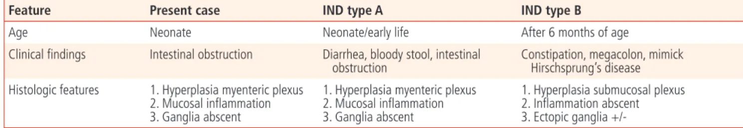
Type A of intestinal neuronal dysplasia is congenital abnormality and is a very rare disease. The ratio of male children-to-female children is 5:3 and it has a tendency to develop more often in male children. The time-to-diagnosis in male and female children is 5 and 3 months after birth and is diagnosed earlier in female children [4]. The clinical symptoms of type B manifest at more than 6 months after birth, and include progressive severe consti- pation. Many cases of type B are clinically indistinguishable from Hirschsprung’s disease (aganglionosis). Type B is commonly as- sociated with Hirschsprung’s disease [5]. Barium enema can help with the differential diagnosis [6]. Clinical symptoms of type A is a functional ileus with hematochezia, with symptoms that progress rapidly. Thus, prompt surgical treatment is needed [7]. Without timely surgical intervention, the condition may deteriorate rapidly and lead to death [8]. The clinicopathologic fi ndings in IND types A & B are shown in the Table 1, as compared to our case. From the above comparison, it can be concluded that our case fi ts in with type A.
Associated anomalies have been reported in 30.5% of patients (29 of 95 patients) with type B intestinal neuronal dysplasia; the most common anomaly is the gastrointestinal system, accounting for
Fig. 2. Micromelia of long bone shortening.A B C
Fig. 3. (A) The mucosa showed a ruptured crypt with acute and chronic infl ammation-arrow (H&E, ×40). (B) The colonic wall showed hypertrophic and tortuous myenteric plexus-arrow (H&E, ×100). (C) Immunohistochemistry for S-100 protein showed strong positivity for hypertrophic nerve bundles- arrow (×100).
67.4% of all anomalies, short stature was reported in the skeletal system [9]. Combined anomalies of IND type A reported infre- quently because of the rarity. Congenital anomalies with colonic obstruction have been reported in patients with IND type A [10].
Congenital anomalies with colonic obstruction have been reported in patients with IND type A [10]. It has been reported that type A intestinal neuronal dysplasia may be associated with colitis or ne- crotic colitis based on histologic evaluation [7,11,12]. In addition to intestinal malrotation, our patient had musculoskeletal anoma- lies (micromelia and polydactyly).
Because clinical features of intestinal neuronal dysplasia are non- specifi c and it is a very rare disease, the diagnosis may be delayed.
All of the reported cases of intestinal neuronal dysplasia were di- agnosed after birth on the basis of symptoms, even though intesti- nal neuronal dysplasia is a congenital disease. Prenatal features of intestinal neuronal dysplasia have not been reported. In our case, at 32 weeks gestation, long, dilated loops of bowel were demon- strated that changed with time. The fi nding of several dilated loops of bowel may exist in patients with small bowel obstruction, the most frequent cause of which is intestinal atresia [13]. The fetal sonographic fi ndings of small bowel obstruction detected during pregnancy are very diverse, and are detected in the third trimester;
moreover, the patterns are not consistent from patient-to-patient and can change with time. The ultrasonographic diagnosis of type A intestinal neuronal dysplasia may also show diverse features like other small bowel obstruction. If several bowel loops that change with time are shown during pregnancy, intestinal neuronal dyspla- sia must be considered in the differential diagnosis, and may be associated with deformities of the skeletal system. Thus, a compre- hensive evaluation of the skeletal system is required.
References
1. Meier-Ruge W. Casuistic of colon disorder with symptoms of
Hirschsprung’s disease (author’s transl). Verh Dtsch Ges Pathol 1971;55:506-10.
2. Fadda B, Maier WA, Meier-Ruge W, Schärli A, Daum R. Neu- ronal intestinal dysplasia. Critical 10-years’ analysis of clinical and biopsy diagnosis. Z Kinderchir 1983;38:305-11.
3. Martucciello G, Caffarena PE, Lerone M, Mattioli G, Barabino A, Bisio G, et al. Neuronal intestinal dysplasia: clinical experience in Italian patients. Eur J Pediatr Surg 1994;4:287-92.
4. Meier-Ruge W. Epidemiology of congenital innervation defects of the distal colon. Virchows Arch A Pathol Anat Histopathol 1992;420:171-7.
5. Kobayashi H, Hirakawa H, Surana R, O’Briain DS, Puri P. Intes- tinal neuronal dysplasia is a possible cause of persistent bow- el symptoms after pull-through operation for Hirschsprung’s disease. J Pediatr Surg 1995;30:253-7.
6. Montedonico S, Acevedo S, Fadda B. Clinical aspects of intesti- nal neuronal dysplasia. J Pediatr Surg 2002;37:1772-4.
7. Schärli AF, Meier-Ruge W. Localized and disseminated forms of neuronal intestinal dysplasia mimicking Hirschsprung’s dis- ease. J Pediatr Surg 1981;16:164-70.
8. Rajalakshmi T, Makhija P, Babu MK, Kini U. Intestinal neuronal dysplasia type A. Indian J Pediatr 2003;70:839-41.
9. Corduk N, Koltuksuz U, Bir F, Karabul M, Herek O, Sarioglu- Buke A. Association of rare intestinal malformations: co- lonic atresia and intestinal neuronal dysplasia. Adv Ther 2007;24:1254-9.
10. Briner J, Oswald HW, Hirsig J, Lehner M. Neuronal intestinal dysplasia: clinical and histochemical fi ndings and its associa- tion with Hirschsprung’s disease. Z Kinderchir 1986;41:282-6.
11. Puri P. Variant Hirschsprung’s disease. J Pediatr Surg 1997;32:149-57.
12. Schofi eld DE, Yunis EJ. What is intestinal neuronal dysplasia?
Pathol Annu 1992;27:249-62.
13. Reyes HM, Meller JL, Loeff D. Neonatal intestinal obstruction.
Clin Perinatol 1989;16:85-96.
Table 1. Clinicopathologic fi nding in our case in comparison with intestinal neuronal dysplasia (IND) type A and B
Feature Present case IND type A IND type B
Age Neonate Neonate/early life After 6 months of age
Clinical fi ndings Intestinal obstruction Diarrhea, bloody stool, intestinal obstruction
Constipation, megacolon, mimick Hirschsprung’s disease Histologic features 1. Hyperplasia myenteric plexus
2. Mucosal infl ammation 3. Ganglia abscent
1. Hyperplasia myenteric plexus 2. Mucosal infl ammation 3. Ganglia abscent
1. Hyperplasia submucosal plexus 2. Infl ammation abscent 3. Ectopic ganglia +/-
다지증과 소지증을 동반한 장신경형성이상 A형의 산전초음파 양상
가톨릭대학교 의과대학 1산부인과학교실, 2병리학교실 손현주1, 조윤성1, 정지한2, 장동규1, 이귀세라1
장신경형성이상(intestinal neuronal dysplasia)은 선천성 내장신경계 이상이며 직장, 대장 혹은 모든 장을 침범하는 드문 질환이다. 저자들 은 산전초음파상 다지증(polydactyly)이 동반되고 장의 확장 소견 양상이 시간적 변화를 보였으며 출생 16일에 장패쇄 증상이 나타나 개 복하에 장절제술을 시행하여 장신경형성이상 A형으로 진단된 증례를 보고하고자 한다.
중심단어: 장신경형성이상, 다지증, 소지증, 초음파