Paraduodenal hernia is the commonest type of inter- nal hernia, accounting for approximately 50% of all such cases. Although internal hernias occur in only 1%
of all patients with intestinal obstruction, 50% of pa- tients with paraduodenal hernia have obstruction (1).
Because associated radiologic reports are rare, paraduo- denal hernia is a condition with which radiologists are not entirely familiar. Failure to recognize the entity may, however, lead to death or to errors in surgical tech- nique, and its early diagnosis and treatment are there- fore essential (2). The CT findings of right paraduodenal hernia have been described (3, 4), but, to our knowl- edge, this is the first case report in which CT findings of acute small bowel obstruction due to right paraduodenal hernia are documented.
Case Report
A 22-year-old man presented with sudden abdominal
pain in the right upper abdomen, vomiting, and a palpa- ble mass in the right upper quadrant. Since childhood, the patient had experienced many episodes of intermit- tent abdominal pain, but no specific management had been undertaken. Laboratory data included an elevated white blood cell count (17,300/mm3), though hemoglo- bin, aminotransferase, and bilirubin levels were normal.
Plain abdominal radiographs demonstrated increased mass-like density in the right middle abdomen, with scanty bowel gas.
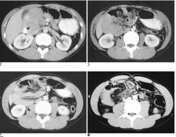
Contrast-enhanced spiral 5-mm axial CT scans of the abdomen were obtained with intravenous power injec- tion of 100 ml nonionic contrast media at a rate of 2.5 ml/sec 30 sec before initiation, and a sac-like mass of di- lated small bowel loop was found to be causing substan- tial lateral displacement of the duedenum (Fig. 1A). The Superior mesenteric vein (SMV) was found to be anterior to the superior mesenteric artery (SMA), while the Inferior vena cava (IVC) and right psoas muscle were se- verely compressed by the hernial sac. Looping of the je- junal branches of the SMA was also demonstrated (Fig.
1B), and the right ureter was laterally displaced (Fig. 1C).
And oral contrast agent, administered 40 minutes before scanning, failed to reach any of the dilated small bowel loops, filling only the stomach and duodenum. Some ex- traluminal fluid and increased mesenteric haziness was also found, suggesting bowel ischemia (Fig. 1D).
J Korean Radiol Soc 2001;44:85-88
─ 85 ─
CT Findings of Right Paraduodenal Hernia Presenting as Acute Small Bowel Obstruction
1Ji Chang Kim, M.D., Man Deuk Kim, M.D., Bong Gak Jeong, M.D., Si Won Kang, M.D., Byung Il Lim, M.D.2
Because it is rare, acute small bowel obstruction due to right paraduodenal hernia is an entity with which radiologists are not entirely familiar. Its clinical importance, how- ever, lies in the fat that delayed diagnosis leads to significantly increased morbidity and mortality rates. We report a case of small bowel obstruction due to right paraduodenal hernia in which all the known characteristic findings were demonstrated.
Index words : Intestines, hernia Intestines, CT
1Department of Radiology, Taejon St Mary’s Hospital, The Catholic University of Korea,
2Department of Radiology, Taejon Armed Forces General Hospital Received July 31, 2000; Accepted October 28, 2000
Address reprint requests to : Man Deuk Kim, M.D., Department of Radiology, Taejon St Mary’s Hospital, The Catholic University of Korea, 520-2 Taehung-dong, Joong-gu, Taejon 301-723, Republic of Korea Tel: 82-42-220-9625 Fax: 82-42-257-0511
E-mail: [email protected]
All these findings were compatible with right paraduo- denal hernia with impending strangulation, and prompt surgical correction was performed. Initial exploration revealed the “empty bowel”sign in the left abdomen, and two thirds of the ileum as well as the jejunum were herniated behind the ascending mesocolon via the fossa of Waldeyer. The remaining small bowel loops were lo- cated in the right lower abdomen, the mesentery of which was twisted. The diameter of the mesenterico- parietal fossa was estimated to be 4×3 cm.
The herniated small bowel loop was dark reddish in color, without normal peristalsis, and thus implying is-
chemia, but after successful manual reduction, normal color were regained and no bowel was resected. The lo- cation of the duodenojejunal junction, to the right of the spine, was abnormally low. During the one-month peri- od following surgery, the patient remained asympto- matic.
Discussion
One-half of internal hernias are paraduodenal in loca- tion and 50% of patients with paraduodenal hernia will have bowel obstruction. The prevalence of these hernias
Ji Chang Kim, et al: CT Findings of Right Paraduodenal Hernia Presenting as Acute Small Bowel Obstruction
─ 86 ─
A B
C D
Fig. 1. Twenty-two-year-old man presenting with acute small bowel obstruction due to right paraduodenal hernia.
A. A sac-like mass of dilated small bowel loop has displaced the duodenum (arrow) far laterally. The superior mesenteric vein (open arrow) is located ventrally in relation to the SMA. Oral contrast agent failed to pass into the more distal loop, implying ob- struction.
B. Looping of jejunal branches of the superior mesenteric artery (arrowheads) is seen. The inferior vena cava (open arrow) and right psoas muscle were compressed by a hernial sac (black arrow).
C. Posterolateral displacement of the right ureter (arrow) is apparent.
D. Extraluminal fluid (open arrows) and increased mesenteric haziness (arrowheads) are demonstrated, implying ischemia.
in the general population is unknown.
A review of reported cases of right paraduodenal her- nia shows that most patients were adults, with a mean age of 36.6 years, and none were younger than eight years (5, 6). Right paraduodenal hernia is a rare congeni- tal anomaly caused by arrest of the second stage of mid- intestinal rotation. As a result of incomplete rotation, the duodenojejunal junction remains on the right of the SMA, with eventual entrapment of the small intestine behind the ascending mesocolon, more accurately rep- resenting a “hernia into the ascending mesocolon”.
Acute intestinal obstruction is frequent, and this may cause chronic intermittent postprandial abdominal pain.
All paraduodenal hernias should be considered poten- tially lethal. Knowledge of this entity is clinically impor- tant because of the significant increase in morbidity and mortality rates that result from delayed diagnosis. In its most malignant form, actual strangulation of the bowel may occur, with a corresponding increase in the mortal- ity rate. Entrance into the hernial sac is most commonly via the mesenterico-parietal fossa (of Waldeyer) which is in the first part of the jejunal mesentery immediately be- hind the SMA and inferior to the third part of the duode- num. The postprandial abdominal pain which can oc- cur, a symptom which may mimic peptic ulcer, chole- cystitis, pancreatitis and gastritis (7), is sometimes re- lieved by changes in position. Initial exploration will of- ten reveal the classic “empty abdomen”sign, with only a segment of ileum present in the abdominal cavity and the remainder of the small bowel encased in the hernial sac. The arteriographic appearance of right paraduode- nal hernias is characteristic, with jejunal arteries arising normally from the left side of the SMA but then revers- ing direction and coursing behind their parent vessel to- wards the right (8). The major CT findings are looping of the SMA and SMV jejunal branches to the right, and posteriorly, in a fashion analogous to the arteriographic findings, and the clustering or apparent encapsulation of
small bowel loops in the right mid-abdomen. Relative to the SMA, the SMV is located further to the left and more ventrally than normal. In the present case, all the char- acteristic findings of right paraduodenal hernia were found. In addition, oral contrast agent failed to pass into the distal small bowel loops, thus indicating obstruction.
To our knowledge, this is the first documented case in which bowel obstruction associated with right paraduo- denal hernia has been disclosed on the by CT scanning.
As well as lateral displacement of the right ureter, previ- ously demonstrated in 1982 by Harbin using intra- venous pyelography (9), CT also revealed that the right psoas muscle and IVC were severely compressed by a hernial sac.
In conclusion, when acute small bowel obstruction due to right paraduodenal hernia is suspected, the pres- ence of certain characteristic CT findings provides the basis for correct disgnosis.
References
1. Berardi RS. Paraduodenal hernias. Collective Review. Surg Gynecol Obstet 1981;152:99-110
2. Bartlett JD, Martel W, Lindenauer SM. Right paraduodenal inter- nal hernia. Surg Gynecol Obstet 1971;132:443-449
3. Yeoman LJ, Patel AG, Michell MJ. Case report: Computed tomogra- phy appearances in a right paraduodenal hernia. Clin Radiol 1994;
49:898-900
4. Warshauer DM, Mauro MA. CT diagnosis of paraduodenal hernia.
Gastrointest Radiol 1992;17:13-15
5. Brown RB, Ross D. Congenital abnormalities of intestinal rotation and mesenteric attachment -A cause of intestinal obstruction in the adult. Ann Surg 1951;134:88-98
6. Murphy AD. Internal hernias in infancy and childhood. Surgery 1964;55:311-316
7. Berens JJ. Small internal hernias in the paraduodenal area. Arch Surg 1963;86:726-732
8. Meyers MA. Paraduodenal hernias. Radiologic and arteriographic diagnosis. Radiology 1970;95:29-37
9. Harbin WP. Computed tomographic diagnosis of internal hernia.
Radiology 1982;143:736 J Korean Radiol Soc 2001;44:85-88
─ 87 ─
Ji Chang Kim, et al: CT Findings of Right Paraduodenal Hernia Presenting as Acute Small Bowel Obstruction
─ 88 ─
대한방사선의학회지 2001;44:85-88
급성 소장 폐쇄로 발현한 우측 십이지장 주위 탈장의 CT 소견1
1가톨릭대학교 의과대학 대전성모병원 진단방사선과
2국군 대전 병원 진단방사선과
김지창・김만득・정봉각・강시원・임병일2
우측 십이지장 주위 탈장으로 인한 급성 소장 폐쇄는 매우 보기 드문 질환이나 진단이 늦어지는 경우 환자에게 치명 적이 될 수 있다. 우측 십이지장 주위 탈장으로 인한 급성 소장 폐쇄는 아직 국내외에 거의 보고된 바가 없다. 이에 저자 들은 특징적인CT 소견으로 진단이 가능했던 1예를 보고한다.