Yonsei Med J http://www.eymj.org Volume 51 Number 5 September 2010 787
Internal hernias are uncommon and difficult to clinically diagnose. They are defined as herniation of a viscus through an intraperitoneal orifice or aperture within the confines of the peritoneal cavity.1Paraduodenal hernias, although rare in clinical practice, represent the most common type of congenital internal her- nia.1-7Presentation can range from acute intestinal obstruction (the most common clinical presentation) to an extended history of vague abdominal pain, often relived by changes in position.2,5,8We present a case of a patient who underwent laparotomy for atypical symptoms due to a left paraduodenal hernia.
A 38-year-old man presented to the emergency department complaining of left flank pain and vomiting. He could not stay in a supine position because of flank pain. He had no remarkable medical history. Specifically, he had no history of abdominal surgery. A physical examination revealed a slightly distended abdo- men and left costovertebral angle tenderness. Blood analysis, urine analysis, and a plain abdominal radiography showed no abnormalities, except for leukocytosis of 11,200 cells/mL. Abdominal ultrasonography showed mild left hydronephrosis with no ureteral obstruction. His pain was initially thought to be caused by acute pyelonephritis. Over the few hours following admission, the patient complained of increased left flank pain and vomiting. Computed tomography showed a cluster of small bowel loops encased in a sac located amidst the stomach, the pancreas, and the left kidney, the latter of which was normal without hydrone- phrosis (Fig. 1). The diagnosis of a left paraduodenal hernia was made using an upper gastrointestinal series with small bowel follow-through, which revealed loops of jejunum clumping over the left upper quadrant of the abdomen (Fig. 2A).
Case Report
DOI 10.3349/ymj.2010.51.5.787pISSN: 0513-5796, eISSN: 1976-2437 Yonsei Med J 51(5):787-789, 2010
Left Paraduodenal Hernia Presenting with Atypical Symptoms
Min Young Yun, Yun Mi Choi, Sun Keun Choi, Sei Joong Kim, Seung Ick Ahn, and Kyung Rae Kim
Department of Surgery, College of Medicine, Inha University, Incheon, Korea.
Paraduodenal hernias are a rare congenital malformation, but they are the most common internal hernias. They develop secondary to a failure in midgut rotation, which may lead to small bowel obstruction or other clinical manifestations. The authors recently experienced a case of a left paraduodenal hernia presenting with unusual symptoms of left flank pain and vomiting.
Key Words: Paraduodenal hernia, small bowel obstruction, left flank pain
Received: October 2, 2008 Revised: February 16, 2009 Accepted: February 16, 2009
Corresponding author: Dr. Kyung Rae Kim, Department of Surgery, College of Medicine, Inha University, 7-206 Sinheung-dong 3-ga, Jung-gu, Incheon 400-103, Korea.
Tel: 82-32-890-3564, Fax: 82-32-890-3097 E-mail: [email protected]
∙The authors have no financial conflicts of interest.
© Copyright:
Yonsei University College of Medicine 2010 This is an Open Access article distributed under the terms of the Creative Commons Attribution Non- Commercial License (http://creativecommons.org/
licenses/by-nc/3.0) which permits unrestricted non- commercial use, distribution, and reproduction in any medium, provided the original work is properly cited.
INTRODUCTION
CASE REPORT
Min Young Yun, et al.
Yonsei Med J http://www.eymj.org Volume 51 Number 5 September 2010 788
These loops were identified in the posterior wall of the stomach (Fig. 2B). With the patient’s left flank pain pro- gressively worsening, a laparotomy was performed via a midline incision. Exploration revealed that about one third of the proximal small bowel was encapsulated in a perito- neal sac formed by a peritoneal flap of the left mesentery.
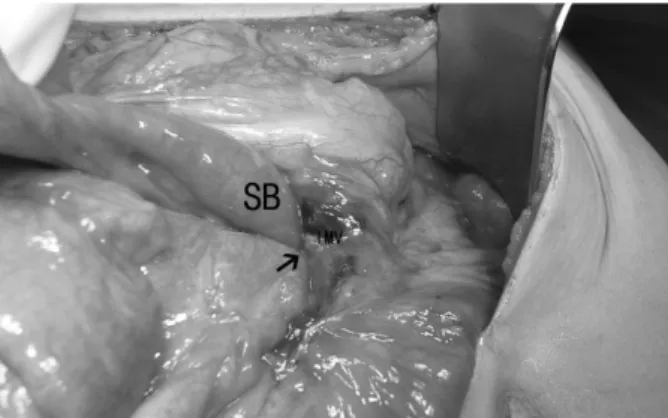
The orifice of the hernia sac was located below the inferior mesenteric vein, with engorgement and crowding (Fig. 3).
The small bowel was viable and was easily reduced after herniotomy. The defects in the left mesocolon and the hernia orifice were closed. Further inspection of the peri- toneal cavity revealed no other abnormality. Postoperati- vely, the patient had an uneventful recovery and was dis- charged on postoperative day 5. During the follow-up period of 1 year, he remained completely free of symptoms.
Andrews first described the currently accepted mechanism of paraduodenal hernia formation as a type of malrota- tion.4As the midgut rotates in the 5th to 11th weeks of gestation, the mesentery becomes fused to the posterior abdominal structures from the ligament of Treitz inferola- terally to the right iliac fossa. This process of attachment may be complete, except for a small zone just below the duodeno-jejunal junction where the former emerges from its retroperitoneal position. The pocket thus formed may extend to the right behind the mesentery, behind the ascend- ing colon, or to the left behind the descending mesocolon and descending colon. When the small bowel enters these two spaces, the resulting lesions are called right or left paraduodenal hernias.1Although paraduodenal hernias are congenital, most patients present between the 4th and 6th decades of life (mean age 38.5 years).1-3Men are com- monly 3 times more affected than women are, and left- sided paraduodenal hernias are more common than right- sided ones are, representing 75% of cases.1-8The most com- mon presentation is acute small bowel obstruction with a background of recurrent, vague abdominal pain. Symp- toms are often postprandial, and pain may be relieved when the patient is supine.5,8However, the present case was characterized by atypical symptoms: left flank pain with vomiting, aggravated in the supine position. Symptoms may result from retroperitoneal mass effect. Although the laboratory findings were inconclusive in this patient, his clinical picture was thought to be consistent with acute pyelonephritis. The clinical symptoms of left paraduodenal hernia formation are variable, as are the gastrointestinal manifestations, such as abdominal pain, nausea, and vomit- ing. The present patient represents a rare case of left para- duodenal hernia presenting with left flank pain.
The diagnosis of paraduodenal hernia formation is often difficult to make due to ambiguous presentation. This makes CT scanning a valuable initial tool for investigation.3The most common radiologic signs of left paraduodenal hernia formation include clustering of small bowel loops, a saclike mass with encapsulation at or above the ligament of Treitz, duodenojejunal junction depression, mass effect on the posterior stomach wall, engorgement and crowding of the mesentery vessels with frequent right displacement of the main mesenteric trunk, and depression of the transverse colon.9Barium contrast studies are most effectively per- formed during a symptomatic period. In contrast to follow-
DISCUSSION
Fig. 2. Small bowel follow-through radiographs showed a separation of the encap- sulated jejunal loops (arrow) from the remaining small intestine (A) and a cluster of jejunum (SB) interposing between the stomach (St) and the spine (Sp) (B).
A B
Fig. 1. Computed tomography showed a saclike mass (arrow) of jejunal loops in the left upper quadrant interposed between the transverse colon and kidney.
Fig. 3. Intraoperative view from the root of the transverse mesocolon reveals the hernia orifice (narrow arrow) through which the small bowel (SB) herniated.
Left Paraduodenal Hernia Presenting with Atypical Symptoms
Yonsei Med J http://www.eymj.org Volume 51 Number 5 September 2010 789
through studies, the small bowel is usually located to the left of the midline with a well-circumscribed edge that cor- responds to the hernia sac.5,9-11There may also be a delay in the passage of contrast through the small bowel loops with changes in the patient’s position. These features were read- ily apparent in our patient. However, because all the above investigations may be normal, especially in chronic inter- mittent cases, the elective diagnosis of a paraduodenal hernia is relatively infrequent.5
Once diagnosed, left paraduodenal hernias should be surgically repaired because they carry an approximately 50% lifetime risk of incarceration, leading to bowel obst- ruction and strangulation.2,3,5,8The mortality rate associated with paraduodenal hernias is not clear, but it has been stated to be 20-50%.5Reports prior to 1981 indicate a high morta- lity,12but recent improvements in radiological facilities have allowed for earlier diagnosis and elective treatment, leading to better outcomes.3,5Surgical treatment of left paraduodenal hernias follows the basic principles of hernia surgery: reduc- tion of the contents, restoration of normal anatomy, and repair of the defect.1-8Recently, there have been reports of laparoscopic left paraduodenal hernia repair.8,13
In conclusion, paraduodenal hernias are an unusual cause of intestinal obstruction, but one with which all sur- geons should be familiar. Because the presentation of a left paraduodenal hernia is ambiguous and variable, it is im- possible to establish a correct diagnosis through physical examination alone. Early surgical intervention should be performed to minimize the morbidity and mortality asso- ciated with this condition.
This work was supported by a Inha University Research Grant.
1. Singh RR, Warren P, Smith P, Wilson W. Image of the month.
Paraduodenal Hernia. Arch Surg 2006;141:711-2.
2. Manji R, Warnock GL. Left paraduodenal hernia: an unusual cause of the small-bowel obstruction. Can J Surg 2001;44:455-7.
3. Huang YM, Chou AS, Wu YK, Wu CC, Lee MC, Chen HT, et al. Left paraduodenal hernia presenting as recurrent small bowel obstruction. World J Gastroenterol 2005;11:6557-9.
4. Dritsas ER, Ruiz OR, Kennedy GM, Blackford J, Hasl D. Para- duodenal hernia: a report of two cases. Am Surg 2001;67:733-6.
5. Tong RS, Sengupta S, Tjandra JJ. Left paraduodenal hernia: case report and review of the literature. ANZ J Surg 2002;72:69-71.
6. Cingi A, Demirkalem P, Manukyan MN, Tuney D, Yegen C.
Left-sided paraduodenal hernia: report of a case. Surg Today 2006;36:651-4.
7. Socas Macías M, Alamo Martín JM, Suárez Grau JM, Suárez Artacho G, Tejada A, Martin Cartes J, et al. Atypical left paraduo- denal hernia. Rev Esp Enferm Dig 2006;98:473-5.
8. Rollins MD, Glasgrow RE. Left paraduodenal hernia. J Am Coll Surg 2004;198:492-3.
9. Blachar A, Federle MP, Dodson SF. Internal hernia: clinical and imaging findings in 17 patients with emphasis on CT criteria.
Radiology 2001;218:68-74.
10. Selçuk D, Kantarci F, Oǧüt G, Korman U. Radiological evalua- tion of internal abdominal hernias. Turk J Gastroenterol 2005;16:
57-64.
11. Martin LC, Merkle EM, Thompson WM. Review of internal her- nia: radiographic and clinical findings. AJR Am J Roentgenol 2006;186:703-17.
12. Brigham RA, Fallon WF, Saunders JR, Harmon JW, d’Avis JC.
Paraduodenal hernia: diagnosis and surgical management. Sur- gery 1984;96:498-502.
13. Uematsu T, Kitamura H, Iwase M, Yamashita K, Orura H, Nakamuka T, et al. Laparoscopic repair of a paraduodenal hernia.
Surg Endosc 1998;12:50-2.