Annals of Surgical Treatment and Research 217
pISSN 2288-6575 • eISSN 2288-6796 http://dx.doi.org/10.4174/astr.2014.86.4.217 Annals of Surgical Treatment and Research
CASE REPORT
Left paraduodenal hernia combined with acute cholecystitis
Seung Eun Lee, Yoo Shin Choi
Department of Surgery, Chung-Ang University College of Medicine, Seoul, Korea
INTRODUCTION
Although paraduodenal hernia is a rare congenital malfor- mation caused by abnormal retroperitoneal fixation of the intestinal mesentery, it represents the most common type of congenital internal hernia [1,2]. These hernias result from small bowel loops herniating through the paraduodenal fossae and should always be considered when a patient with no previous surgical history presents with an intestinal obstruction [3]. We present a case of a patient who was diagnosed with con comitant acute cholecystitis and left paraduodenal hernia based on computed tomography and underwent laparoscopic reduction of a herniated bowel and closure of the hernial orifice.
CASE REPORT
A 74-year-old woman presented to the Emergency Depart- ment with persistant right upper quadrant pain that began 3 hours prior to presentation. She had an unremarkable me- dical history, but asymptomatic gallstones were detected during routine check-up. More specifically, she had no history of abdominal surgery or abdominal pain prior to this visit.
Physical examination revealed a thin woman (height, 156 cm; weight, 49 kg) with a blood pressure of 110/70 mmHg, a pulse of 64 beats/min, and a body temperature of 36.6oC. The abdomen was tender in the right upper quadrant. No guarding and rebound tenderness were noted. The laboratory data showed neutrophilia (white blood cells, 9,710 / mm3 with 81.4%
segmented neutrophils). Other blood chemistry parameters including liver function test were unremarkable. CT revealed
Received August 13, 2013, Revised October 22, 2013, Accepted October 24, 2013
Corresponding Author: Seung Eun Lee
Department of Surgery, Chung-Ang University Hospital, Chung-Ang University College of Medicine, 102 Heukseok-ro, Dongjak-gu, Seoul 156- 755, Korea
Tel: +82-2-6299-3121, Fax: +82-2-824-7869 E-mail: selee508@cau.ac.kr
Copyright ⓒ 2014, the Korean Surgical Society
cc Annals of Surgical Treatment and Research is an Open Access Journal. All articles are distributed under the terms of the Creative Commons Attribution Non- Commercial License (http://creativecommons.org/licenses/by-nc/3.0/) which permits unrestricted non-commercial use, distribution, and reproduction in any medium, provided the original work is properly cited.
Paraduodenal hernia is a rare congenital malformation. Management consists of reduction of the herniated intestine and repair of the defect. A 74-year-old woman presented to the Emergency Department with persistent right upper quadrant pain that began 3 hours ago. Physical examination revealed tenderness at right upper quadrant of abdomen. Computed tomography revealed multiple gallstones with gallbladder wall thickening, marked dilatation of stomach and duodenum and a sac-like mass of small bowel loops to left of ligament of Treitz suggesting acute cholecystitis and left paraduodenal hernia. Laparoscopic exploration of abdomen was performed and cholecystectomy, bowel reduction, and closure of defect with intracorporeal interrupted suturing were performed. For left paraduodenal hernia without bowel necrosis, laparoscopic reduction of incarcerated bowel and closure of hernial orifice are technically feasible and may be the surgical method of choice because of its minimal invasiveness and aesthetic advantage.
[Ann Surg Treat Res 2014;86(4):217-219]
Key Words: Paraduodenal, Hernia, Laparoscopic, Repair
218
Annals of Surgical Treatment and Research 2014;86(4):217-219
multiple gallstones with gallbladder wall thickening, marked dilatation of stomach and duodenum and a sac-like mass of small bowel loops to the left of the ligament of Treitz (Fig. 1) suggesting acute cholecystitis and left paraduodenal hernia.
After performing CT, the patient developed bilous vomiting without left abdominal pain. We proceeded to perform laparoscopic exploration of the abdomen with cholecystectomy.
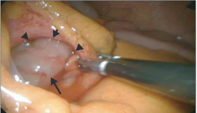
The defect was located at the Treitz ligament where proximal jejunal loops were noted to be herniating through the defect (Fig. 2). About 50 cm of jejunal loops were easily reduced and the bowel appeared viable. The 3-cm defect was closed using 3-0 Vicryl intracorporeal interrupted sutures. Laparoscopic cholecystectomy was then performed. The total operation time was 105 minutes. The postoperative course was uneventful and the patient was discharged on postoperative day 4. During the 6 months follow-up period, the patient remained completely free of symptoms.
DISCUSSION
Paraduodenal hernia is the most common form of internal hernias, accounting for more than 50% of all reported cases [1]. Paraduodenal hernias result from abnormal rotation of the midgut during embryonic development and can be divided into two subtypes, left and right paraduodenal hernias. Left paraduodenal hernia (hernia of Lanzert) is about three times more common than its right counterpart (Walayer’s hernia) [4]. Left paraduodenal hernias arise from the fossa of Lanzert, a congenital defect that is presents in approximately 2% of the population and located at the inferior mesenteric vein and left branches of the middle colic artery [1,5]. Although paraduodenal hernias are congenital, most patients present between the 4th and 6th decades of life (median age, 47 years) with a male to female ratio of 3:1 [6]. The most common presentation is
acute small bowel obstruction in the setting of recurrent vague abdominal pain [2,6]. Approximately 50% of patients with paraduodenal hernias have episodes of intestinal obstruction at certain periods in their lives [2,6]. The symptoms observed in these cases range from temporary colicky abdominal pain to signs of intestinal obstruction. Our patient was unusual in that she presented at an advanced age of 74, without prior history of abdominal pain or other gastrointestinal symptoms.
The diagnosis of paraduodenal hernia formation is often difficult to make due to its ambiguous presentation. Therefore, CT scanning is a valuable initial tool for investigation. The most common radiologic signs of left paraduodenal hernia formation include clustering of small bowel loops, a sac-like mass with encapsulation at or above the ligament of Treitz, duodeno-jejunal junction depression, mass effect on the posterior stomach wall, engorgement and crowding of the mesentery vessels with frequent right displacement of the main mesenteric trunk, and depression of the transverse colon.
Once diagnosed, left paraduodenal hernias should be sur- gically treated because they carry a risk of incarceration, with the potential for bowel obstruction and strangulation. Surgical management essentially consists of reduction of the herniated small bowel loops and closure of the hernia orifice. Care must be taken not to damage the left colic artery or inferior mesen- teric vessels, which are often found anterior to the hernia opening. Laparoscopy is indicated when there are no signs of bowel necrosis or dilatation of the incarcerated bowel loops [7].
A recent small case series comparing laparoscopy to open repair of paraduodenal hernias showed that the laparoscopic approach resulted in shorter hospital stay, earlier intake of a soft diet and lower rate of postoperative ileus [8].
In conclusion, left paraduodenal hernias are rare cause of intestinal obstruction; however, in the setting of recurrent small bowel obstruction and no previous surgical history, it is crucial to consider internal hernias in the differential diagnosis.
Furthermore, timely surgical intervention should be performed Fig. 1. Computed tomography showed a sac-like mass of
jejunal loops (arrows) in the left upper quadrant.
Fig. 2. Intraoperative view demonstrating a loop of small bowel prolapsing (arrow) through Landzert’s fossa (arrowheads).
Annals of Surgical Treatment and Research 219 1. Berardi RS. Paraduodenal hernias. Surg
Gynecol Obstet 1981;152:99-110.
2. Tong RS, Sengupta S, Tjandra JJ. Left para- duodenal hernia: case report and review of the literature. ANZ J Surg 2002;72:69- 71.
3. Hirasaki S, Koide N, Shima Y, Nakagawa K, Sato A, Mizuo J, et al. Unusual variant of left paraduodenal hernia herniated into the mesocolic fossa leading to jejunal strangulation. J Gastroenterol 1998;33:
734-8.
4. Khan MA, Lo AY, Vande Maele DM. Para- duodenal hernia. Am Surg 1998;64:1218- 22.
5. Willwerth BM, Zollinger RM Jr, Izant RJ Jr. Congenital mesocolic (paraduodenal) hernia. Embryologic basis of repair. Am J Surg 1974;128:358-61.
6. Al-Khyatt W, Aggarwal S, Birchall J, Row- lands TE. Acute intestinal obstruction secondary to left paraduodenal hernia: a case report and literature review. World J Emerg Surg 2013;8:5.
7. Fukunaga M, Kidokoro A, Iba T, Sugiyama K, Fukunaga T, Nagakari K, et al. Laparo- scopic surgery for left paraduo denal hernia. J Laparoendosc Adv Surg Tech A 2004;14:111-5.
8. Jeong GA, Cho GS, Kim HC, Shin EJ, Song OP. Laparoscopic repair of paraduodenal hernia: comparison with conventional open repair. Surg Laparosc Endosc Per- cutan Tech 2008;18:611-5.
REFERENCES
Seung Eun Lee and Yoo Shin Choi: Left paraduodenal hernia
to minimize the morbidity and mortality associated with this condition. When there is no evidence of bowel necrosis, laparoscopic surgery may be the surgical method of choice because of its minimal invasiveness and aesthetic advantage.
CONFLICTS OF INTEREST
No potential conflict of interest relevant to this article was reported.
ACKNOWLEDGEMENTS
This research was supported by the Chung-Ang University Research Grants in 2010.
