ISSN 0378-6471 (Print)⋅ISSN 2092-9374 (Online)
http://dx.doi.org/10.3341/jkos.2016.57.8.1222
Original Article
망막분지정맥폐쇄 환자에서 맥락막 두께의 변화
Changes in Choroidal Thickness in Branch Retinal Vein Occlusion
송미령1⋅김재석1⋅최 진1⋅박정현2
Miryoung Song, MD1, Jaesuk Kim, MD, PhD1, Jin Choi, MD, PhD1, Jung Hyun Park, MD, PhD2
인제대학교 의과대학 상계백병원 안과학교실1, 인제대학교 의과대학 서울백병원 안과학교실2 Department of Ophthalmology, Sanggye Paik Hospital, Inje University College of Medicine1, Seoul, Korea
Department of Ophthalmology, Seoul Paik Hospital, Inje University College of Medicine2, Seoul, Korea
Purpose: To compare the choroidal thickness of a branch retinal vein occlusion (BRVO) lesion and that of other areas in the eyes.
Methods: Patients who visited the Ophthalmologic Clinic of Inje University Sanggye Paik Hospital for BRVO between March 2015 and October 2015 were reviewed retrospectively. We performed basic ophthalmologic exam and enhanced depth imaging optical coherence tomography in 48 eyes of 24 patients with BRVO. The choroidal thickness was compared in a total of 4 places, the branch retinal vein occlusion lesion, the symmetric site in the same eye, and the equivalent sites in the fellow eye by paired t-test. All measurements were performed by 2 independent observers.
Results: Choroidal thickness had strong inter-observer correlation. Choroidal thickness of the BRVO lesion was significantly thicker than that in the symmetric site of same eye, the equivalent site of lesion, and the equivalent site of the symmetric site to lesion in the fellow eye.
Conclusions: Choroidal thickness in acute BRVO lesions was thicker than choroidal thickness in other areas of the eyes. It is thought that both hydrostatic pressure and the effects of vascular endothelial growth factor influence choroidal thickness in the acute phase of BRVO.
J Korean Ophthalmol Soc 2016;57(8):1222-1227
Keywords: Branch retinal vein occlusion, Choroidal thickness, Spectral domain-optical coherence tomography (SD-OCT)
■Received: 2016. 1. 28. ■ Revised: 2016. 6. 23.
■Accepted: 2016. 7. 14.
■Address reprint requests to Jaesuk Kim, MD, PhD
Department of Ophthalmology, Inje University Sanggye Paik Hospital, #1342 Dongil-ro, Nowon-gu, Seoul 01757, Korea Tel: 82-2-950-1096, Fax: 82-2-935-6904
E-mail: [email protected]
ⓒ2016 The Korean Ophthalmological Society
This is an Open Access article distributed under the terms of the Creative Commons Attribution Non-Commercial License (http://creativecommons.org/licenses/by-nc/3.0/) which permits unrestricted non-commercial use, distribution, and reproduction in any medium, provided the original work is properly cited.
망막분지정맥폐쇄는 동정맥 교차로에서 딱딱한 동맥혈 관벽이 정맥혈관 내경을 좁게 하여 폐쇄가 발생하며 분지 정맥 영역에 망막출혈이 나타나고 황반부종이나 비관류에 의하여 시력장애가 발생하는 질환이다.1 망막분지정맥폐쇄 의 급성기에는 출혈과 황반부종 등이 시력을 저하시키는 주요 원인이며, 황반출혈이 흡수된 뒤의 만성기에는 황반
허혈과 지속적인 황반부종, 신생혈관녹내장 등이 원인이 되어 시력을 저하시킨다. 이 중 실제적으로 시력장애를 유 발하는 대부분의 경우는 황반부종과 황반부 허혈이 원인이 된다. 분지정맥의 임상적 경과는 위치와 범위, 곁순환 배출 능력의 영향을 받게 되는데 곁순환은 망막의 다른 부분으 로 우회로를 형성하기 위해 혈관이 폐쇄된 부위 근처의 정 맥에서 정맥으로 이측 솔기를 통과하여 발생하게 된다.2
정맥폐쇄가 있는 쪽의 황반하 맥락막의 두께가 반대안의 황반하 맥락막 두께보다 두껍고, 유리체강내 베바시주맙 주사 시행 후 감소하였다는 보고가 있다.3,4 맥락막이 두꺼 워지는 것은 여러 가지 원인이 있다. 먼저, 외측 망막의 정 상적인 대사 요구량을 충족시키기 위해 맥락막 혈류가 증 가되어 있어 두께가 증가하였을 수 있다. 또한 망막저산소
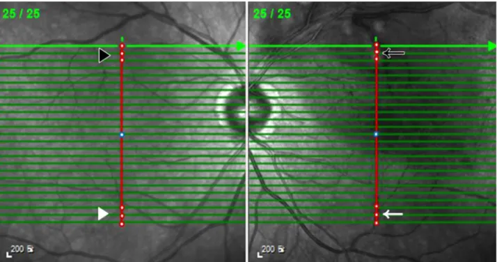
Figure 1. Method of measuring the choroidal thickness. Cross sectional image centered in macula (blue circle) was taken in the en-
hanced depth imaging mode with suprerior branch retinal vein occlusion (BRVO) in left eye. Black arrow points the lesion in BRVO. W hite arrow points the symmetrical site to lesion in BRVO. Black arrowhead points the equivalent site to lesion in fellow eye, and white arrowhead points the equivalent site of symmetrical site to lesion in fellow eye. Choroidal thickness was measured in 3 points (red circles) of each site. And the average choroidal thickness was calculated.증으로 인해 혈관내피세포성장인자가 발현되고 이로 인해 혈관 확장과 혈류가 증가되며 대사산물로 발생하는 산화질 소가 혈관의 과투과성을 증가시켰기 때문으로도 볼 수 있 다.5 Optical coherence tomography (OCT)의 발달로 맥락막 두께에 대한 접근이 가능해 졌으며6-11 특히 spectral domain- OCT (SD-OCT)의 enhanced depth imaging (EDI) 기능은 맥락막을 시각적으로 구분할 수 있게 해 주었다. 이 기능을 활용한 망막정맥 폐쇄에 대해 환측과 건측의 황반하 맥락 막 두께를 비교하는 연구는 여럿 보고되어 있으나4,12 이 연 구들은 황반근처의 중심부에 대해 연구하였고 병변 부위를 직접적으로 측정한 연구 결과는 없었다. 망막분지정맥폐쇄 환자의 시력 예후는 병변이 발생한 위치와 정도에 따라 달 라지므로 폐쇄된 부위에 대한 평가는 임상적으로 중요하기 에 부위에 따른 두께변화에 대해서는 알려진 바가 거의 없 다. 이에 망막분지정맥폐쇄 환자의 환측안의 병변과 병변 대칭 부위, 또 건측안의 병변 대응 부위와 병변 대칭 부위 의 대응 부위의 맥락막 두께를 비교해 보고자 하였다.
대상과 방법
2015년 3월부터 2015년 10월까지 인제대학교 상계백병 원 안과 외래 방문 환자 중 망막분지정맥폐쇄를 진단 받은
환자들을 후향적으로 분석하였다. 본 연구는 헬싱키 선언 에 입각한 인제대학교 상계백병원 의학연구윤리심의위원 회의 승인 아래 진행되었다(승인 번호: 2015-06-029-001).
망막분지정맥폐쇄는 안저검사상 망막의 상이측 혹은 하이 측 일사분면에 국한된 망막내 출혈, 망막정맥의 울혈과 사 행, 그리고 삼출물 등 검안경을 이용한 안저소견과 함께 형 광안저혈관 조영검사에서 폐쇄된 혈관에 형광충만이 지연 되는 것을 통해 진단하였다. 모든 환자의 성별, 연령, 과거 력을 조사하였고, 증상이 나타나거나 진단된 지 6개월 이내 인 환자를 대상으로 하였으며 망막분지정맥폐쇄 이외의 다 른 유리체 망막병변을 가지고 있는 환자는 제외하였다. 또 한 망막에 영향을 줄 수 있는 6 diopter 이상의 고도근시, 당뇨, 심부전 등의 기저질환이 있는 환자들은 제외하였다.
Spectralis-OCT (Heidelberg Engineering, Heidelberg, Germany) 를 이용하여 EDI-OCT를 시행하였다. 맥락막의 두께는 EDI mode를 이용하여 황반을 중심으로 8.9 × 7.4 mm에 해당하 는 부위에 대해 0.296 μm 간격으로 25개의 cross sectional image를 얻은 후 황반을 기준으로 수직선상에서 가장 멀리 떨어진 병변 부위의 3지점을 선택하여 망막색소상피인 고 반사선의 외측에서 맥락막-공막의 경계(chorioscleral inter- face)까지의 수직 거리를 내장된 캘리퍼를 이용하여 측정 후 평균값을 구하였다. 환측안에서 병변 대칭 부위와 건측
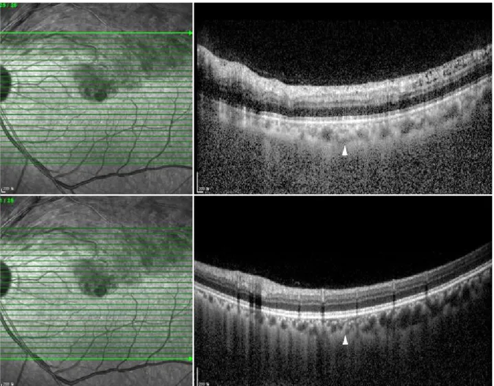
Figure 2. Method of measuring the choroidal thickness. Cross sectional image centered in macula taken in the enhanced depth imag-
ing mode with suprerior branch retinal vein occlusion in left eye. W hite arrowheads point the choroid-scleral junction.Table 1. Characteristics of retinal vein occlusion patients
Characteristics Subjects (n = 24)
Mean age (years, range) 60.95 ± 11.53 (43-79)
Sex (M:F) 11:13
Duration from onset (months) 0.74 ± 0.36
Laterality (right:left) 12:12
Location (n, %)
Superotemporal 15 (62)
Inferotemporal 9 (38)
Hypertension 9 (38)
Values are presented as mean ± SD or n (%) unless otherwise indicated.
안에서 병변 대응 부위와 병변 대칭 부위의 대응 부위에 대 해서도 같은 방법으로 맥락막 두께를 구하였다(Fig. 1, 2).
각각의 측정치는 눈가림된 2명의 저자가 독립적으로 측정 한 뒤 평균치를 이용하였다. 통계학적 분석은 SAS software program version 6.01 (SAS Institute, Cary, NC, USA)을 이 용하였다. 대응표본 t-검정을 이용하여 맥락막의 두께를 비 교하였으며, 유의확률 p<0.05인 경우 통계학적으로 유의한 것으로 보았다.
결 과
망막분지정맥폐쇄 환자 24명 48안을 대상으로 하였으며 대상군 중 남자는 11명이었고, 여자는 13명이었다. 대상군 의 평균나이는 60.95 ± 11.53세였다. 망막분지정맥폐쇄가 발생한 눈은 우안이 12안이었고, 좌안 12안이었으며 발생 한 위치는 상이측 15안이었고, 하이측 9안이었다(Table 1).
48안에서 맥락막 두께의 두 연구자 간 분석 결과는 전반 적으로 높은 수준의 일관성을 보였고(r=0.93, p<0.001), 각 위치별 결과는 다음과 같았다(Table 2). 환측안의 병변 부 위 평균 맥락막 두께는 261.72 ± 58.17 μm였고, 환측안의 병변 대칭 부위의 평균 맥락막 두께는 203.63 ± 48.47 μm
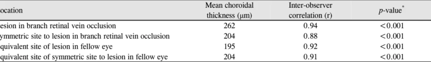
Table 2. Mean choroidal thickness and inter-observer correlation of measurements
Location Mean choroidal
thickness (μm)
Inter-observer
correlation (r) p-value*
Lesion in branch retinal vein occlusion 262 0.94 <0.001
Symmetric site to lesion in branch retinal vein occlusion 204 0.88 <0.001
Equivalent site of lesion in fellow eye 195 0.92 <0.001
Equivalent site of symmetric site to lesion in fellow eye 204 0.91 <0.001
*Paired t-test.
Figure 3. Comparison of choroidal thickness. Lesion in branch
retinal vein occlusion was significantly thicker than others. All p-value by paired t-test.였으며 건측안의 병변 대응 부위의 평균 맥락막 두께는 195.87 ± 52.94 μm였고, 건측안의 병변 대칭 부위의 대응 부위의 평균 맥락막두께는 204.83 ± 48.60 μm였다. 환측안 의 병변 부위 평균 맥락막 두께는 환측안 병변대칭 부위, 건측안의 병변 대응 부위와 건측안 병변 대칭 부위의 대응 부위의 평균 맥락막 두께와 유의한 차이가 있었다(p<0.001, p<0.001, p<0.001). 환측안의 병변 대칭 부위 평균 맥락막 두께는 건측안의 병변 대응 부위 평균 맥락막과 건측안의 병변 대칭 부위의 대응 부위 평균 맥락막 두께와 유의한 차 이가 없었으며(p=0.205, p=0.811), 건측안의 병변 대응 부 위의 평균 맥락막 두께와 건측안의 병변 대칭 부위의 대응 부위의 평균 맥락막 두께 또한 유의한 차이가 없었다 (p=0.180) (Fig. 3).
고 찰
본 연구에서 EDI-OCT를 통해 측정한 환측안 병변하의 평균 맥락막 두께는 환측안 병변 부위에서 더 높게 측정되 었으며 병변이 발생하지 않은 부위의 평균 맥락막 두께와 유의한 차이가 있었다. 황반하 맥락막의 두께에 대해서는
여러 연구들이 있었다. Tsuiki et al4은 망막중심정맥폐쇄 환자에게서 황반하 맥락막의 두께가 영향을 받지 않은 눈 에 비해 증가해 있다고 보고하였으나, Du et al12이 시행한 대규모 연구인 Beijing Study에서는 망막정맥폐쇄가 없는 환자보다 망막정맥폐쇄 환자의 황반하 맥락막의 두께가 얇 다고 보고하였다. 이러한 차이는 연구 설계의 차이 때문으 로 생각되며 Beijing Study와 같은 대규모 연구에서는 망막 정맥폐쇄가 발생한지 오래된 환자까지 포함하여 급성기의 맥락막 변화를 반영하지 못하였기 때문으로 생각된다. 망 막분지정맥발생의 초기 변화를 알아보기 위해 본 연구에서 는 증상이 발생하거나 망막분지정맥폐쇄가 진단된 지 6개 월 이내의 환자들을 대상으로 하였다. 과거의 연구들은 황 반하 맥락막 두께에 국한하여 연구하였으며 망막분지정맥 폐쇄가 발생한 부위에서의 맥락막 두께를 측정하지 않아 본 연구에서는 분지정맥폐쇄가 발생한 부위와 발생하지 않 은 부위의 맥락막 두께를 비교해 보았다.
망막분지정맥폐쇄 환자에게서 맥락막 두께가 증가하는 원인은 혈관내피세포성장인자의 발현이 라고 여러 논문에 서 밝힌 바 있다.13,14 망막분지정맥폐쇄안에서 망막 저산소 증이 나타나면, 저산소증은 망막색소상피에서 혈관내피세 포성장인자의 발현을 증가시킨다.15 혈관내피세포성장인자 는 혈관 확장을 유발시키며 산화질소의 생산을 증가시켜 안내 혈류량을 증가시키고, 안구혈관의 투과성을 높인
다.16-18 안구혈관의 투과성의 높아지면 맥락막 두께가 증가
할 수 있다.13,14 혈관내피세포성장인자가 망막 저산소증이 있는 부위에 국소적으로 증가한다면 국소적인 맥락막 두께 증가를 일으킬 수 있다. 토끼를 이용한 동물실험 모델에서 망막정맥폐쇄가 발생한 부위에만 혈관내피세포성장인자의 mRNA 농도가 상승되어 있다는 보고가 있었다.15 이는 혈 관내피세포성장인자가 망막저산소증 부위에 국소적으로 증가할 수 있음을 시사하며, 국소적으로 증가한 혈관내피 세포성장인자는 저산소 상태의 망막에 인접한 맥락막의 확 장과 두꺼워짐을 유발할 수 있다. 그러나 토끼를 이용한 동 물실험에서는 국소적인 맥락막의 두께를 측정하지 않아 혈 관내피세포성장인자와 국소적인 맥락막 두께 사이의 직접 적인 인과관계는 밝혀지지 않았다.
한편, 국소적인 맥락막 두께의 증가를 유발할 수 있는 또 다른 원인으로 망막 내 정수압의 증가를 생각해 볼 수 있 다. 중심장액맥락망막병증에서 맥락막 투과성이 증가한 곳 에만 국소적으로 맥락막의 두께가 증가해 있고, 맥락막 내 정수압의 증가가 망막색소상피와 망막에 구조적인 변화를 일으켜 망막색소상피박리를 발생시켰다는 보고가 있었다.19 구조적인 변화로 액체성분의 이동이 발생할 수 있다는 것 을 고려해 볼 때, 망막분지정맥폐쇄에서 망막의 정수압 증 가도 맥락막 두께의 증가를 유발할 수 있다고 유추해 볼 수 있다. 망막분지정맥의 폐쇄가 발생하면 혈관내피세포들이 손상되고, 혈관확장과 혈관폐쇄로 인해 안내 혈류가 증가 하게 된다. 이로 인해 혈관 외에 액체성분이 쌓이게 되고 정수압이 높아지면서 바깥혈관-망막 장벽이 손상을 받게 되고, 망막 내 액체성분이 폐쇄정맥에 인접한 맥락막으로 새어나가 맥락막을 두껍게 만들었다고 가설을 세워볼 수 있다.19
본 연구의 한계는 다른 대규모 망막 연구들에 비해 적은 수의 표본을 대상으로 하였으며 혈관내피세포성장인자의 망막폐쇄부위 내 농도를 측정하지 못하여 정확한 인과관계 를 밝히지 못한 점이다. 결론적으로, 망막분지정맥폐쇄가 발생한 부위에서만 맥락막 두께가 증가하는 것은 국소적인 혈관내피세포성장인자의 발현과 망막 내 정수압의 증가 두 가지가 모두 영향을 미쳤을 수 있으며 이는 망막분지정맥 폐쇄의 병태생리에 대한 이해에 도움을 줄 수 있을 것으로 생각된다.
REFERENCES
1) Weinberg D, Dodwell DG, Fern SA. Anatomy of arteriovenous crossings in branch retinal vein occlusion. Am J Ophthalmol 1990;
109:298-302.
2) Christoffersen NL, Larsen M. Pathophysiology and hemody- namics of branch retinal vein occlusion. Ophthalmology 1999;
106:2054-62.
3) Uemoto R, Nakasato-Sonn H, Kawagoe T, et al. Factors associated with enlargement of chorioretinal atrophy after intravitreal bev- acizumab for myopic choroidal neovascularization. Graefes Arch Clin Exp Ophthalmol 2012;250:989-97.
4) Tsuiki E, Suzuma K, Ueki R, et al. Enhanced depth imaging optical coherence tomography of the choroid in central retinal vein occlusion.
Am J Ophthalmol 2013;156:543-7.e1.
5) Shin YU, Lee MJ, Lee BR. Choroidal maps in different types of macular edema in branch retinal vein occlusion using swept-source optical coherence tomography. Am J Ophthalmol 2015;160:328-34.e1.
6) Chung SE, Kang SW, Lee JH, Kim YT. Choroidal thickness in pol- ypoidal choroidal vasculopathy and exudative age-related macular degeneration. Ophthalmology 2011;118:840-5.
7) Fujiwara T, Imamura Y, Margolis R, et al. Enhanced depth imaging optical coherence tomography of the choroid in highly myopic eyes. Am J Ophthalmol 2009;148:445-50.
8) Imamura Y, Fujiwara T, Margolis R, et al. Enhanced depth imaging optical coherence tomography of the choroid in central serous chorioretinopathy. Retina 2009;29:1469-73.
9) Maruko I, Iida T, Sugano Y, et al. Subfoveal choroidal thickness in fellow eyes of patients with central serous chorioretinopathy.
Retina 2011;31:1603-8.
10) Maruko I, Iida T, Sugano Y, et al. Subfoveal choroidal thickness af- ter treatment of Vogt-Koyanagi-Harada disease. Retina 2011;31:
510-7.
11) Maruko I, Iida T, Sugano Y, et al. Subfoveal retinal and choroidal thickness after verteporfin photodynamic therapy for polypoidal choroidal vasculopathy. Am J Ophthalmol 2011;151:594-603.e1.
12) Du KF, Xu L, Shao L, et al. Subfoveal choroidal thickness in retinal vein occlusion. Ophthalmology 2013;120:2749-50.
13) Yuan A, Ahmad BU, Xu D, et al. Comparison of intravitreal ranibi- zumab and bevacizumab for the treatment of macular edema sec- ondary to retinal vein occlusion. Int J Ophthalmol 2014;7:86-91.
14) Campochiaro PA, Sophie R, Pearlman J, et al. Long-term outcomes in patients with retinal vein occlusion treated with ranibizumab:
the RETAIN study. Ophthalmology 2014;121:209-19.
15) Aiello LP, Northrup JM, Keyt BA, et al. Hypoxic regulation of vas- cular endothelial growth factor in retinal cells. Arch Ophthalmol 1995;113:1538-44.
16) Ku DD, Zaleski JK, Liu S, et al. Vascular endothelial growth factor induces EDRF-dependent relaxation in coronary arteries. Am J Physiol 1993;265(2 Pt 2):H586-92.
17) Tilton RG, Chang KC, LeJeune WS, et al. Role for nitric oxide in the hyperpermeability and hemodynamic changes induced by in- travenous VEGF. Invest Ophthalmol Vis Sci 1999;40:689-96.
18) Ferrara N. Vascular endothelial growth factor: molecular and bio- logical aspects. Curr Top Microbiol Immunol 1999;237:1-30.
19) Jirarattanasopa P, Ooto S, Tsujikawa A, et al. Assessment of mac- ular choroidal thickness by optical coherence tomography and an- giographic changes in central serous chorioretinopathy. Ophthalmology 2012;119:1666-78.
= 국문초록 =
망막분지정맥폐쇄 환자에서 맥락막 두께의 변화
목적: 망막분지정맥폐쇄가 발생한 환자에게서 병변 부위와 병변 이외의 부위의 맥락막의 두께를 비교하고자 하였다.
대상과 방법: 2015년 3월부터 2015년 10월까지 인제대학교 상계백병원 안과 외래에 내원한 망막분지정맥폐쇄 환자 24명 48안을 대상 으로 기본 안과 검사와 Enhanced depth imaging optical coherence tomography를 시행하였다. 환측안의 병변 부위와 병변 대칭 부위 맥락막 두께 및 건측안에서 각각 대응하는 부위의 맥락막 두께를 구하여 대응표본 t-검정 방법을 통해 총 4곳의 맥락막 두께를 비교하였다. 2명의 독립적인 눈가림을 한 연구자가 측정을 시행하였다.
결과: 맥락막 두께의 연구자 간 일치도는 매우 높았으며 분지정맥폐쇄가 있는 환자에게서 병변 부위의 맥락막 두께는 환측안의 병변 대칭 부위, 건측안의 병변 대응 부위, 건측안의 병변 대칭 부위의 대응 부위의 맥락막 두께보다 유의하게 높게 측정되었다.
결론: 망막분지정맥폐쇄 환자에게서 병변이 발생한 부위는 병변이 발생하지 않은 부위에 비해 맥락막의 두께가 더 높게 측정되는 것으로 보아 급성기의 맥락막 두께 변화에 혈관내피세포성장인자의 영향과 정수압에 모두 영향을 미칠 것으로 생각된다.
<대한안과학회지 2016;57(8):1222-1227>