INTRODUCTION
Cervical cancer is a major cause of death, and the second most frequent cancer in women worldwide.1 Many studies have indicated a causal relation between genital human papillomavirus (HPV) infections and cervical cancer. High risk HPV genotypes have been detected in almost 100% of
all cervical cancers, and the process of HPV mediated carcinogenesis has been partly clarified.
Because HPV infections are so widespread in the general population and because HPV-immortalized cell lines are generally not tumorigenic, other factors in addition to HPV infections, such as the host immune response to HPV infections, are thought to have a role in controlling both HPV infections and HPV-related neoplasm.2 This is supported by the observation that an ineffective cellular immune response, as immunocompromised individuals with human immunodeficiency virus, is associated with an in-
No association between genetic polymorphisms of the Interleukin-4 Receptor α gene and cervical cancer in Korean population
Sang Eun Lee, Jae Weon Kim, Noh Hyun Park, Yong Sang Song, Soon Beom Kang, Hyo Pyo Lee
Department of Obstetrics and Gynecology, Cancer Research Institute, Seoul National University, Seoul, Korea
Objective:Genetic variants of IL-4Rα polymorphisms of Ile50Val were known to upregulate receptor response to IL-4. IL-4 was found in cervical cancer cell lines and known to promote cervical carcinogenesis and the progression from cervical intraepithelial neoplasia to cervical cancer. So, we aim to explore whether the Ile50Val polymorphisms of Interleukin-4 receptor α (IL-4Rα) gene increase cervical cancer risk, which could serve as useful genetic markers for assessing the risk of the development and progression of cervical cancer in Korean population.
Methods:The blood samples of 228 cervical cancer patients who were diagnosed at Seoul National University Hospital from 1999 to 2002 and 204 subjects who had screened at the health care system of Seoul National University Hospital and confirmed as non-cancer controls, were obtained. PCR amplification and TagMan assay were used. We used the chi-square test to evaluate whether the distribution of genotypes varied significantly between cervical cancer and controls. Odds Ratio and 95% confidence intervals were calculated using logistic regression test after age adjusting.
Results:The distribution of homozygotes and heterozygotes closely approximated the expected values under Hardy-Weinberg equilibrium in cases and controls (p=0.33, chi-square=0.94; p=0.15, chi-square=2.04). In cervical cancer group, allele frequency of Ile was 46.1%, in comparison with 43.4% in control group which showed no significant difference statistically (p=0.52). Using subject with the Val/Val homozygote as a reference group, we found no association between the Ile/Val and Ile/Ile genotypes and the risk of cervical cancer with age adjusted regression analysis (aOR=1.09, 95% CI=0.70-1.72, p=0.7; aOR=1.21, 95% CI=0.67-2.19, p=0.52). Subanalyses of the cervical cancer according with clinical stage, histologic type, lymph node status and parametrial invasion status showed no statistically significant association with these polymorphisms.
Conclusion:The polymorphisms of the IL-4Rα gene are neither associated with increasing risk of cervical cancer nor more vulnerable for cervical cancer progression in Korean population.
Key Words : IL-4, IL-4Rα gene, Ile50Val polymorphisms, Cervical cancer
논문접수일:2005년 10월 7일
교신저자:김재원, 110-744 서울시 종로구 연건동 28번지 서울대학교 의과대학 산부인과학교실 전화:02) 2072-3511․전송:02) 762-3599 E-mail:[email protected]
creased incidence of HPV-related disorders.3
Type 1 cytokines, such as interferon gamma and IL-2, increase cell mediated immune responses and are considered to be beneficial for antitumor immunity. Type 2 cytokines, such as IL-4, IL-5, and IL-10, inhibit Type 1 responses and promote humoral responses.2,4 Of these Type 2 cytokines, IL-4 is intriguing in that high affinity IL-4R is expressed on a variety of solid tumor cells, including renal cell carci- noma, squamous cell carcinoma of head and neck, lung, and gastric carcinoma.5-9 IL-4 was found in cervical cancer cell lines and known to promote cervical carcinogenesis and over- expressed parallel to the progression from cervical intra- epithelial neoplasia to cervical cancer.2,10 Signaling through the receptors of IL-4 has been suggested to have some biologic effects on theses tumor cells, such as production of IL-6.5-8 IL-4Rα gene has several types of polymorphic sites.
Genetic variants of IL-4Rα polymorphisms of Ile50Val were known to upregulate receptor response to IL-4 which is associated with atopy.11 The result supports the idea that Ile50Val polymorphisms of IL-4Rα gene may be associ- ated with the cervical cancer progression through the in- creased response of IL-4R to IL-4, which could serve as useful genetic markers for assessing the risk of the develop- ment and progression of cervical cancer. The aim of this study was to explore an association between the Ile50Val polymorphisms of IL-4Rα gene and cervical cancer in Korean population.
MATERIALS AND METHODS
1. Subjects
The blood samples of 228 cervical cancer patients who
underwent surgery or concurrent chemoradiotherapy at Seoul National University Hospital from 1999 to 2002 were collected. Final diagnoses were made on the basis of path- ology reports of the specimens obtained by diagnostic bio- psy or surgery. As a control, blood samples of 204 subjects, who had screened at the health care system of Seoul National University Hospital and confirmed as non-cancer controls, were also obtained. All of the subjects in the control group took cervical cytology examination in order to confirm the lack of cervical intraepithelial neoplasia.
Cervical cancer group and control group were all Korean meaning the same ethnics. Informed consent and 10 ml of peripheral blood were obtained from each of them.
2. PCR Amplification
Amplification reactions (5μl) were carried with 50ng of template DNA, 1×TagMan Universal Master Mix buffer (Applied Biosystems, Foster City, CA), 20 pM of each primer, and 5 pM of each fluorogenic probe. Thermal cy- cling was initiated with 2 minutes incubation at 50oC, fol- lowed by first denaturation step of 10 minutes at 95oC, and then by 40 cycles of 15 seconds at 95oC and 1 minute at 60oC.
3. TagMan assay
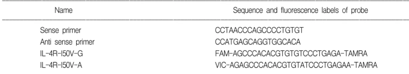
The sequences of the primer and probe used in this study are listed in Table 1. Primer and probe were designed using the Primer Express software version 1.5 (Applied Bio- systems, Foster City, CA). Detection system was ABI PRISM 7900 HT.
Table 1. Sequences of primer and probe
ꠏꠏꠏꠏꠏꠏꠏꠏꠏꠏꠏꠏꠏꠏꠏꠏꠏꠏꠏꠏꠏꠏꠏꠏꠏꠏꠏꠏꠏꠏꠏꠏꠏꠏꠏꠏꠏꠏꠏꠏꠏꠏꠏꠏꠏꠏꠏꠏꠏꠏꠏꠏꠏꠏꠏꠏꠏꠏꠏꠏꠏꠏꠏꠏꠏꠏꠏꠏꠏꠏꠏꠏꠏꠏꠏꠏꠏꠏꠏꠏꠏꠏꠏꠏꠏꠏꠏꠏꠏꠏꠏꠏꠏꠏꠏꠏꠏꠏꠏꠏꠏꠏꠏꠏꠏꠏꠏꠏꠏꠏꠏꠏ
Name Sequence and fluorescence labels of probe
ꠏꠏꠏꠏꠏꠏꠏꠏꠏꠏꠏꠏꠏꠏꠏꠏꠏꠏꠏꠏꠏꠏꠏꠏꠏꠏꠏꠏꠏꠏꠏꠏꠏꠏꠏꠏꠏꠏꠏꠏꠏꠏꠏꠏꠏꠏꠏꠏꠏꠏꠏꠏꠏꠏꠏꠏꠏꠏꠏꠏꠏꠏꠏꠏꠏꠏꠏꠏꠏꠏꠏꠏꠏꠏꠏꠏꠏꠏꠏꠏꠏꠏꠏꠏꠏꠏꠏꠏꠏꠏꠏꠏꠏꠏꠏꠏꠏꠏꠏꠏꠏꠏꠏꠏꠏꠏꠏꠏꠏꠏꠏꠏ
Sense primer CCTAACCCAGCCCCTGTGT
Anti sense primer CCATGAGCAGGTGGCACA
IL-4R-I50V-G FAM-AGCCCACACGTGTGTCCCTGAGA-TAMRA
IL-4R-I50V-A VIC-AGAGCCCACACGTGTATCCCTGAGAA-TAMRA
ꠏꠏꠏꠏꠏꠏꠏꠏꠏꠏꠏꠏꠏꠏꠏꠏꠏꠏꠏꠏꠏꠏꠏꠏꠏꠏꠏꠏꠏꠏꠏꠏꠏꠏꠏꠏꠏꠏꠏꠏꠏꠏꠏꠏꠏꠏꠏꠏꠏꠏꠏꠏꠏꠏꠏꠏꠏꠏꠏꠏꠏꠏꠏꠏꠏꠏꠏꠏꠏꠏꠏꠏꠏꠏꠏꠏꠏꠏꠏꠏꠏꠏꠏꠏꠏꠏꠏꠏꠏꠏꠏꠏꠏꠏꠏꠏꠏꠏꠏꠏꠏꠏꠏꠏꠏꠏꠏꠏꠏꠏꠏꠏ
4. Statistical Analysis
We used the chi-square test to evaluate whether the distribution of genotypes varied significantly between cer- vical cancer group and controls. Odds Ratio (OR) and 95%
confidence intervals (CI) were calculated using logistic re- gression test after age adjusting. Clinicopathological para-
meters were dichotomized as follows: stage (stage Ia vs Ib- IV), nodal statu (>1 vs no positive lymph node), and parametrial invasion status (involvement vs no invol- vement).
RESULTS
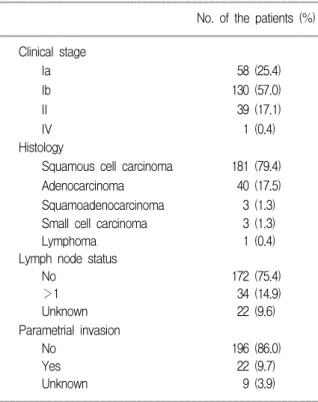
In cancer group, 58 patients were stage Ia, 130 patients were stage Ib, 39 patients were stage II, and 1 patient was stage IV. Concerning the histology, 181 patients (79.4%) were squamous cell carcinoma, 40 (17.5%) were adenocar- cinoma, 3 (1.3%) were squamousadenocarcinoma, 3 (1.3%) were small cell carcinoma, and 1 (0.4%) were lymphoma.
Thirty four patients showed lymph node metastasis and 172 patients did not. Twenty two patients showed parametrial invasion and 196 patients did not (Table 2). Age distribution was significantly different in the cervical cancer group and control group, 53.1±11.1 (range, 30-79) years in cervical cancer group and 47.6±10.6 (range, 21-77) years in control group (p=0.00).
The distribution of homozygotes and heterozygotes clo- sely approximated the expected values under Hardy-Wein- berg equilibrium in cases and controls (p=0.33, chi-square=
0.94; p=0.15, chi-square=2.04). In cervical cancer group, allele frequency of Ile was 46.1%, in comparison with 43.4% in control group which showed no significant di-
Table 3. Allele frequency and genotype distribution in cervical cancer patients and controls
ꠏꠏꠏꠏꠏꠏꠏꠏꠏꠏꠏꠏꠏꠏꠏꠏꠏꠏꠏꠏꠏꠏꠏꠏꠏꠏꠏꠏꠏꠏꠏꠏꠏꠏꠏꠏꠏꠏꠏꠏꠏꠏꠏꠏꠏꠏꠏꠏꠏꠏꠏꠏꠏꠏꠏꠏꠏꠏꠏꠏꠏꠏꠏꠏꠏꠏꠏꠏꠏꠏꠏꠏꠏꠏꠏꠏꠏꠏꠏꠏꠏꠏꠏꠏꠏꠏꠏꠏꠏꠏꠏꠏꠏꠏꠏꠏꠏꠏꠏꠏꠏꠏꠏꠏꠏꠏꠏꠏꠏꠏꠏꠏ
Genotype Cervical Cancer (n=228) Controls (n=204) aOR (95% CI)* p
ꠏꠏꠏꠏꠏꠏꠏꠏꠏꠏꠏꠏꠏꠏꠏꠏꠏꠏꠏꠏꠏꠏꠏꠏꠏꠏꠏꠏꠏꠏꠏꠏꠏꠏꠏꠏꠏꠏꠏꠏꠏꠏꠏꠏꠏꠏꠏꠏꠏꠏꠏꠏꠏꠏꠏꠏꠏꠏꠏꠏꠏꠏꠏꠏꠏꠏꠏꠏꠏꠏꠏꠏꠏꠏꠏꠏꠏꠏꠏꠏꠏꠏꠏꠏꠏꠏꠏꠏꠏꠏꠏꠏꠏꠏꠏꠏꠏꠏꠏꠏꠏꠏꠏꠏꠏꠏꠏꠏꠏꠏꠏꠏ Allele frequency
Val 246 (53.9) 231 (56.6) 1
Ile 210 (46.1) 177 (43.4) 1.10 (0.83-1.15) 0.52
H-W† NS NS
Genotype distribution
Val/Val (%) 61 (26.8) 62 (30.4) 1
Ile/Val (%) 124 (54.4) 107 (52.5) 1.09 (0.70-1.72) 0.70
Ilel/Ile (%) 43 (26.8) 35 (17.2) 1.21 (0.67-2.19) 0.52
Ile/Val or Ilel/Ile (%) 167 (81.2) 142 (69.7) 1.12 (0.73-1.73) 0.60 ꠏꠏꠏꠏꠏꠏꠏꠏꠏꠏꠏꠏꠏꠏꠏꠏꠏꠏꠏꠏꠏꠏꠏꠏꠏꠏꠏꠏꠏꠏꠏꠏꠏꠏꠏꠏꠏꠏꠏꠏꠏꠏꠏꠏꠏꠏꠏꠏꠏꠏꠏꠏꠏꠏꠏꠏꠏꠏꠏꠏꠏꠏꠏꠏꠏꠏꠏꠏꠏꠏꠏꠏꠏꠏꠏꠏꠏꠏꠏꠏꠏꠏꠏꠏꠏꠏꠏꠏꠏꠏꠏꠏꠏꠏꠏꠏꠏꠏꠏꠏꠏꠏꠏꠏꠏꠏꠏꠏꠏꠏꠏꠏ
*Age adjusted odds ratio with 95% confidence interval, †Goodness of fit to the Hardy-Weinberg equilibrium for genotype distribution (NS, not significant)
Table 2. Clinicopathological characteristics of the cervical cancer patients
ꠏꠏꠏꠏꠏꠏꠏꠏꠏꠏꠏꠏꠏꠏꠏꠏꠏꠏꠏꠏꠏꠏꠏꠏꠏꠏꠏꠏꠏꠏꠏꠏꠏꠏꠏꠏꠏꠏꠏꠏꠏꠏꠏꠏꠏꠏꠏꠏꠏꠏꠏꠏꠏ No. of the patients (%) ꠏꠏꠏꠏꠏꠏꠏꠏꠏꠏꠏꠏꠏꠏꠏꠏꠏꠏꠏꠏꠏꠏꠏꠏꠏꠏꠏꠏꠏꠏꠏꠏꠏꠏꠏꠏꠏꠏꠏꠏꠏꠏꠏꠏꠏꠏꠏꠏꠏꠏꠏꠏꠏ
Clinical stage
Ia 58 (25.4)
Ib 130 (57.0)
II 39 (17.1)
IV 1 (0.4)
Histology
Squamous cell carcinoma 181 (79.4) Adenocarcinoma 40 (17.5) Squamoadenocarcinoma 3 (1.3) Small cell carcinoma 3 (1.3)
Lymphoma 1 (0.4)
Lymph node status
No 172 (75.4)
>1 34 (14.9)
Unknown 22 (9.6)
Parametrial invasion
No 196 (86.0)
Yes 22 (9.7)
Unknown 9 (3.9)
ꠏꠏꠏꠏꠏꠏꠏꠏꠏꠏꠏꠏꠏꠏꠏꠏꠏꠏꠏꠏꠏꠏꠏꠏꠏꠏꠏꠏꠏꠏꠏꠏꠏꠏꠏꠏꠏꠏꠏꠏꠏꠏꠏꠏꠏꠏꠏꠏꠏꠏꠏꠏꠏ
fference statistically (p=0.52). Using subject with the Val/
Val homozygote as a reference group, we found no asso- ciation between the Ile/Val and Ile/Ile genotypes and the risk of cervical cancer with age adjusted regression analysis (aOR=1.09, 95% CI=0.70-1.72, p=0.70; aOR=1.21, 95% CI=
0.67-2.19, p=0.60). The prevalence of Ile50Val was higher in cervical cancer group (81.2%) than in control group (69.7%) without significance (Table 3).
Subanalyses of the patients with cervical cancer were conducted according with clinical stage, histologic type, lymph node status, and parametrial invasion status (Table 4). There was no statistically significant interaction between these polymorphisms and these parameters.
DISCUSSION
Although we sought to determine whether IL-4Rα gene polymorphisms of Ile50Val influenced cervical cancer risk, we did not observe any association of Ile50Val with cervical cancer risk.
Some studies have showed the association of IL-4 and cervical cancer. A shift in cytokine, especially IL-4, pro- duction patterns was reported to occur during the progre- ssion from CIN to invasive cervical cancer.12 An increased immunohistochemical expression of intratumoral IL-4 in HGL compared with LGL or normal cervix, and IL-4
mRNA was more expressed in cervical cancer biopsies than in normal or CIN biopsies without significant.13,14 In many cancers, including cervical cancer, ovarian cancer, renal cell carcinoma or colorectal carcinoma, IL-4 expression is also detected in biopsies.4,15-17
With respect to serum concentrations of interleukins, me- dian serum IL-4 in healthy women was higher than in pa- tients with cervical cancer.10 The serum level of IL-4 was not different between patients with CIN and the healthy women.18,19 However, serum IL-6, IL-8, and IL-10 were elevated in patients with cervical cancer and correlate with progression of the disease.20 However, the production of IL-6 may be associated with the signaling through the receptors of IL-4.5-8
The switch form type 1 to type 2 cytokines described in many human cancers mainly depends on upregulation of IL-6 and IL-10 and not IL-4.21 In CIN, the level of IL-10 was increased compared to the controls.17,22 The increased serum level of IL-6 was observed both in CIN and cervical cancer. Furthermore, IL-6 itself has a role in carcinogenesis of uterine cancer.23,24
The Hardy-Weinberg equilibrium suggested no deviation in recruiting the controls. However, the result of this study had low calculation power and large numbers of samples were needed to confirm the lack of association of polymor- phisms of IL-4Rα and risk of cervical cancer. Comparing Table 4. Association of Ile/Val or Val/Val genotype with clinical parameters in cervical cancer patients
ꠏꠏꠏꠏꠏꠏꠏꠏꠏꠏꠏꠏꠏꠏꠏꠏꠏꠏꠏꠏꠏꠏꠏꠏꠏꠏꠏꠏꠏꠏꠏꠏꠏꠏꠏꠏꠏꠏꠏꠏꠏꠏꠏꠏꠏꠏꠏꠏꠏꠏꠏꠏꠏꠏꠏꠏꠏꠏꠏꠏꠏꠏꠏꠏꠏꠏꠏꠏꠏꠏꠏꠏꠏꠏꠏꠏꠏꠏꠏꠏꠏꠏꠏꠏꠏꠏꠏꠏꠏꠏꠏꠏꠏꠏꠏꠏꠏꠏꠏꠏꠏꠏꠏꠏꠏꠏꠏꠏꠏꠏꠏꠏ
Clinical parameter Val/Val Ile/Val or Ile/Ile p-value
ꠏꠏꠏꠏꠏꠏꠏꠏꠏꠏꠏꠏꠏꠏꠏꠏꠏꠏꠏꠏꠏꠏꠏꠏꠏꠏꠏꠏꠏꠏꠏꠏꠏꠏꠏꠏꠏꠏꠏꠏꠏꠏꠏꠏꠏꠏꠏꠏꠏꠏꠏꠏꠏꠏꠏꠏꠏꠏꠏꠏꠏꠏꠏꠏꠏꠏꠏꠏꠏꠏꠏꠏꠏꠏꠏꠏꠏꠏꠏꠏꠏꠏꠏꠏꠏꠏꠏꠏꠏꠏꠏꠏꠏꠏꠏꠏꠏꠏꠏꠏꠏꠏꠏꠏꠏꠏꠏꠏꠏꠏꠏꠏ Clinical stage
Ia 13 44 0.47
Ib-IV 48 123
Histologic type
Squamous cell carcinoma 46 135 0.55
Others 14 33
Lymph node status
0 49 123 0.55
>1 8 26
Parametrial invasion
No 49 147 0.11
Yes 9 13
ꠏꠏꠏꠏꠏꠏꠏꠏꠏꠏꠏꠏꠏꠏꠏꠏꠏꠏꠏꠏꠏꠏꠏꠏꠏꠏꠏꠏꠏꠏꠏꠏꠏꠏꠏꠏꠏꠏꠏꠏꠏꠏꠏꠏꠏꠏꠏꠏꠏꠏꠏꠏꠏꠏꠏꠏꠏꠏꠏꠏꠏꠏꠏꠏꠏꠏꠏꠏꠏꠏꠏꠏꠏꠏꠏꠏꠏꠏꠏꠏꠏꠏꠏꠏꠏꠏꠏꠏꠏꠏꠏꠏꠏꠏꠏꠏꠏꠏꠏꠏꠏꠏꠏꠏꠏꠏꠏꠏꠏꠏꠏꠏ
the equilibrium of Ile50Val between the controls of this study and those of Japanese and German studies, we found different genotype distributions of Val/Val, Val/Ile, and Ile/Ile (p=0.06; p=0.00).5,25 However, the genotype distribu- tion of this study did not show difference in comparison with other Korean population study (p=0.89).26 Ethnic diffe- rence could make disappointed results of this study.
In addition to the ethnic difference which influences the genetic distribution, other risk factors are also important to the vulnerability of cervical cancer. Although HPV infection is a key factor of cervical cancer, especially squamous cell carcinoma, other environment factors such as cigarette smo- king or other immunocompromised status must be thought as causes of cervical cancer.
Smoking itself can suppress the immune system and alter the cytokine expression. Smoking was thought to modulate immune response by a direct effect on the cells that produce the type 2 cytokines involved in asthma and allergy, including IL-4. Evidence for this mechanism is supported by the findings of higher level of IL-4 in smokers compared to nonsmokers and in endothelial cell lines stimulated by cigarette smoke condensate.27,28 However, no significant differences in IL-4 level by cigarette consumption were observed in the study with nonasthmatic monozygotic twins.29 Concerning the IL-4, changes of the level of IL-4 after exposure to smoking were not constant. Increased level of IL-6 was observed in the cell line study and the production of IL-6 may be stimulated by smoking in the cervix.29,30 Coinfection of HIV and HPV could alter the cytokine expression compared to HPV infection alone, which increased numbers of cells expressing IL-4, IL-6, and IL-8.31 Analyzing the significance of genetic variants of IL-4Rα polymorphisms of Ile50Val without considering above factors could lead false results.
There was no association between variants of genotypes and clinical parameters such as stage, histology types, lymph node involvement, and parametrial invasion. There- fore, with these results, we may postulate that polymor- phisms of IL-4Rα did not promote the progress of cervical cancer. In a previous study, although no clear correlation
was observed between the levels of IL-4 mRNA expression and the clinical stage of patients with detectable cytokine mRNA expression, patients with undetectable cytokine mRNA expression more often presented with advanced stage cervical carcinoma. Down regulation of IL-4 in advanced stage cancers was considered to be a tumor escape me- chanism.4
Ile50Val can signal increased Stat 6-dependent transcrip- tional activity, which is known to be correlated with an increased risk of atopic disease.11,32 Aberrant activation of Stat-signaling gives rise to different pathological event.
Generally, one group of Stat proteins including Stat 2, Stat 4, and Stat 6, is thought to be activated by a small number of cytokines and play a distinct role in the development of T-cells and in IFNγ signaling. The other group including Stat 1, Stat 3, and Stat 5, plays an important role in controlling cell-cycle progression and apoptosis and thus contributes to oncogenesis.33,34 However, in the viewpoint of immunosurveillance of tumor, Stat 6-deficiency was sug- gested as a potent strategy for immunotherapy.35
This is the first article which explored the association between the polymorphisms of IL-4Rα gene, Ile50Val, and cervical cancer. In summary, we did not provide any evi- dence that Korean women with the polymorphisms of Ile50Val in IL-4Rα genes had an altered risk of develop- ment and progression of cervical cancer.
REFERENCES
1. Bekkers RA, Massuger LF, Bulten J, Melchers WJ. Epidemio- logy and clinical aspects of human papillomavirus detection in the prevention of cervical cancer. Rev Med Viol 2004; 14:
95-105.
2. Hazelbag S, Fleuren GJ, Baelde JJ, Schuuring E, Kenter GG, Gorter A. Cytokine profile of cervical cancer cells. Gynecol Oncol 2001; 83: 235-43.
3. Petry KU, Scheffel D, Bode U, Gabrysiak T, Kochel H, Kups- ch E, et al. Cellular immunodeficiency enhances the progre- ssion of human papillomavirus-associated cervical lesions. Int J Cancer 1994; 57: 836-40.
4. Gey A, Kumari P, Sambandam A, Lecuru F, Cassard L, Ba- doual C, et al. Identification and characterisation of a group of cervical carcinoma patients with profound downregulation of intratumoral Type 1 (IFNγ) and Type 2 (IL-4) cytokine
mRNA expression. Eur J Cancer 2003; 39: 595-603.
5. Nakamura E, Megumi Y, Kobayashi T, Kamoto T, Ishitoya S, Terachi T, et al. Genetic polymorphisms of the Inter- leukin-4 receptor α gene are associated with an increasing risk and a poor prognosis of sporadic renal cell carcinoma in a Japanese population. Clin Cancer Res 2002; 8: 2620-5.
6. Strome SE, Kawakami K, Alejandro D, Voss S, Kasperbauer JL, Salomao D, et al. Intereleukin 4 receptor-directed cyto- toxin therapy for human head and neck squamous cell car- cinoma in animal models. Clin Cancer Res 2002; 8: 281-6.
7. Kawakami M, Kawakami K, Stepensky VA, Maki RA, Robin H, Muller W, et al. Interleukin 4 receptor on human lung cancer: a molecular target for cytotoxin therapy. Clin Cancer Res 2002; 8: 3503-11.
8. Essner R, Huynh Y, Nguyen T, Rose M, Kojimi M, Hoon DS. Functional interleukin-4 receptor and I terleukin-2 recep- tor common gamma chain in human gastric carcinoma: a possible mechanism for cytokine-based therapy. J Gastrointest Surg 2001; 5: 81-90.
9. Kawakami K, Kawakami M, Leland P, Puri RK. Internali- zation property of Interleukin-4 receptor α chain increases cytotoxic effect of interleukin-4 receptor-targeted cytotoxin in cancer cells. Clin Cancer Res 2002; 8: 258-66.
10. Lebrechet A, Hefler L, Tempfer C, Koelbl H. Serum cytokine concentrations in patients with cervical cancer: interleukin-4, interferon-γ, and monocyte chemoattractant protein-1. Gyne- col Oncol 2001; 3: 170-1.
11. Mitsuyasu H, Izuhara K, Mao XQ, Gao PS, Arinobu Y, Eno- moto T, et al. Ile50Val variant of IL4R alpha upregulates IgE synthesis and associates with atopic asthma. Nat Genet 1998;
19: 119-20.
12. Clerici M, Shearer GM, Clerici E. Cytokine dysregulation in invasive cervical carcinoma and other human neoplasias: time to consider the TH1/TH2 paradigm. J Natl Cancer Inst 1998;
90: 261-3.
13. al-Saleh W, Giannini SL, Jacobs N, Moutschen M, Doyen J, Boniver J, et al. Correlation of T-helper secretory differenti- ation and types of antigen-presenting cells in squamous intraepithelial lesions of the uterine cervix. J Pathol 1998; 184:
283-90.
14. Gruijil TD, Bontkes HJ, Muysenberg AJC, Oostveen JW, Stu- kart MJ, Verheijen RHM, et al. Differences in cytokine mRNA profile between premalignant and malignant lesions of the uterine cervix. Eur J Cancer 1999; 35: 490-7.
15. Rabinowich H, Suminami Y, Reichert TE, Crowley-Nowick P, Bell M, Edwards R, et al. Expression of cytokine genes or proteins and signaling molecules in lymphocytes associated with human ovarian carcinoma. Int J Cancer 1996; 68: 276-84.
16. Nakagomi H, Pisa P, Pisa EK, Yamamoto Y, Halapi E, Backlin K, et al. Lack of interleukin-2 (IL-2) expression and selective expression of IL-10 mRNA in human renal cell carcinoma. Int J Cancer 1995; 63: 366-71.
17. Piancatelli D, Romano P, Sebastiani P, Adorno D, Casciani CU. Local expression of cytokines in human colorectal car-
cinoma: evidence of specific inteleukin-6 gene expression. Int Immunother 1999; 22: 25-32.
18. Jacobs N, Giannini SL, Doyen J, Baptista A, Moutschen M, Boniver J, et al. Inverse modulation of IL-10 and IL-12 in the blood of women with preneoplastic lesions of the uterine cervix. Clin Exp Immunol 1998; 111: 219-24.
19. Pardo-Govea T, Callejas D, Nunez-Troconis J, Araujo M, Co- sta L, Pons H, et al. Gamma interferon (IFN-gamma), tumor necrosis factor alpha (TNF-alpha) and interleukins 2, 4 and 6 (IL-2, IL-4, IL-6) in cervical-uterine cells of intraepithelial neoplasia: a preliminary report. Invest Clin 2005; 46: 5-13.
20. Chopra V, Dinh TV, Hannigan EV. Circulating serum levels of cytokines and angiogenic factors in patients with cervical cancer. Cancer Invest 1998; 16: 152-9.
21. Gastl GA, Abrams JS, Nanus DM, Oosterkamp R, Silver J, Liu F, et al. Interleukin-10 production by human carcinoma cell lines and its relationship to interleukin-6 expression. Int J Cancer 1993; 55: 96-101.
22. Azar KK, Tani M, Yasuda H, Sakai A, Inoue M, Sasagawa T. Increased secretion patterns of interleukin-10 and tumor ne- crosis factor-alpha in cervical squamous intraepithelial lesions.
Hum Pathol 2004; 35: 1376-84.
23. Wei LH, Kuo ML, Chen CA, Chou CH, Lai KB, Lee CN, et al. Interleukin-6 promotes cervical tumor growth by VEGF- dependent angiogenesis via a STAT3 pathway. Oncogene 2003; 22: 1517-27.
24. Wei LH, Kuo ML, Chen CA, Chou CH, Cheng WF, Chang MC, et al. The anti-apoptotic role of interleukin-6 in human cervical cancer is mediated by up-regulation of Mcl-1 through a PI 3-K/Akt pathway. Oncogene 2001; 20: 5799-809.
25. Klein W, Tromm A, Griga T, Fricke H, Fowaczny C, Hocke M, et al. Interleukin-γ and interleukin-4 receptor gene poly- morphisms in inflammatory bowel diseases. Genes Immun 2001; 2: 287-9.
26. Lee SB, Kim BS, Kim JH, Lee SY, Lee SY, Choi SO, et al. Gene-gene interaction between interleukin-4 and inter- leukin-4 receptor α in Korean children with asthma. Clin Exp Allergy 2004; 34: 1202-8.
27. Byron KA, Varigos GA, Wootton AM. IL-4 production is increased in cigarette smokers. Clin Exp Immunol 1994; 95:
333-6.
28. Nordskog BK, Fields WR, Hellmann GM. Kinetic analysis of cytokine response to cigarette smoke condensate by human endothelial and monocytic cells. Toxicology 2005; 212: 87-97.
29. Cozen W, Diaz-Sanchez D, Gauderman WJ, Zadnick J, Cock- burn MG, Gill PS, et al. Th1 and Th2 cytokines and IgE levels in identical twins with varying levels of cigarette consumption.
J Clin Immunol 2004; 24: 617-22.
30. Wolfgang E, Christof W, Peter F, Mamood M, Klaus C. The influence of cotinine on interleukin 6 expression in smokers with cervical preneoplasia. Acta Obstet Gynecol Scand 2000;
79: 1105-11.
31. Nicol AF, Fernandes AT, Grinsztejn B, Russomano F, Silva JRE, Tristao A, et al. Distribution of immune cell subsets and
cytokine-producing cells in the uterine cervix of human papil- lomavirus (HPV)-infected women: influence of HIV-1 coin- fection. Diagn Mol Pathol 2005; 14: 39-47.
32. Stephenson L, Johns MH, Woodward E, Mora AL, Boothby M. An IL-4Rα allelic variant, 150, acts as a gain-of function variant relative to V50 for Stat6, but nor Th2 differentiation.
J Immunol 2004; 173: 4523-8.
33. Bromberg JF. Stat proteins and oncogenesis (review). J Clin Invest 2002; 109: 1139-42.
34. Calo V, Migliavacca M, Bazan V, Macaluso M, Buscemi M, Gebbia N, et al. Stat proteins: from normal control of cellular events to tumorigenesis. J Cell Physiol 2003; 197: 157-68.
35. Ostrand-Rosendberg S, Sinha P, Clements V, Dissanayake SI, Miller S, Davis C, et al. Signal transducer and activator of transcription 6 (Stat6) and CD1: inhibition of immunosur- veillance against primary tumors and metastatic disease. Can- cer Immunol Immunother 2004; 53: 86-91.
Interleukin-4수용체α 유전자 다형성과 자궁경부암 발생위험도 및 임상인자 사이의 연관성 분석
서울대학교 의과대학 산부인과학교실, 암연구소 이상은․김재원․박노현․송용상․강순범․이효표
목적:Interleukin (IL)-4수용체α의 다형성인 Ile50Val 유전 변이는 IL-4에 대한 IL-4수용체의 반응을 증가시킨다.
IL-4는 자궁경부세포주에서 발현되고 자궁경부상피내암에서 자궁경부암으로의 진행하는 발암과정에 작용을 하는 것으 로 알려져 있다. 따라서, 우리는 본 연구를 통해서 IL-4수용체α 유전자의 Ile50Val 다형성이 한국 여성의 자궁경부암 의 발생과 진행의 위험을 증가시키는지의 여부를 알아보고자 하였다.
연구 방법:1999년부터 2002년까지 서울대학교병원에서 자궁경부암을 진단받은 228명과 서울대학교병원 강남센터 에서 자궁경부암이 없는 것을 확인한 204명의 대조군의 혈액을 사용하였다. 그리고 PCR 증폭과 Taqman 분석을 이용 하였다. 자궁경부암과 대조군의 유전형이 차이를 보기 위해서 chi-square 검사를 하였다. 나이를 보정한 후에 선형회귀 분석을 통해서 교차비와 95%신뢰구간을 계산하였다.
결과:동종접합체와 이형접합체의 분포는 환자군과 대조군에서 Hardy-Weinberg 평형을 이루었다(p=0.33, chi-squre=
0.94; p=0.15, chi-square=2.04). 자궁경부암환자에서 Ile 대립유전자의 빈도가 46.1%이고, 대조군에서는 43.4%로 의미있 는 차이를 보이지 않았다(p=0.52). Val/Val 동종접합체를 참고로 비교하면, 연령보정 선형회귀분석을 통해 Ile/Val와 Ile/Ile 유전자가 자궁경부암의 발생위험과 관련이 없다는 것을 알 수 있었다(aOR=1.09, 95% CI=0.70-1.72, p=0.7;
aOR=1.21, 95% CI=0.67-2.19, p=0.52). 자궁경부암을 임상병기, 조직병리, 림프절 전이, 자궁방 침범의 상태에 따라서 분 석을 했을 때, 이러한 유전자의 다형성과 관련이 없었다.
결론:IL-4수용체α 유전자의 다형성은 한국인에서 자궁경부암의 발생 위험을 증가시키지 않고 자궁경부암의 진행 에 영향을 주지 않는다.
중심단어:IL-4, IL-4수용체α 유전자, Ile50Val 다형성, 자궁경부암