하악 구치부위에 식립한 358개 임플란트의 생존율에 대한 3년간 후향적 연구
서울 보훈병원 치과센터
윤이권․이기․이동운․최주영․유정아․박필규․김정희
최근 무치악 부위를 대신하여 광범위하게 식립되고 있는 치과용 임플란트는 높은 임상 성공률을 보이고 있으나 실패 가능성은 늘 존재하며 이는 예기치 않은 경우가 많다. 임플란트의 실패과 관련하여 여러 가지 요인들이 거론 되고 있으나, 아직 논란의 여지가 있다. 본연구에서는 서울 보훈병원에서 2005-2006년에 하악 구치부위에 임플란 트를 식립한 212명의 환자, 총 358개의 임플란트를 대상으로 환자의 연령, 임플란트 식립 부위, 시스템, 직경과 길이, 골이식 유무에 따른 생존율을 비교하였다. 각 요소에 따른 생존율을 SPSS chi-square test를 이용한 multi-variable analysis를 시행하여 관련성을 검증하였다. 연구 결과 3년간 98.3% 의 누적 생존율을 보였으며, 조사 한 요인 중 임플란트 직경만이 임플란트 생존율과 관련성이 있는 것으로 나타났다. 이는 넓은 직경의 임플란트가 주로 대체(rescue) 임플란트 혹은 골질이 좋지 않은 부위에 사용되는 경우가 많은 것이 원인으로 작용한 것으로 사료된다. 향후 임플란트의 성공과 관련하여 명확한 지표를 제공할 수 있는 지속적인 연구가 필요할 것이다.
주요어: 생존율, 실패요인, 임플란트, 직경, 하악구치부 (구강회복응용과학지 2010:26(1):59~68)
서 론
임플란트는 1985년 Brandmark에 의해 개발된 이후로1 최근 십년간 부분 또는 완전 무치악 환 자의 치아를 대치하는 치료방법으로 널리 쓰이 고 있다. 치료 결과에 대한 수많은 임상적 연구 들은2,3 임플란트 시스템의 종류와 상관없이 91-99% 정도로 높은 성공률을 보고하고 있다.4,5 Testori 등은 하악 구치부에서 누적 성공률이 99.4%로 매우 높았다고 보고하였으며, 이밖에도 대부분의 연구에서 하악 구치부는 상악 구치부 에 비해 높은 생존율을 나타내는 것으로 보고되
교신저자:김정희
서울보훈병원 치과센터, 서울시 강동구 둔촌동, 134-791, 대한민국 팩스: +82-2-2225-1659 이메일: [email protected]
원고접수일: 2009년 11월 05일, 원고수정일: 2009년 12월 20일, 원고채택일: 2010년 03월 25일
고 있다.6,7 그러나 일부 임상 연구에서는 이와는 정반대의 결과를 보여주고 있으며,8,9 저자들은 하악 구치부에서 하악관의 위치로 인해 임플란 트 길이가 제한되며 bicortical stabilization을 얻기 어려운 점을 그 원인으로 꼽았다.
높은 성공률에도 불구하고 실제 임상에서 임 플란트 실패가능성은 항상 존재하며 예기치 않 은 경우가 많다.10,11 임플란트의 실패는 골유착이 일어나기 전의 초기 실패와, 보철적 하중을 가한 후에 발생하는 후기 실패로 나눌 수 있으며,12실 패 원인으로 임플란트 자체의 특성, 부적합한 골 양이나 골질, 골삭제시 과도한 열발생으로 인한
골괴사, 감염 등의 수술적 합병증, 교합 과부하, 임플란트 주위염 등이 거론되고 있다.13,14
실패요인을 분석하고 양호한 예후를 위한 치 료지침을 확립하는데 있어서 임플란트 시술 후 의 평가는 필수적이며, 따라서 각 기관 나름대로 의 술 후 분석이 의의가 있다하겠다. 본 연구에 서는 서울 보훈병원에서 2005-2006년에 임플란 트를 식립한 환자들을 대상으로 환자의 나이, 식 립위치, 임플란트 직경, 길이, 골이식에 따른 후 향적 평가를 시행하여 각 조건에 따른 생존율을 비교하여, 향후 임플란트 치료시 높은 성공률을 위한 지표를 제시하고자 한다.
연구재료 및 방법 1. 실험 대상
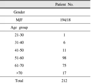
2005년부터 2006년까지 서울 보훈병원 치과진 료처에 내원하여 하악 구치부위에 임플란트를 식립한 총 212명의 환자를 대상으로 조사를 시 행하였다. 총 212명의 환자중 남자 194명, 여자 18명이며, 연령층은 26세부터 78세까지, 평균나 이는 59세였다.(Table Ⅰ)
Patient No.
Gender
M/F 194/18
Age group
21-30 1
31-40 6
41-50 11
51-60 98
61-70 75
>70 17
Total 212
Table Ⅰ. Distribution of Patients
하악 구치부에 식립된 임플란트의 개수는 총 358로, 사용된 임플란트 시스템은 Paragon (Zimmer dental, USA), Biohorizons(Biohorizons,
Implant No.
Implant system
replace 42
paragon 127
camlog 115
bio-horizons 74
Position
1st premolar 21
2nd premolar 58
1st molar 161
2nd molar 118
Implant length
8.0-9.5 14
10.0-11.5 165
12-13 179
Implant diameter
3.0-3.9 62
4.0-4.9 213
>5.0 83
Surgical stage
1 stage 149
2 stage 209
bone graft
nagative 237
augmented 121
Total No. 358
Table Ⅱ. Distribution of Implants
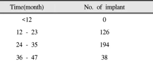
USA), Replace(Nobel-Biocare, sweden), Camlog (Alteatec, Germany) 4가지 였으며, 특별한 선별 기준은 적용되지 않았고 본원에서 일반적으로 식립된 시스템으로 선정하였다. 시스템에 따른 임플란트 표면 유형은 Paragon, Biohorizons의 경 우 HA coated type, Camlog의 경우 etched type, Replace의 경우 oxide type이었다. 모든 임플란트 가 Internal hexa type이었으며 0.5-1.5mm의 machined collar를 포함하였다.(Table Ⅱ) 추적기 간은 17-41개월이었으며 평균 추적기간은 27.3개 월이었다.(Table Ⅲ)
모든 임플란트는 다음과 같은 요건을 만족하 였다.
1) 부분 무치악 부위에 식립
2) 하악 소구치와 대구치 부위에 식립 3) 단일 혹은 다수의 임플란트 4) 고정성 보철물로 수복됨 5) 임시 보철물은 사용되지 않음 2. 술식 및 변수
1) 치유 기간: 모든 임플란트는 보철적 하중을 가 하기 전 3-9개월의 치유 기간을 거쳤다. 149개 의 임플란트는 식립 당일 치유 지대주를 연결 하였으며 대부분의 209개에서 3개월 이후에 이차 수술을 시행하였다.
2) 식립 위치: 하악 제1소구치, 제2소구치, 제1대 구치, 제2대구치 부위에 각각 21개(5.9%), 58 개(16.2%), 161개(45.0%), 118개(33.0%)의 임플 란트가 식립되었다.
Time(month) No. of implant
<12 0
12 - 23 126
24 - 35 194
36 - 47 38
Table Ⅲ. No. of Patient Followed in Study
3) 임플란트 직경: 임플란트 직경은 3.5-6.0mm이 며, 5.0mm 이상의 넓은 직경 임플란트는 83개 (23.2%)였다.
4) 임플란트 길이: 9-13mm의 임플란트가 식립되 었으며 10mm 미만 14개(3.9%), 10-11.5mm 165개(46.1%), 12-13mm 179개(50%)였다.
5) 골이식: 전체 358개 임플란트 중 골이식은 121 개(33.8%)에서 시행되었다. 이식 재료는 자가 골, 동종골, 이종골로 다양하였으며, 이 중 45 개(12.6%)는 차폐막 없이 골이식만을 시행하 였으며, 76개 임플란트(21.2%)에서는 차폐막 과 함께 골이식을 시행하였다.
3. 임플란트 생존의 기준
임상적, 방사선학적 방법을 통해 생존여부를 평가하였으며, 그 기준은 다음과 같다.15 1) 동요도가 존재하지 않음
2) 동통이나 감각이상 등의 임상증상이 존재하 지 않음
3) 방사선 사진상 임플란트 주위 방사선 투과성 병소가 존재하지 않음
4. 통계학적 방법
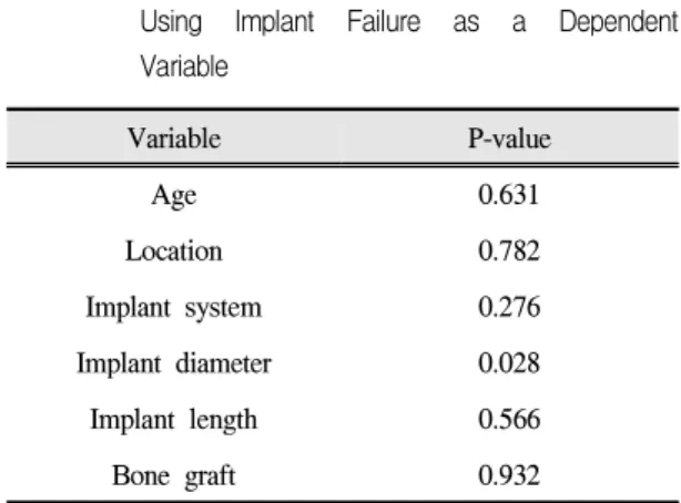
SPSS 프로그램의 chi-square test를 이용하여 각 항목과 임플란트 생존율과의 상관관계에 대하여 multi-variable analysis를 시행하였으며, P-values
<0.05의 범위에서 통계학적으로 유의한 것으로 간주하였다. 관찰 기간 중 Kaplan-Meier 생존분 석을 시행하였다.
결 과
1. 생존율 비교
총 358개 임플란트 중 6개의 임플란트가 제거 되었으며, 3년간 누적생존율은 98.3%였다. 임플 란트 생존율과 환자의 연령, 수술부위, 임플란트
직경과 길이, 골이식 시행여부 등의 상관관계를 Chi-square test를 이용한 multi-variable analysis를 이용하여 분석하였다.
1) 연령
연구에 포함된 총 212명의 환자 중 6명의 환자 에서 임플란트 실패가 발생하였으며 모든 실패 가 60세 이전의 환자에게서 발생하였다. 30대, 40 대, 50대에서 각각 1명,1명,4명의 환자에게서 임 플란트가 제거되었으나 임플란트의 생존율에 있 어서 연령에 따른 상관관계는 나타나지 않았다.
2) 수술부위
제1소구치, 제2소구치, 제1대구치, 제2대구치 부위에서 각각 0/1/2/3개의 임플란트가 제거되었 다. 부위에 따른 생존율은 각각 100%, 98.3%, 98.8%, 97.5%로 제2대구치 부위에서 가장 낮게 나타났으나 통계학적으로 유의한 차이는 없었다.
3) 임플란트 시스템, 직경, 길이
하악 구치부위에 식립된 총 358개의 임플란트 중 Biohorizons, Camlog, Paragon 제품군에서 각각 3개, 2개, 1개의 임플란트가 제거되었다.
Biohorizons 임플란트가 6개중 3개로 가장 개수 가 많았으며 95.9%의 생존율을 보였으나 임플란 트 생존율에 있어 시스템간 유의한 통계학적 차 이는 없었다.
임플란트 직경에 따른 생존율을 살펴보면, 직 경 5.0mm 이상의 넓은 직경의 임플란트에서 83 개 중 4개의 임플란트가 제거되어(4.8%), 273개 중 2개의 임플란트가 제거된(0.7%) 표준 직경의 임플란트에 비해 임플란트의 생존율이 유의성있 게 낮았다.(p=0.025)
길이 10mm 미만의 짧은 길이의 임플란트에서 는 실패가 관찰되지 않았으며 실패한 6개의 임 플란트 모두 길이 10mm 이상이었다.(10-13mm) 길이 10mm 이상의 임플란트의 생존율은 98.3%
로 짧은 임플란트에 비해(100%) 낮았으나 통계 학적 유의성은 없었으며, 길이 10mm 미만의 짧
은 임플란트 개수가 총 14개로 매우 적었다.
4) 골이식 유뮤
골이식을 시행하지 않은 총 237개의 임플란트 중 4개의 임플란트가 제거되어 98.3%의 생존율 을 보였다. 골이식을 시행한 121개의 임플란트 중 2개의 임플란트가 제거되었으며(생존율 98.3%) 1개의 임플란트는 이종골(Bio-Oss, Osteohealth, NY, US)을 이용한 골이식을 시행하 였고, 다른 1개의 임플란트는 자가골(block bone graft)과 이종골(Bio-Oss)을 이용하여 골이식을 시 행하고 차폐막(Bio-Gide, Geistlich Biomaterials, Wohlhusen, Switzerland)을 적용하였다. 골이식을 시행한 경우와 시행하지 않은 경우에 있어서의 임플란트 생존율은 두 경우 모두에서 98.3%로 차이를 보이지 않았다.
조사 결과 여러 가지 요소 중 임플란트 직경만 이 임플란트 생존율과 관련이 있는 것으로 나타 났으며(p<0.05), 환자의 연령, 수술 부위, 임플란 트 길이, 골이식 시행여부와 임플란트 생존율과 의 관련성은 없는 것으로 나타났다.(Table Ⅳ) 2. 실패의 유형과 시기
6개의 임플란트 중 5개의 임플란트가 기능 부 하 전 제거되어 골유착의 실패에 기인한 초기실
Variable P-value
Age 0.631
Location 0.782
Implant system 0.276
Implant diameter 0.028
Implant length 0.566
Bone graft 0.932
Table Ⅳ. Multivariate Logistic Regression Analysis Using Implant Failure as a Dependent Variable
패가 많은 것으로 나타났다. 1개의 임플란트만이 기능 부하 이후에 제거되었다.(Table Ⅴ) 초기 실 패한 임플란트의 평균 생존 기간은 12.2주였고,
Total No.
placed
Total No. of early failure
Total No. of late failure
Survival rate
358 5 1 98.3%
Table Ⅴ. Cumulative Survival Rate of Implants
Interval (month)
Failed implant
Interval survival rate
Cumulative survival rate
1 to 6 4 98.9 98.9
7 to 12 2 99.4 98.3
13 to 18 0 100 98.3
19 to 24 0 100 98.3
25 to 30 0 100 98.3
31 to 36 0 100 98.3
Table Ⅵ. Interval/ Cumulative Survival Rate of Implants
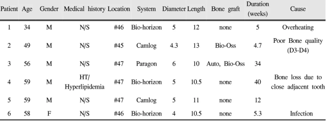
Patient Age Gender Medical history Location System Diameter Length Bone graft Duration
(weeks) Cause
1 34 M N/S #46 Bio-horizon 5 12 none 5 Overheating
2 49 M N/S #45 Camlog 4.3 13 Bio-Oss 4.7 Poor Bone quality
(D3-D4)
3 56 M N/S #47 Paragon 6 10 Auto, Bio-Oss 34
4 59 M HT/
Hyperlipidemia #47 Bio-horizon 5 10.5 none 40 Bone loss due to close adjacent tooth
5 59 M N/S #47 Camlog 5 11 none 12
6 58 F N/S #46 Bio-horizon 4 10.5 none 5.3 Infection
Table Ⅶ. Details of Failed Implants
후기 실패한 임플란트의 생존 기간은 40주였 다.(Table Ⅵ), (Fig. 1)
실패원인은 식립시의 과도한 열방생으로 인한 골괴사, 감염 등의 수술적 합병증과 식립위치나 식립각도 불량으로 인한 골소실, 골질 불량 (D3-D4)으로 인한 골유착의 실패 등으로 추정되 었다.(Table Ⅶ)
Fig 1. Kaplan-Meier Survival analysis
총괄 및 고안
악골의 총 미네랄 함량, 골양 및 골질은 나이 가 증가함에 따라 감소하며16 혈액공급 및 창상 치유 또한 느려진다.17 이러한 변화로 인해 60세 이상의 고령의 환자에게서는 임플란트 골유착이 더 어려울 것으로 예상되지만, 고령의 환자에서 도 임플란트 골유착의 성공 유무 및 성공률은 큰 차이가 없다는 연구결과가 있으며, 여러 임상 연 구들도 이와 일치하는 결과를 보여주고 있다.18,19 60세 이상의 고령의 환자에게서 임플란트 성공 률은 94~97%로 보고되고 있으며,20,21 Jemt 등은 80~90세의 환자군에서 보철 수복 1년 후 평균 골 소실 양이 0.2~0.3mm로 중년환자 그룹에 비해 높지 않다고 보고하였다.22 이번 연구의 결과에 서도 환자의 연령은 임플란트 생존율과 무관한 것으로 나타났다.
그러나 오랜 무치악 기간으로 인한 치조제의 퇴축, 관절염이나 골다공증과 같은 근골격계 질 환, corticosteroid, bispphosphate 등의 약물 복용으 로 인한 골의 성질의 변화와 이 밖에 여러가지 전신질환과 관련된 위험인자들이 고령의 환자군 에서는 존재할 가능성이 높으며 따라서 임플란 트를 시행할 경우 주의 깊은 접근이 필요하다.
하악구치 부위는 후방으로 갈수록 교합력의 크 기가 증가하고 치조제의 흡수, 하치조신경의 위 치, 골밀도의 감소 등 해부학적 한계점이 존재한 다. 이로인해 bicortical stabilization을 얻기가 어 렵고, 임플란트 길이나 직경이 제한될 수 있으며 이는 곧 임플란트 성공에 영향을 미치게 된
다.8,9,23,24 부위에 따른 차이로는 소구치 부위가
대구치 부위에 비해 하중을 적게 받으면서 골질 이 양호하기 때문에 대구치 부위보다 일반적으 로 예후가 양호한 것으로 보고되고 있다.25Block 등은 하악 대구치 부위에서의 단일 임플란트 성 공률이 제1대구치 부위에서 78.5%, 제2대구치 부 위에서 71.8%로 매우 낮았다고 보고하였으며,26 Misch는 하악 제2대구치 부위의 골이 덜 치밀하 며 수술시 시야확보와 접근이 어렵고 하악관의
위치로 인해 충분한 길이의 임플란트 식립이 어 려우며 교합력이 제1대구치에 비해 10%정도 더 발생하고 측방 균형 간섭이 빈번하다는 점 등 여 러 가지 단점이 있다고 하였다.27그러나 이번 연 구에서는 식립부위간 임플란트 생존율의 차이는 없었으며, 모든 부위에서 높은 생존율을 보였다.
임플란트 직경이 증가하면 임플란트의 표면적이 증가하게 되며 이는 치밀골과 더 넓은 면적에서 접촉이 일어나게 한다. Matsushita 등이 임플란트 직경과 응력분산에 대해 조사한 결과, 임플란트 직경이 증가할수록 상부 치조골에서의 응력이 감소하였다는 사실을 보고한 바 있으며,28Friberg 등의 연구에 따르면 3.75mm와 4.0mm 직경의 임 플란트군에 비해 5.0mm 직경의 그룹에서 더 적 은 변연골소실을 보였는데, 보철 1년 후 3.75/4.0/5.0mm 직경의 임플란트에서 각각 1.2/1.3/0.8mm의 골소실이 나타났으며, 보철 3년 후 0.2/0.1/0.1mm의 골소실이 나타난 것으로 보고 되었다.29Rabbit tibia를 이용한 동물 실험 논문에 서도 임플란트 직경이 증가할수록 제거할 때 드 는 힘(removal torque value)이 통계적으로 유의성 있게 컸다.30 이 밖에도 넓은 직경의 임플란트가 표준 직경의 임플란트에 비해 좀 더 좋은 예후를 보인다는 여러 임상보고가 있었으나, 추적 기간 이 비교적 짧았다.31,32
이에 반해 Ivanoff 등의 연구에서는 5.0mm 직 경의 임플란트에서 18%, 4.0mm 직경의 임플란 트에서 3%, 3.75mm 직경의 임플란트에서 5%의 실패율을 보고하고 있어 정반대의 결과를 나타 낸다.33 이와 유사하게 넓은 직경의 임플란트가 더 높은 실패율을 보인다는 여러 임상 연구 결과 가 있었는데, 저자들은 그 이유를 넓은 직경의 임플란트가 주로 대체(rescue) 임플란트 혹은 골 질이 좋지 않은 부위에 사용되는 경우가 많으며, 임플란트 형태의 변화와 초기의 시행착오 때문 인 것으로 해석하였다.34,35 넓은 직경의 임플란트 의 경우 많은 실패가 기능부하 전에 일어났으며 대부분 골유착의 실패에서 기인하였다.
이번 논문의 결과에서도 5.0mm 이상의 넓은
직경의 임플란트에서 임플란트의 3년간 생존율 이 유의성있게 낮았으며, (p=0.025) 초기 실패율 이 높게 나타났다.(75%) 이번 연구의 결과는 Ivanoff가 이전에 언급한 원인과 관련이 있을 것 으로 생각되며, 여러 가지 조절되지 않은 요소들 이 존재하여 엄격한 비교가 이루어 지지 않았다 는 한계점을 보인다. 임상적으로 구치부에서 넓 은 직경의 임플란트 사용은 표면적의 증가, 상부 치조골에서의 응력분산, 유리한 치경 외형 등 명 백한 장점을 갖으며36 적절한 증례에서 적합한 외과적 술식으로 식립된다면 양호한 임상적 결 과를 보일 것으로 사료된다.
임플란트 길이가 성공에 미치는 영향에 대해 서는 아직 명확히 밝혀지지 않았다. 길이 10mm 이하의 짧은 임플란트가 긴 임플란트에 비해 높 은 실패율을 보인다는 보고가 있는 반면37,38임플 란트 길이에 따른 성공률이 큰 차이가 없으며, 임플란트의 실패는 길이와 무관하다는 주장도 제기되었다.39,40 Boster 등은 임플란트 길이에 따 른 8년간 누적 성공률을 조사하였는데 길이 8mm 임플란트에서 91.4%, 10mm와 12mm 임플 란트에서 각각 93.4%, 95.0%의 성공률을 보고하 였다. 일반적으로 길이 10mm 이상의 임플란트의 사용이 추천되고 있으며, 일단 최소한의 임플란 트 길이에서 초기 고정이 확립되면, 직경이 길이 보다 더 중요한 요인으로 작용하는 것으로 알려 져 있다.41,42 최근 임플란트의 표면처리나 여러 물리적 성질의 개선으로 짧은 길이의 임플란트 에서도 높은 성공률이 보고되고 있으나 아직 연 구 기간이 짧으며 보다 충분한 연구가 필요하다 고 생각된다.43,44
Becktor와 Fugazzotto는 골유도재생술에 의해 적절한 골이 형성된다면 임플란트의 생존율은 골이식이나 골유도재생술의 유무에 의해 크게 영향을 받지 않는다고 하였다.45,46이번 연구에서 도 골이식을 동반한 경우와 그렇지 않은 경우에 있어서 유의한 생존율의 차이는 보이지 않았다.
본 연구에서는 임플란트의 생존율만을 평가하였 으나, 임플란트의 성공률이 높아짐에 따라 더 엄
격한 기준이 요구되고 있으며 환자의 만족도 등 의 주관적인 요소도 중요하게 부각되고 있다. 또 한 3년으로 추적기간이 비교적 짧으며 후향적 방법으로 조사가 이루어졌다는 한계가 있다. 향 후 이에 대해 좀 더 엄격한 기준을 적용한 지속 적인 연구가 필요하리라 사료된다.
결 론
2005년에서 2006년까지 서울 보훈병원에 내원 하여 하악 구치부위에 식립한 358개의 임플란트, 212명의 환자를 대상으로 3년간 후향적 조사를 시행하여 다음과 같은 결론을 얻었다.
1. 하악 구치부에서 3년간의 임플란트 생존율은 98.3%로 높게 나타났다.
2. 연령에 따른 임플란트의 누적 생존율은 유의 한 차이가 없었으며, 60세 이상 고령의 환자 군에서도 높은 생존율을 보였다.(p>0.05) 3. 임플란트 식립 부위에 따른 생존율의 차이는
없었다.(p>0.05)
4. 5.0mm 이상의 넓은 직경 임프란트에서의 생 존율이 유의성 있게 낮았다.(p<0.05)
5. 임플란트 길이에 따른 생존율의 차이는 없었 다.(p>0.05)
6. 골이식의 유무에 따른 생존율의 차이는 없었 다.(p>0.05)
참 고 문 헌
1. Smith GC. Surgical principles of the Branema가 osseointegration implant system. Aust Prosthodont Soc Bull. 1985;15:37-40
2. Branemark PI: An introduction to osseointegration.
Tissue integrated prostheses: Osseointegration in Clinical Dentistry. Edited by: Branemark P-I.
Albrektsson T. Chicago: Quintes-sence; 1985:11-53 3. Attard NJ. Zard GA: Long-term treatment outcomes
in edentulous patients with implant-fixed prostheses:
the Toronto study. Int J Prosthodont 2004 17:417-424 4. Esposito M, Worthington HV, Coulthard P.
Interventions for replacing missing teeth: Treatment of perimplantitis. Cochrane Database Syst Rev.
2004;4:CD004970.
5. Scolozzi P, Jaques B. Treatment of midfacial defects using proshteses supported by ITI dental implants.
Plast Reconstr Surg. 2004;114:1395-1404
6. Testori T, Wiseman L, Woolfe S, Porter SS. A prospective Multicenter clinical study of the Osseotite implant: Four year interim report. Int J oral Maxillofac Implants 2001;16:193-200
7. Mithridade D, Henry M, Daniel E. A prospective evaluation of 1,583 3i implants: 1 to 5 year data. Int J oral Maxillofac Implants 2002;17:820-828 8. Bahat O, Handelsman M. Use of wide implants and
double implants in the posterior jaw: A clinical report. Int J Oral Maxillofac Implants 1996;11:
379-386
9. Graves SL, Jansen CE, Siddiqui AA, Beaty KD.
Wide diameter implants: Indications. considerations and preliminary results over a two-year period. Aust Prosthodont J 1994;8:31-37
10. Fugazzotto PA. Success and failure rates of osseointegrated implants in function in regenerated bone for 72 to 133 months. Int J Oral Maxillofac Implants. 2005;20:77-83
11. Ellen RP. Microbial colopnization of the peri-implant environment and its relevance to long-term success of osseointegrated implants. Int J Prosthodont. 1998;11:
433-441.
12. Esposito M, Hirsch J, Lekholm U, et al. Differential diagnosis and treatment strategies for biologic complications and failing oral implants: A review of the literature. Int J Oral Maxillofac Implants. 1999;
14:473-490
13. Guy Huynh-Ba, J.R. Frriedberg, Vogiatzi, Effie loannidou. Implant failure predictors in the Posterior Maxilla: A retrospective study of 273 consecutive implants. Periodontol 2008;79:2256-2261
14. Porter JA, von Fraunhofer JA. Success or failure of dental implants? A literature review with treatment considerations. Gen Dent. 2005;53:423-432; quiz 433, 446
15. Alberktsson T, Zard G, Worthington P, Eriksson AR.
The long-term efficacy of currently used dental implants. A review and and proposed criteria of success. Int J Oral Maxillofac Implants 1986;1;11-25 16. Southard KA, Southard TE. Comparison of digitized radiographic alveolar features between 20- and 70-year-old women: A preliminary study. Oral Surg Oral Med Oral Pathol 1992;74:111-117
17. Holm-Pedersen P, Loe H. Wound healing in the gingiva of young and old individuals. Scand J Dent Res 1971;79:40-53
18. Albrektsson T, Zarb GA, Worthington P, Eriksson AR. The long-term efficacy of currently used dental implants: A review and proposed criteria of success.
Int J Oral Maxillofac Implants 1986;1:11-25 19. S. Ross Bryant. The effects of age, jaw site, and bone
condition on oral implant outcomes. Int J Prosthodont 1998;11;470-490
20. Zrab GA, Schmitt A. Osseointegration for elderly patients:The Toronto study. J Prosthet Dent 1994;72:
559-568
21. Ochi S, Morris HF, Winkler S. Patient demographics and implant survival at uncoverting: Dental implant clinical research group: Interim report No. 6. Implant Dent 1994;3:247-251
22. Jemt T. Implant treatment in elderly patients. Int J Prosthodont 1993;6:456-461
23. Bass SL, Triplett RG. The effects of preoperative resorption and jaw anatomy on implant success. A report of 303 cases. Clin Oral implants Res 1991:2:
193-198.
24. Bryant SR. The effects of age, jaw site, and bone condition on oral implant outcome. Int J Prosthodont 1998:11:470-490
25. Young-Suk Kim, Dong-Keun Lee, Seung-Ki Min.
Clinical study on success of osteointegrated dental implants. Kor J Oral Maxillofac Sug 2002;24;137- 147
26. Block MS, Gardiner D, kent JN, Misiek DJ, Israel MF, LG. Hydroxyapatite-coated cylindrical implants in the posterior mandible: 10-year observations. Int J Oral Maxillofac Implants 1996;11:626-633
27. Aquilino SA, Shugars DA, Barder JD, et al. Ten year survival rates of teeth adjacent to treated and
untreated posterior bounded edentulous spaces. J Prosthet Dent 2001;85:455-460
28. Mastsushita Y, Kitoh M, Mizuta K, Ikeda H, Suetsugu T. Two-dimensional FEM analysis of hydroxyapatite implants: Diameter effects on stress distribution. J Oral Implantol 1990;16:6-11 29. Bertil Friberg, Annika Ekestubbe, Lars Sennerby.
Clinical outcome of Brandmark system implants of various diameters: retrospective study. Int J Oral Maxillofac Implants 2002;17:671-677
30. Ivanoff C-J, Sennerby L. Johansson C, Rangert B, Lekholm U. Influence of implant diameters on the integration of screw implants. An experimental study rabbits. Int J Oral Maxillofac Surg 1997;26:141-148 31. Langer B, Langer L, Herrmann I. Jorneus L. The wide fixture. A solution for special bone situations and a rescue for the compromised implant. Part
Ⅰ.Int J Oral Maxillofac Implants 1993;8:400-408 32. Bahat O, Handelsman M. Use of wide implants and
double implants in the posterior jaw: A clinical report. Int J Oral Maxillofac Implants 1996;11:379- 386
33. Carl-Johan Ivanoff, Kerstin Grondhl, Lars Sennerby.
Influnce of variations in implant diameters: A 3 to 5 year retrospective clinical report. Int J Oral Maxillofac Implants 1999;14:173-180
34. Aparico C, Orozco P. Use of 5-mm diameter implants: periotest values related to a clinical and radiographic evaluation. Clin Oral Impl Res 1998;9:
398-406
35. Eckert SE, Meraw SJ, Weaver AL, Lohse CM. Early experience with wide-platform MK Ⅱ implants. Part
Ⅰ: implant survival. Part Ⅱ: evaluation of risk factors involving implant survival. Int J Oral Maxillofac Implants 2001;16:208-216
36. Langer B, Langer L, Herrmann I. Jorneus L. The wide fixture. A solution for special bone situations and a rescue for the compromised implant. Part
Ⅰ.Int J Oral Maxillofac Implants 1993;8:400-408
37. Teixeira ER, Wadamoto M, Akagawa Y, Kimoto T.
Clinical application of short hydroxylapatite-coated dental implants to the posterior mandible: a 5-year survival study. J Prosthet Dent 1997;78:166-71 38. Gunne J, Astrand P, Lindh T, Borg K, Olsson M.
Tooth-implant and implant-supported fixed partial dentures: a 10-year report. Int J Oral Maxillofac Implants 2001;16:193-200
39. Nedir R, Bischof M, Briaux JM, Beyer S, Szmukler Moncler S, Bernard JP. A 7-year life table analysis from a prospective study on ITI implants with special emphasis on the use of short implants. Results from a private practice. Clin Oral Implants Res 2004:15:150-157
40. Renouard F, Nisand D. Impact of implant length and diameter on survival rates. Clin Oral Implants Res 2006;17(Suppl. 2):35-51
41. Tarnow Dp, Emtiaz S, Classi A. Immediate loading of threaded implants at stage 1 surgery in edentulous arches: Ten consecutive case reports with 1-to-5-year data. Int J Oral Maxillofac Implants 1997;12:319-324 42. Misch CE. Contemporary Implant Dentistry. 2nd ed.
Mosby, 1998. p.91-123
43. Misch CE, Steignga J, Barboza E, et al: Short dental implants in posterior partial edentulism: A multicenter retrospective 6-year case series study. J Periodontal 77:1340,2006
44. Higuchi KW, Folmer T, Kultje C: Implant survival rates in partially edentulous patients: A 3-year prospective multicenter study. J Oral Maxillofac Surg 53:264, 1995
45. Becktor JP, Isaksson S, Sennerby L. Survival analysis of endosseous implants in grafted and nongrafted edentulous maxillae. Int J Oral Maxillofac Implants 2004;19:107-115.
46. Fugazzotto PA. Success and failure rates of osseointegrated implants in function in regenerated bone for 72 to 133 months. Int J Oral Maxillofac Implants 2005;20:77-83
Evaluation of 358 Mandibular Posterior Implants: A 3-year Retrospective Study
I-Kwon Yoon, Gi-Lee, Dong-Un Lee, Ju-Young Choi, Jeong-A Yu, Pil-Gyu Park, Jeong-Hee Kim Department of Dentistry, Seoul Veterans Hospital
Recently, dental implants extensively inserted on edentulous area show highly clinical success rate. However, clinicians cannot exclude the possibility of failure and it often unexpectively occures. Many possible factors associated with failure of dental implants have been reported but controversy exists over the extent to them.
In this study, we collected 212 patients who had been inserted 358 dental implants on mandibular premolar and molar area from 2005 to 2006. The survival rate of fixtures was recorded according to age of patients, implantation site, implant system, diameter and length of fixtures. Multi-variable analysis using SPSS chi-square test was operated to verify relation of each factors and survival rates. Accumulative survival rate was 98.3%
for 3 years. Only diameter of fixtures was related to the implant survival rate. This may be thought that wider fixtures had been chosen to rescue implants or used in sites of poor bone quality. Further continuous study will be needed for direct guidance associated with survival rate of implants.
Key words: dental implant, failure factor, implant diameter, mandibular posterior tooth, survival rate
Correspondence to : Dr. Jeong Hee Kim Department of Dentistry, Seoul Veterans Hospital, Dunchondong, Kangdongku, Seoul, 134-791, South Korea Fax: +82-2-2225-1659. Email: [email protected]
Received: November 05, 2009, Last Revision: December 20, 2009, Accepted: March 25, 2010