서론
Brånemark 등1에 의해 골과 티타늄 간의 골유착 개념 이 확립되었으며, 임플란트의 골유착이 장기간 성공적으 로 유지된다는 후세의 많은 연구들에 의해 임플란트 술 식은 신뢰성 있는 치료로 각광받게 되었다. 그러나 부족
한 골량과 불량한 골질, 보철 후 부적절한 하중 분배 등 다양한 이유로 임플란트의 실패가 발생할 수 있다. 골유 착성 임플란트의 성공을 평가하기 위해 1975년 Swedish National Board of Health and Welfare2에서 최초의 평가 를 시행한 이후 1977년 Brånemark 등3의 치근형 골유착 성 임플란트의 10년 임상 관찰 및 1981년 Adell 등4의 15
*Correspondence to: Young-Deok Chee, DDS
Department of Oral and Maxillofacial Surgery, Sanbon Dental Hospital, College of Dentistry, Wonkwang University, 1142, Sanbon-dong, Gunpo, 435-040, Republic of Korea
Tel: +82-31-390-2875, Fax: +82-31-390-2777, E-mail: [email protected] Received: August 15, 2014/Last Revision: November 11, 2014/Accepted:
November 14, 2014
retrospective study on survival rate of 2158 osseointegrated implants placed in 770 patients in Sanbon dental hospital of Wonkwang
university
Hwa-Gyeong Seon, Young-Deok Chee*
Derpartment of Oral and Maxillofacial Surgery, Sanbon Dental Hospital, College of Dentistry, Wonkwang University, Gunpo, Republic of Korea
Purpose: The aim of the study was to evaluate preprosthetic initial survival rate and factors associated with survival of osseointe-
grated implants placed in edentulous area of maxilla and mandible and to suspect the possible causes leading to failure. Materialsand Methods: A total of 2158 endosseous implants that had been inserted between 2004 through 2013 were placed in 770 patients.
The clinical comparisons were performed to evaluate implant loss in relation to age and gender of patients, position, system, length and diameter of implant, and bone graft technique. Results: According to position, the survival rates were 98.23% in maxillary anterior site, 96.98% in maxillary posterior site, 97.85% in mandibular anterior site and 98.76% in mandibular posterior site (P
< 0.05). According to diameter of implant, the survival rates were 100% under 3.0 mm, 97.09% between 3.0 to 3.5 mm, 98.19%
between 3.5 to 4.0 mm and 98.29% between 4.0 to 4.5 mm but relatively lower survival rate was 75% in 5.0 mm-over (P < 0.05). The survival rates of implants were 89.51%, 98.28%, 98.34% and 99.27% in the group with isolated sinus graft , with isolated GBR, with sinus graft and GBR simultaneously and without bone graft, especially (P < 0.05). Conclusion: This study establishes a relationship between survival rate of implant and position, diameter of implant system and bone graft technique. In conclusion, there were low survival rates in maxillary posterior site, in dental implants with wide diameter of 5 mm-over, and in the group with isolated sinus graft. (J Dent Rehabil Appl Sci 2014;30(4):278-88)
Key words: dental implant; osseointegration; implant survival rate
Copyright© 2014 The Korean Academy of Stomatognathic Function and Occlusion.
It is identical to Creative Commons Non-Commercial License.
cc
ISSN 2233-4084
년 장기 임상 관찰 및 보고 등 최근까지도 수많은 연구 가 이루어졌다. 2002년 Kim 등5은 임플란트의 식립 유 형에 따른 성공률 연구에서 평균 3년의 임플란트 성공 률이 94%라고 보고하였다. 또한 이러한 환경 속에서 다 양한 특성의 임플란트 제품이 생산되고 있으며 초기에 비해 최근에는 형태, 표면처리 기법, 기타 품질 등의 향 상으로 근래 연구에서는 제품 간의 차이가 거의 없다고 보고되고 있다.6,7 현재 다양하게 출시되어 있는 임플란 트 중에서 치과의사는 각 임플란트 제품들이 갖고 있는 장단점을 파악하고 치료계획에 맞춰 적절한 임플란트를 선택해야 한다.
임플란트의 생존율(survival rate)은 어떤 시기에 임플 란트를 제거했거나 제거하기로 결정하기 전까지 구강 내 남아있는 임플란트의 비율로 정의되며 이는 특정 시 간이 경과한 후 성공 기준에 부합하는 임플란트의 비율 을 말하는 성공률(success rate)과는 다른 의미이다.8,9 본 연구에서는 관찰 시 기간이 한정되어 성공률을 조사하 기 어려웠으며 이에 매식체의 동요 및 치유 지대주 연결 시 매식체 회전으로 인해 골유착이 실패한 경우, 방사선 사진 촬영 시 임플란트 길이의 50% 이상의 골소실이 관 찰된 경우, 치유되지 않는 지속적인 임플란트 주위 염이 존재하는 경우, 임플란트가 구강 외로 탈락한 경우를 실 패로 정하여 구강 내 잔존 임플란트의 생존율을 조사하 였다.10
다수의 임플란트의 실패가 임플란트 식립 초기에 발 생하는 경우가 관찰되었는데 Adell 등4은 임플란트 식 립 이후 초기 2년간 가장 높은 실패율을 보인다는 것을 보고하였으며, Zarb와 Schmitt11의 골유착과 관련된 연 구에서 임플란트 실패는 매식 후 1년 사이에 나타나기 쉽다고 하였다. 1998년 Esposito 등12은 보철물 장착 시 기를 기준으로 초기 실패와 후기 실패로 분류하였을 때 Brånemark 임플란트의 보철 전에 나타나는 초기 실패 가 전체 실패의 47% 가량을 차지한다고 보고하였다. 따 라서 본 연구에서는 임플란트 식립 후 높은 초기 실패율 을 참고하여 임플란트의 최종 보철 전 초기 식립 시기의 임플란트 생존율을 관찰하고 또한 생존율에 영향을 줄 수 있는 요인들을 알아보기로 하였다.
본 연구의 목적은 원광대학교 산본치과병원 구강악안 면외과에서 임플란트를 식립한 770명의 환자를 대상으 로 환자의 성별 및 연령, 식립 부위, 임플란트의 종류 및 표면처리 기법, 임플란트의 길이와 직경, 임플란트 식립 시 골이식 여부에 따라 임플란트의 보철 전 초기 생존율
을 조사하였고, 여러 위험 요인들과 각 조건에 따른 생 존율의 상관관계를 알아보고자 하는 것이다.
연구 재료 및 방법
1. 연구 대상
2004년 7월부터 2013년 5월까지 원광대학교 산본치 과병원 구강악안면외과에서 4명의 술자에게 임플란트 식립술을 받은 770명(남자 386명, 여자 384명)의 환자, 2158개의 임플란트를 대상으로 하였다. 본 연구는 원 광대학교 임상윤리 위원회의 심의를 거쳐 진행되었다 (W1403-004-001).
임플란트의 생존 기준은 매식체 식립 후 최종 보철물 이 완성된 시기까지의 관찰 기간 중 다음의 원인에 의해 임플란트를 제거한 경우를 실패로 분류하여 임플란트의 생존률을 분석하였다.7
1) 골유착의 실패(치유 지대주 연결 시 매식체의 동요 및 회전)
2) 방사선 사진 상 임플란트 길이의 50% 이상의 골소 실이 관찰된 경우
3) 치유되지 않는 지속적인 임플란트 주위 염이 존재 하는 경우
4) 임플란트가 구강 외로 탈락한 경우 2. 연구 방법
총 770명의 임플란트 식립 환자를 평균 7.84개월 간 추적 조사하여 다음과 같은 분류로 임플란트의 생존율 을 조사하였다.
1) 성별과 연령에 따른 생존율
2) 임플란트 식립 위치에 따른 생존율
식립 위치를 상악 전치부, 상악 구치부, 하악 전치부, 하악 구치부의 네 부위로 분류하여 부위별 생존율을 비 교하였다.
3) 임플란트 종류 및 표면처리 기법에 따른 생존율 임플란트의 종류는 본 병원에서 사용한 임플란트로써 제조회사, 표면처리 기법, 임플란트의 디자인 등에 따라 분류하였다(Table 1).
4) 임플란트 직경에 따른 생존율
임플란트의 직경을 각각 3.0 mm 이하, 3.0 - 3.5 mm, 3.5 - 4.0 mm, 4.0 - 4.5 mm, 4.5 - 5.0 mm, 5.0 mm 초과 로 분류하여 각각에 해당하는 임플란트의 생존율을 조 사하였다.
5) 임플란트 길이에 따른 생존율
임플란트의 길이를 각각 7 mm 미만, 7 - 9 mm, 9 - 11 mm, 11 - 13 mm, 13 - 15 mm, 15 mm 이상으로 분류하 여 각각에 해당하는 임플란트의 생존율을 조사하였다.
6) 임플란트 식립 시 골이식 여부에 따른 생존율 임플란트를 식립할 당시 골유도 재생술을 단독으로 시행한 경우, 상악동 골이식술을 단독으로 시행한 경우, 골유도 재생술과 상악동 골이식술을 모두 시행한 경우 그리고 어떠한 골이식술도 시행하지 않은 경우로 분류 하여 이에 따른 임플란트의 생존율을 조사하였다.
3. 통계학적 분석
우선 진료 기록부에서 자료를 도출한 후 결과 분석 시 분석 대상을 정리하여 각각의 조건에 따른 임플란트의 생존율을 계산하였다. 또한 환자의 성별, 나이, 식립 부 위, 임플란트의 종류 및 표면 처리 기법, 임플란트의 직
경 및 길이, 골이식 여부 및 방법에 따른 임플란트 생존 율에 통계학적 유의성을 분석하기 위해서 통계 프로그 램 SPSS version 12.0 (SPSS Inc., Chicago, IL, USA)을 사용하여 각각의 요인별로 카이제곱 검정을 시행하였 다. 분석 결과 얻어진 확률 값이 0.05 이하일 경우 통계 학적 유의성이 있다고 판정하였다.
결과
이번 연구에서 환자의 평균 연령은 약 50세(연령 범위 17 - 83세)였고, 평균 관찰 기간은 임플란트를 식립한 후 최종 보철물을 완성하기까지 7.84개월이었으며 상악은 9.49개월, 하악은 6.97개월의 시간이 소요되었다. 전체 임플란트에 대한 생존율은 97.96% (2114 / 2158)였으 며 임플란트 식립 시 조건에 따른 생존율은 아래와 같이 조사되었다.
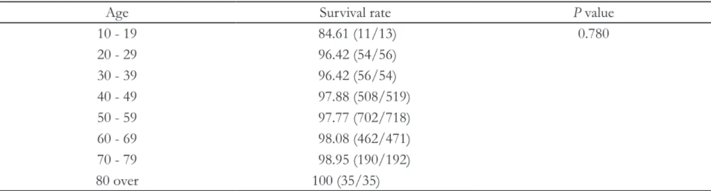
1. 성별 및 연령에 따른 임플란트 생존율
총 770명의 환자 중 남자는 386명, 여자는 384명이었 으며 남자 환자에서는 총 1135개를 식립하여 1101개가 생존한 97.00%의 생존율을, 여자 환자에서는 총 1023 개를 식립하여 1013개가 생존한 99.02%의 생존율을 보였다. 연령별로는 10대 84.61%, 20대 96.42%, 30대
Table 1. Classification of implant system and surface treatment
Implant system
Company Country Surface
A1 Osstem® Korea AO/RBM
A2 Osstem® Korea RBM
A3 Osstem® Korea RBM
A4 Osstem® Korea RBM
B1 Dentium® Korea AO/SLA
B2 Dentium® Korea SLA
C Dio® Korea RBM
D Friadent® Germany SLA
E Camlog® Germany SLA
F Friadent® Germany SLA
G1 Nobel biocare® Sweden AO
G2 Nobel biocare® Sweden AO
H Astra Tech® Sweden TiOblastTM
AO, anodizing oxidation; RBM, resorbable blast media; SLA, sandblasted large-grit acid-etched.
98.7%, 40대 97.88%, 50대 97.77%, 60대 98.08%, 70대 98.95%, 80대 이상 100%로 나타났으며 성별 및 연령에 따른 통계학적으로 유의한 차이는 없었다(Table 2, 3).
2. 임플란트 식립 위치에 따른 생존율
상악 전치부에서 98.23%, 상악 구치부에서 96.97%, 하악 전치부에서 97.85%, 하악 구치부에서 98.75%의 임플란트 생존율을 보였으며 이는 통계학적으로 유의할 만한 차이를 보였다(P < 0.05, Table 4).
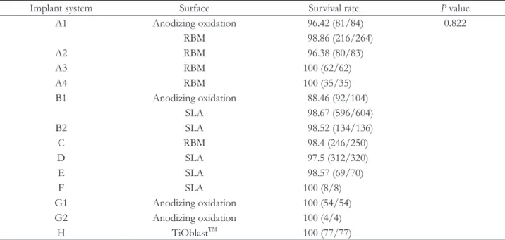
3. 임플란트 종류에 따른 생존율
양극 산화처리(anodizing oxidation)를 시행한 A1 제 품군에서 96.42%, RBM 표면처리를 시행한 A1 제품
군에서 98.86%, A2 제품군에서 96.38%, 양극산화처리 (anodizing oxidation)를 시행한 B1제품군에서 88.46%, SLA 표면처리를 시행한 B1 제품군에서 98.67%, B2 제 품군에서 98.52%, C 제품군에서 98.4%, D 제품군에 서 97.5%, E 제품군에서 98.57%의 생존율을 보였으며, A3, A4, F, G1, G2, H 제품군에서 모두 100%의 임플란 트의 생존율을 보였다. 그러나 임플란트의 종류 및 표면 처리에 따라 통계학적으로 유의한 차이를 보이지 않았 다(Table 5).
4. 임플란트 직경에 따른 생존율
임플란트의 직경이 3.0 mm 이하에서 100%, 3.0 - 3.5 mm 97.09%, 3.5 - 4.0 mm에서 98.19%, 4.0 - 4.5 mm에 서 98.29%, 4.5 - 5.0 mm에서 97.36%의 임플란트 생존
Table 2. Survival rate of implants according to gender (%, Number of survival implants/Number of total implants)
Sex Male Female Total P value
Survival rate 97.00 (1101/1135) 99.02 (1013/1023) 97.96 (2114/2158) 0.849
*Statistically significant difference (P < 0.05).
Table 3. Survival rate of implants according to age (%, Number of survival implants/Number of total implants)
Age Survival rate P value
10 - 19 84.61 (11/13) 0.780
20 - 29 96.42 (54/56)
30 - 39 96.42 (56/54)
40 - 49 97.88 (508/519)
50 - 59 97.77 (702/718)
60 - 69 98.08 (462/471)
70 - 79 98.95 (190/192)
80 over 100 (35/35)
*Statistically significant difference (P < 0.05).
Table 4. Survival rate of implants according to implant site (%, Number of survival implants/Number of total implants)
Site Mx. ant Mx. post Mn. ant Mn. post P value
Survival rate 98.23 (223/227) 96.97 (802/827) 97.85 (137/140) 98.75 (952/964) 0.024*
*Statistically significant difference (P < 0.05).
Table 6. Survival rate of implants according to diameter
of implant (%, Number of survival implants/Number of total implants)Diameter (mm) Survival rate P value
≤ 3.0 100 (12/12) 0.003*
3.0 - 3.5 97.09 (234/241) 3.5 - 4.0 98.19 (1253/1276) 4.0 - 4.5 98.29 (461/469) 4.5 - 5.0 97.36 (148/152) 5.0 > 75.0 (6/8)
*Statistically significant difference (P < 0.05).
Table 7. Survival rate of implants according to length of
implant (%, Number of survival implants/Number of total implants)Length (mm) Survival rate P value 7.0 - 9.0 96.26(129/134) 0.745 9.0 - 11.0 98.12(679/692)
11.0 - 13.0 98.17(967/985) 13.0 - 15.0 97.66(293/300) 15.0 ≥ 97.87(46/47)
*Statistically significant difference (P < 0.05).
율을 나타냈으며, 5.0 mm 초과 직경을 가진 넓은 폭경 의 임플란트는 8개를 식립한 것 중 2개가 실패하여 75%
의 낮은 생존율을 보였다. 직경에 따른 임플란트의 생존 율은 통계적학으로 유의할 만한 차이를 나타내었다(P <
0.05, Table 6).
5. 임플란트의 길이에 따른 생존율
임플란트의 길이별로 7 - 9 mm에서 96.26%, 9 - 11 mm에서 98.12%, 11 - 13 mm에서 98.17%, 13 - 15 mm 에서 97.66%, 15 mm 이상에서 97.87%의 임플란트 생
존율을 나타냈으며 이는 통계학적으로 유의할 만한 차 이는 없었다(Table 7).
6. 임플란트 식립 시 골 이식 여부에 따른 생존율
임플란트의 식립 시 상악동 골이식술을 단독으로 시 행한 경우 89.05%, 골유도재생술을 단독으로 시행한 경 우 98.28%, 상악동 골이식술과 골유도 재생술을 동시에 시행한 경우 98.34%, 골이식술을 시행하지 않은 경우에 는 99.28%의 생존율을 보였으며 이는 통계학적으로 유 의한 차이를 보였다(P < 0.05, Table 8).
Table 5. Survival rate of implants according to implant systems and surface characteristics (%, Number of survival
implants/Number of total implants)Implant system Surface Survival rate P value
A1 Anodizing oxidation 96.42 (81/84) 0.822
RBM 98.86 (216/264)
A2 RBM 96.38 (80/83)
A3 RBM 100 (62/62)
A4 RBM 100 (35/35)
B1 Anodizing oxidation 88.46 (92/104)
SLA 98.67 (596/604)
B2 SLA 98.52 (134/136)
C RBM 98.4 (246/250)
D SLA 97.5 (312/320)
E SLA 98.57 (69/70)
F SLA 100 (8/8)
G1 Anodizing oxidation 100 (54/54) G2 Anodizing oxidation 100 (4/4)
H TiOblastTM 100 (77/77)
*Statistically significant difference (P < 0.05).
RBM, resorbable blast media; SLA, sandblasted large-grit acid-etched.
고찰
임플란트 식립 후 2차 수술 전까지 기간 동안 임플란 트의 실패가 발생하는 원인은 다양하다.13-15 성공적인 골유착은 임플란트의 안정성을 향상시키며 이는 임플 란트 치료 시 중요한 요소이다.16 Schwartz-Arad 등17과 Goodacre 등18은 보철물 장착 전에 임플란트의 실패가 많이 발생한다고 보고하였고, Kim 등19은 초기 안정성 이 높을수록 임플란트의 생존율이 높으며 이는 통계학 적으로 유의한 차이를 보였다고 보고하였다. 일단 골유 착이 성공적으로 시행된 임플란트의 보철 치료 시 장기 적인 안정성을 가지며 이는 임플란트 식립 초기의 골유 착이 중요한 것으로 간주되었다.20,21
이번 연구의 목적은 임플란트 식립 이후 최종 보철물 을 시적하기 전까지의 초기 식립 기간의 임플란트 생존 율을 조사하고, 위험 변수와 임플란트 생존율 사이의 연 관성을 확인하기 위함이다. 이번 연구에서는 97.96%의 임플란트 생존율을 관찰하였으며, 주로 이차 수술 도중 에 또는 이차 수술 이후 보철물 제작 과정에서 발생하였 다. 이러한 결과는 1996년 Hass 등22이 1920개의 IMZ 임플란트에서 연구한 97.71%의 생존율과 거의 차이가 없으며, 1998년 Scrurria 등23이 보고한 94.7%의 식립 초 기 임플란트 생존율보다 높은 수치를 나타냈다.
이번 연구에서는 임플란트의 식립 부위, 임플란트 매 식체의 직경, 골 이식 여부 및 방법에 따라 임플란트 생 존율 간에 통계학적으로 유의한 차이가 있음을 확인하 였다. 식립 부위에 따른 임플란트의 생존율은 상악 전치 부에서 98.23%, 상악 구치부에서 96.97%, 하악 전치부 에서 97.85%, 하악 구치부에서 98.75%로 상악 구치부 에서 가장 낮게 나타났으며 이는 통계학적으로 유의한 차이를 보였다. 이러한 결과는 Hass 등22이 보고한 상악 95.9%, 하악 98.8%의 생존율, Kim 등24이 보고한 상악 96.4%, 하악 97.0%의 생존율 그리고 Park25 등이 보고한
상악 93.6%, 하악 96.3% 등 여러 문헌에서 보고된 결과 와 비슷하였다. 상악의 임플란트 생존율이 하악에 비해 낮게 보고되고 있는데 이는 부적당한 골질 및 골량과 관 련성이 있다. 부위에 따른 임플란트 생존율은 다수의 문 헌을 통해 상악이 하악보다, 구치부가 전치부보다 임플 란트 실패율이 높으며, 하악 전치부의 실패율이 가장 낮 다고 보고되었다.26
임플란트 직경에 따른 생존율을 관찰하였을 때, 3.0 - 3.5 mm의 임플란트에서 97.09%, 3.5 - 4.0 mm의 임 플란트에서 98.19%, 4.0 - 4.5 mm의 임플란트에서 98.29%의 생존율을 보였으며 이는 통계학적으로 유의 한 차이를 나타냈다. Ivanoff 등27은 임플란트의 직경 이 생존 및 실패와 관련성이 있다고 보고하였고, Jang 등28은 3.75 - 4.5 mm의 직경을 가진 임플란트 그룹에 서 높은 생존율을 관찰한다고 보고하였으며 이는 본 연 구의 결과와 비슷한 양상을 나타냈다. 그러나 Friberg 등29 은 3.75, 4.0, 5.0 mm 직경의 임플란트 실패율이 각각 5.5%, 3.9%, 4.5%로 나타난 것과 같이 임플란트의 직 경과 실패 간의 관련성이 없다고 보고하였으며 2013년 Mijiritsky 등30은 3년간 내원한 787명의 환자에서 식립 초기 2년간 생존율을 후향적으로 연구하였을 때 임플란 트 직경은 생존율에 대해 영향을 끼치는 통계학적으로 유의한 차이를 가지지 않는 등 다수의 문헌에서도 상반 된 결과가 보고되고 있다.
5.0 mm 이상의 넓은 직경을 가진 임플란트 식립 후 생존율은 75%로 가장 낮은 생존율을 나타냈다. 이는 주 로 넓은 직경의 임플란트가 임플란트가 실패한 부위에 서 재식립을 할 때 사용되거나, 발치 후 즉시 식립 또는 불량한 골질, 가용골이 부족한 부위, 이 외에 이갈이 등 의 악습관이 있는 환자 등 불리한 구강 내 환경에서 주 로 식립되는 경우가 많고 이는 높은 실패율을 나타내는 주요 원인이다.31,32 Shin 등33은 넓은 직경의 임플란트와 표준 임플란트를 각각 구치부에 식립 후 5년간 누적 성
Table 8. Survival rate of implants according to technique of bone graft (%, Number of survival implants/Number of total
implants)Technique Sinus graft GBR Sinus graft and GBR Non P value Survival rate 89.05 (122/137) 98.28 (1317/1340) 98.34 (119/121) 99.28 (556/560) 0.019*
*Statistically significant difference (P < 0.05).
GBR, guided bone regeneration.
공률을 조사하였을 때 각각 80.9%와 96.8%로 넓은 직 경의 임플란트에서 낮은 성공률을 보였으며 이는 임플 란트 식립 시 다량의 해면골이 제거되어 임플란트에 비 해 잔존 골량의 비율이 작아져 결정적으로 임플란트 실 패에 영향을 주어 넓은 직경의 임플란트 자체가 생존율 을 저해할 수 있다고 제시하였다. 따라서 넓은 직경의 임플란트 식립 후 골조직 재형성에 의한 후기 안정성을 얻기 위해 충분한 치유 기간을 가지는 것이 생존율을 향 상시키는데 있어 주요한 요소이다.34,35
본 연구에서는 임플란트 식립을 위한 골이식술 기법 에 따른 임플란트 생존율은 통계학적으로 유의한 차이 를 나타냈다. Becktor 등36과 Fugazzotto37은 골유도 재 생술에 의해 형성된 적절한 골에서 임플란트의 성공율 은 골유도 재생술의 여부에 따른 영향이 크지 않을 것이 라고 하였으며, Corinaldesi 등38은 titanium mesh를 동반 한 치조제 골이식술을 시행한 후 증대된 부위에 임플란 트를 식립하였을 때 장기간 임플란트의 실패없이 만족 스러운 결과를 관찰하였다고 보고하였다. 본 연구에서 는 상악동 골이식술을 단독으로 시행한 경우 89.05%의 낮은 생존율을 보였으나 골유도 재생술을 단독으로 시 행하였거나 상악동 골이식술과 동시에 치조골 결손부에 골유도 재생술을 시행한 부위, 골이식을 하지 않은 부위 에서의 생존율은 각각 98.28%, 98.34%와 99.28%로 통 계적으로 유의한 차이를 보이지 않았다.
상악동 골이식술 시 89.05%로 가장 낮은 임플란트 생 존율을 관찰하였으며 이는 주로 상악동 함기화 및 상악 치조골의 빠른 흡수로 인해 잔존골 고경이 낮은 부위에 서 상악동 골이식술이 시행되는 것과 관계가 있다.39,40 Jensen42은 술전 잔존 골의 고경을 각각 4 mm 이하, 5 - 6 mm, 7 mm 이상인 경우에 따라 임플란트 생존율을 평가하였을 때 각각 85.7%, 96.0%, 96.4%로 잔존골 고 경에 따른 생존율의 차이가 있다고 하였다. Toffler42와 Kim과 Lee43도 4 mm 이하의 잔존골 고경을 가지는 경 우 임플란트 생존율이 각각 73.3%와 69.8%로 보고하였 으며 이는 4 mm 이상의 잔존골 고경을 가진 군과 비교하 였을 때 통계학적으로 유의하게 낮은 생존율을 보였다.
상악동저 거상술은 크게 Boyne과 James44에 의해 보 고된 측방 개창법과 Summers45에 의해 보고된 치조정 접근법으로 나눌 수 있다. 1998년 Zitzmann과 Scharer46 는 상기 두 방법 모두 높은 임플란트 성공률을 보였으 며, 술전 상악 구치부 잔존골 고경 및 초기 고정력의 정 도에 따라 각각의 적응증에 맞게 술식을 선택해야 한다
고 제시하였다.
또한 골이식재의 종류에 따라 상악동 골이식술 부위 에 식립한 임플란트의 생존율에 대한 연구도 최근 이루 어졌다. Tong 등47은 상악동 골이식술 시 자가골 및 합성 골을 단독으로 사용하였을 때보다 여러 가지 골이식재 를 혼용하여 사용하였을 경우 더 높은 임플란트 생존율 을 보인다고 보고하였다.
본 연구에서는 상악동 거상술이 거의 대부분 측방 접 근법을 통해 동결 건조 동종골을 단독으로 또는 동결 건 조 동종골과 수산화인회석-합성인산칼슘 합성골을 혼 용하여 이식한 후 즉시 또는 지연된 임플란트 식립을 시 행하였으며, 수술 방법, 잔존골의 높이, 이식재의 종류 등 추가적인 조건에 따른 임플란트 생존율에 대한 부가 적인 연구가 필요할 것으로 사료된다.
결론
총 770명의 환자에서 2158개의 임플란트를 대상으로 임플란트 식립 후 최종 보철물을 시적하기까지의 평균 7.84개월의 기간 동안 관찰한 임플란트의 보철 전 생존 율에 관한 연구에서 다음과 같은 결론을 얻었다.
1. 2158개의 임플란트 중 2114개가 생존하여 총 97.96%의 생존율을 보였다.
2. 임플란트 식립 위치에 따라 상악 전치부에서는 98.23%, 상악 구치부에서는 96.97%, 하악 전치부 에서는 97.85%, 하악 구치부에서는 98.75%의 임플 란트 생존율을 보였다(P < 0.05).
3. 임플란트 매식체의 직경에 따라 3.0 mm 이하의 임 플란트에서 100%, 3.0 - 3.5 mm의 임플란트에서 97.09%, 3.5 - 4.0 mm의 임플란트에서 98.19%, 4.0 - 4.5 mm의 임플란트에서 98.29%의 생존율을 보 였으며, 5.0 mm 초과 직경을 가진 넓은 폭경의 임 플란트에서 75%의 비교적 낮은 생존율을 보였다 (P < 0.05).
4. 상악동 골이식술을 단독으로 시행한 부위에서 임 플란트 생존율은 89.05%, 골유도 재생술을 단독으 로 시행한 경우 98.28%, 상악동 골이식술과 골유도 재생술을 동시에 시행한 경우 98.34%, 골이식술이 동반되지 않은 경우 99.28%의 생존율을 보였다(P
< 0.05).
본 연구에서 최종적인 총 임플란트 생존율은 97.96%
로 조사되었으며, 임플란트의 식립 위치, 임플란트 고정
체의 직경, 골이식 여부 및 방법과 같은 조건들이 임플 란트 보철 전 초기 생존율에 영향을 주는 요인으로 나 타났다. 또한 각각 상악 구치부, 5.0mm 이상의 넓은 임 플란트, 그리고 상악동 골이식술을 단독으로 시행한 부 위에서 현저하게 낮은 보철 전 초기 생존율을 확인할 수 있었다.
Acknowledgements
이 논문은 2013년 원광대학교의 교비 지원에 의해서 수행됨.
References
1. Brånemark PI, Adell R, Breine U, Hansson BO, Lindström J, Ohlsson A. Intra-osseous anchorage of dental prostheses. I. Experimental studies. Scand J Plast Reconstr Surg 1969;3:81-100.
2. Bergman B. Evaluation of the results of treat- ment with osseointegrated implants by the Swedish National Board of Health and Welfare. J Prosthet Dent 1983;50:114-5.
3. Brånemark PI, Hansson BO, Adell R, Breine U, Lindström J, Hallén O, Ohman A. Osseointegrated implants in the treatment of the edentulous jaw.
Experience from a 10-year period. Scand J Plast Reconstr Surg Suppl 1977;16:1-132.
4. Adell R, Lekholm U, Rockler B, Brånemark PI. A 15-year study of osseointegrated implants in the treatment of the edentulous jaw. Int J Oral Surg 1981;10:387-416.
5. Kim YS, Lee DK, Min SK, Lee J, Moon C. Clini- cal study on success rate of osseointegrated dental implants. J Korean Assoc Maxillofac Plast Reconstr Surg 2002;24:137-47.
6. Esposito M, Grusovin MG, Coulthard P, Thomsen P, Worthington HV. A 5-year follow-up compara- tive analysis of the efficacy of various osseointe- grated dental implant systems: a systematic review of randomized controlled clinical trials. Int J Oral Maxillofac Implants 2005;20:557-68.
7. Hong JY, Chae GJ, Jung UW, Kim CS, Cho KS, Chae JK, Kim CK, Choi SH. Retrospective studies of dental implant placement at each intraoral site
and situation. J Korean Acad Periodontol 2007;37:
805-24.
8. Albrektsson T, Brånemark PI, Hansson HA, Lind- ström J. Osseointegrated titanium implants. Re- quirements for ensuring a long-lasting, direct bone- to-implant anchorage in man. Acta Orthop Scand 1981;52:155-70.
9. van Steenberghe D, Quirynen M, Naert I. Survival and success rates with oral endosseous implants. In:
Lang NP, Karring T, Lindhe J editors. Proceedings of the 3rd European Workshop on Periodontology.
Berlin; Quintessence Publishing Co.; 1999. p. 242- 54.
10. Misch CE, Perel ML, Wang HL, Sammartino G, Galindo-Moreno P, Trisi P, Steigmann M, Rebaudi A, Palti A, Pikos MA, Schwartz-Arad D, Chouk- roun J, Gutierrez-Perez JL, Marenzi G, Valavanis DK. Implant success, survival, and failure: the International Congress of Oral Implantologists (ICOI) Pisa Consensus Conference. Implant Dent 2008;17:5-15.
11. Zarb GA, Schmitt A. The longitudinal clinical ef- fectiveness of osseointegrated dental implants: the Toronto study. Part III: problems and complica- tions encountered. J Prosthet Dent 1990;64:185-94.
12. Esposito M, Hirsch JM, Lekholm U, Thomsen P.
Biological factors contributing to failures of os- seointegrated oral implants. (I). Success criteria and epidemiology. Eur J Oral Sci 1998;106: 527-51.
13. Sussman HI. Endodontic pathology leading to im- plant failure: a case report. J Oral Implantol 1997;
23:112-15.
14. el Askary AS, Meffert RM, Griffin T. Why do den- tal implants fail? Part I. Implant Dent 1999;8:173- 85.
15. Piattelli A, Scarano A, Piattelli M, Podda G. Im- plant periapical lesions: clinical, histologic, and his- tochemical aspects. A case report. Int J Periodon- tics Restorative Dent 1998;18:181-7.
16. Lioubavina-Hack N, Lang NP, Karring T. Signifi- cance of primary stability for osseointegration of dental implants. Clin Oral Implants Res 2006;17:
244-50.
17. Schwartz-Arad D, Laviv A, Levin L. Failure causes, timing, and cluster behavior: an 8-year study of
dental implants. Implant Dent 2008;17:200-7.
18. Goodacre CJ, Kan JY, Rungcharassaeng K. Clinical complications of osseointegrated implants. J Pros- thet Dent 1999;81:537-52.
19. Kim S, Kim SJ, Lee KW, Han DH. The effects of local factors on the survival of dental implants: a 19 year retrospective study. J Korean Acad Prostho- dont 2010;48:28-40.
20. Lazzara RJ, Testori T, Trisi P, Porter SS, Weinstein RL. A human histologic analysis of osseotite and machined surfaces using implants with two oppos- ing surfaces. Int J Periodontics Restorative Dent 1999;19:117-29.
21. Davies JE. Mechanism of endosseous integration.
Int J Posthodont 1998;11:391-401.
22. Haas R, Mensdorff-Pouilly N, Mailath G, Watzek G.
Survival of 1920 IMZ implants followed for up to 100 months. Int J Oral Maxillofac Implants 1996;
11:581-8.
23. Scurria MS, Morgan ZV 4th, Guckes AD, Li S, Koch G. Prognostic variables associated with im- plant failure: a retrospective effectiveness study. In J Oral Maxillofac Implants 1998;13:400-6.
24. Kim JS, Chang HH, Chang CH, Rhyu SH, Kang JH. Preprosthetic stage dental implant failure. J Ko- rean Assoc Oral Maxillofac Surg 2001;27:178-83.
25. Park KA, Jeong CW, Ryoo GH, Park KB, Kim YJ.
Retrospective study of wide-diameter implants in maxillary & mandibular molar regions. J Korean Acad Periodontol 2007;37:825-38.
26. Schwartz-Arad D, Laviv A, Levin L. Failure causes, timing, and cluster behavior: an 8-year study of dental implants. Implant Dent 2008;17:200-7.
27. Ivanoff CJ, Gröendahl K, Sennerby L, Bergström C, Lekholm U. Influence of variation in implant diam- eters: a 3- to 5-year retrospective clinical report. Int J Oral Maxillofac Implants 1999;14:173-80.
28. Jang JW, Ryoo KH, Chung HJ. Survival analysis of dental implants in maxillary and mandibular molar regions; a 4-5 year report. J Korean Acad Periodon- tol 2007;37:165-80.
29. Friberg B, Ekestubbe A, Sennerby L. Clinical out- come of Brånemark system implants of various di- ameters: a retrospective study. Int J Oral Maxillofac Implants 2002;17:671-7.
30. Mijiritsky E, Mazor Z, Lorean A, Levin L. Implant diameter and length influence on survival: interim results during the first 2 years of function of im- plants by a single manufacturer. Implant Dent 2013;22:394-8.
31. Langer B, Langer L, Herrmann I, Jorneus L. The wide fixture: a solution for special bone situations and a rescue for the compromised implant. Part 1.
Int J Oral Maxillofac Implants 1993;8:400-8.
32. Renouard F, Nisand D. Impact of implant length and diameter on survival rates. Clin Oral Implants Res 2006;17 Suppl 2:35-51.
33. Shin SW, Bryant SR, Zarb GA. A retrospective study on the treatment outcome of wide-bodied implants. Int J Prosthodont 2004;17:52-8.
34. Anner R, Better H, Chaushu G. The clinical effec- tiveness of 6 mm diameter implants. J Periodontol 2005;76:1013-5.
35. Krennmair G, Waldenberger O. Clinical analysis of wide-diameter frialit-2 implants. Int J Oral Maxil- lofac Implants 2004;19:710-5.
36. Becktor JP, Isaksson S, Sennerby L. Survival analy- sis of endosseous implants in grafted and non- grafted edentulous maxillae. Int J Oral Maxillofac Implants 2004;19:107-15.
37. Fugazzotto PA. Success and failure rates of os- seointegrated implants in function in regenerated bone for 72 to 133 months. Int J Oral Maxillofac Implants 2005;20:77-83.
38. Corinaldesi G, Pieri F, Sapigni L, Marchetti C. Eval- uation of survival and success rates of dental im- plants placed at the time of or after alveolar ridge augmentation with an autogenous mandibular bone graft and titanium mesh: a 3- to 8-year retrospective study. Int J Oral Maxillofac Implants 2009;24:1119- 28.
39. Mish CE. Bone character: second vital implant cri- terion. Dent Today 1998;7:39-40.
40. Friberg B, Sennerby L, Roos J, Lekholm U. Identifi- cation of bone quality in conjunction with insertion of titanium implants. A pilot study in jaw autopsy specimens. Clin Oral Impants Res 1995;6:213-9.
41. Jensen OT. The sinus bone graft. 2nd ed. Chicago;
Quintessence Publishing Co.; 2006. p. 178-81.
42. Toffler M. Minimally invasive sinus floor elevation
procedures for simultaneous and staged implant placement. N Y State Dent J 2004;70:38-44.
43. Kim BJ, Lee JH. The retrospective study of surviv- al rate of implants with maxillary sinus floor eleva- tion. J Korean Assoc Oral Maxillofac Surg 2010;36:
108-18.
44. Boyne PJ. James RA. Grafting of the maxillary sinus floor with autogenous marrow and bone. J Oral Surg 1980;38:613-6.
45. Summers RB. A new concept in maxillary implant surgery: the osteotome technique. Compendium
1994;15:152, 154-6, 158 passim; quiz 162.
46. Zitzmann NU, Schärer P. Sinus elevation proce- dures in the resorbed posterior maxilla. Compari- son of the crestal and lateral approaches. Oral Surg Oral Med Oral Pathol Oral Radiol Endod 1998;85:
8-17.
47. Tong DC, Rioux K, Drangsholt M, Beirne OR.
A review of survival rates for implants placed in grafted maxillary sinuses using meta-analysis. Int J Oral Maxillofac Implants 1998;13:175-82.
*교신저자: 지영덕
(435-040) 경기도 군포시 산본동 1142 원광대학교 치과대학 산본치과병원 구강악안면외과 Tel: 031-390-2875|Fax: 031-390-2777|E-mail: [email protected]
접수일: 2014년 8월 15일|수정일: 2014년 11월 11일|채택일: 2014년 11월 14일
원광대학교 산본치과병원에서 770명의 환자에 식립한 2158개의 골유착성 임플란 트의 보철 전 초기 생존율에 관한 후향적 연구
선화경, 지영덕*
원광대학교 치과대학 산본치과병원 구강악안면외과
목적: 이 연구의 목적은 상악 및 하악의 완전 무치악 및 부분 무치악 부위에 식립된 골유착성 임플란트의 보철 전 초기 생존율 및 생존과 관련된 요소를 평가하고 실패를 야기하는 요인에 대해 알아보는 것이다.
연구 재료 및 방법: 2004년부터 2013년까지 770명의 환자에서 총 2158개의 골내 임플란트를 식립하였다. 임플란트의 소실과 환자의 연령 및 성별, 식립 위치, 임플란트 시스템, 길이 및 직경, 그리고 골이식 방법과의 연관성을 평가하기 위 해 임상적 비교를 시행하였다.
결과: 임플란트 식립 위치에 따라 상악 전치부에서 98.23%, 상악 구치부 96.97%, 하악 전치부 97.85%, 하악 구치부 98.75%의 임플란트 생존율을 보였다(P < 0.05). 임플란트의 종류 및 표면처리 기법은 임플란트 생존율과 특이성을 보 이지 않았다. 임플란트 매식체의 직경에 따라 3.0 mm 이하의 임플란트에서 100%, 3.0 - 3.5 mm 97.09%, 3.5 - 4.0 mm 98.19%, 4.0 - 4.5 mm 98.29%의 생존율을 보였고, 5.0 mm 이상의 직경을 가진 넓은 폭경의 임플란트에서 75%의 비교 적 낮은 생존율을 보였다(P < 0.05). 임플란트 매식체의 길이에 따라 9 - 11 mm 및 11 - 13 mm의 길이를 가진 임플란 트에서 각각 98.12% 및 98.17%의 높은 생존율을 보였으나 통계학적 유의성을 가지진 못하였다. 골이식술의 방법에 따 라 상악동 골이식술을 단독으로 시행한 부위에서 임플란트 생존율은 89.05%, 골유도 재생술을 단독으로 시행한 경우 98.28%, 상악동 골이식술과 골유도 재생술을 동시에 시행한 경우 98.34%, 골이식술이 동반되지 않은 경우 99.28%의 생존율을 보였다(P < 0.05).
결론: 이번 연구에서 임플란트 식립 후 최종 보철물 시적 전 초기단계에서의 임플란트 생존율은 임플란트의 식립위치, 임플란트 매식체의 직경, 그리고 골이식 방법과 관련이 있었다. 이러한 결과에 따라 각각 상악 구치부, 5.0 mm 이상의 넓은 임플란트, 그리고 상악동 골이식술을 단독으로 시행한 부위에서 현저하게 낮은 생존율을 확인할 수 있었다.
(구강회복응용과학지 2014;30(4):278-88)
주요어: 치과 임플란트; 골유착; 임플란트 생존율